Transcription
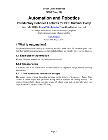
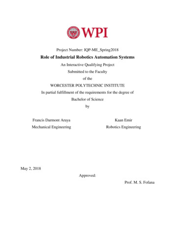
Proteomics 2006, 6, 0000–00001DOI 10.1002/pmic.200600060REVIEWAutomation, parallelism, and robotics for proteomicsGil Alterovitz1, 2, 3, Jonathan Liu4, Jijun Chow4 and Marco F. Ramoni1, 2, 31Division of Health Sciences and Technology (HST), Harvard Medical School and MassachusettsInstitute of Technology, Boston, MA, USA2Children’s Hospital Informatics Program at HST, Boston, MA, USA3Harvard Partners Center for Genetics and Genomics, Harvard Medical School, Boston, MA, USA4Department of Biology, Massachusetts Institute of Technology, Cambridge, MA, USAThe speed of the human genome project (Lander, E. S., Linton, L. M., Birren, B., Nusbaum, C.et al., Nature 2001, 409, 860–921) was made possible, in part, by developments in automation ofsequencing technologies. Before these technologies, sequencing was a laborious, expensive, andpersonnel-intensive task. Similarly, automation and robotics are changing the field of proteomicstoday. Proteomics is defined as the effort to understand and characterize proteins in the categories of structure, function and interaction (Englbrecht, C. C., Facius, A., Comb. Chem. HighThroughput Screen. 2005, 8, 705–715). As such, this field nicely lends itself to automation technologies since these methods often require large economies of scale in order to achieve cost andtime-saving benefits. This article describes some of the technologies and methods being appliedin proteomics in order to facilitate automation within the field as well as in linking proteomicsbased information with other related research areas.Received: January 24, 2006Revised: April 6, 2006Accepted: April 7, 2006Keywords:Multiplexing / Proteomics methods / Protein chips1Proteomics and automationRobotics and intelligent systems technologies can help overcome technological hurdles such as speed, cost, and precision in large-scale biological endeavors. High-throughputautomation is also useful to capture higher quality snapshotsof cellular activity. Researchers are becoming more reliant onmodern automated systems and robotics to find therapeutictargets in the proteome. Robotics increases laboratory efficiency by reducing contamination and human error (seeFig. 1). It also can help in making sense of raw data, allowingresearchers to concentrate on other tasks [1, 2]. The need forapplying automation in proteomics is increasing daily as largCorrespondence: Dr. Gil Alterovitz, Bioinformatics Core, HarvardMedical School, New Research Building, Room 250, 77 Ave LouisPasteur, Boston, MA 02115, USAE-mail: ga@alum.mit.eduFax: 11-617-525-4488Figure 1. Automated robot used to mount and align proteincrystals at Berkeley Lab Advanced Light Source. Reproduced,with permission, from Thomas Earnest (LBL) [40].Abbreviations: CD, compact disk; LIMS, Laboratory InformationManagement Systems; SPR, surface plasmon resonance; Y2H,yeast two-hybrider and more complex proteome datasets are being generated.For example, innovation among drug companies has progressed to the point where automated methods of targeting 2006 WILEY-VCH Verlag GmbH & Co. KGaA, Weinheimwww.proteomics-journal.com
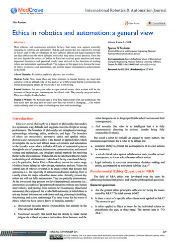
2G. Alterovitz et al.and screening have replaced traditional, manual techniques[1]. Moreover, in order to make whole-organism proteomebased experiments a reality, machine learning techniques,such as predicting the functional implications of certain posttranslational alteration, are becoming an indispensable partof the research [1]. Thus, automation is permeating into manyareas of laboratory research: from assays to statistical analysis.This article focuses on automated technologies in thethree areas of proteomics research: (i) 2-DE and MS, uses traditional tools in new applications, providing comprehensiveanalysis of proteins; (ii) array-based proteomics builds uponthe same concept behind the widely used cDNA microarraytechnology in genomic research [3]; (iii) protein structure andimaging, uses information about 3-D structure and interaction to provide a complete picture of protein character [4].These technologies are currently being applied to studycellular processes and regulations in high-throughput formats. Unfortunately, many current automation schemesgenerate large quantities of unwieldy data. Researchers aretherefore relying on Laboratory Information ManagementSystems (LIMS) to assist them with the extraction of usefulinformation [5–7].Proteomics 2006, 6, 0000–0000Improvements have been made since the first generations of LIMS used in genomics research. Newer incarnations of LIMS have attracted considerable commercial interest such as Decodon with its Protecs and Bruker Daltronicswith its ProteinScape database. Genologics, with its ProteusLIMS system is comprised of four capabilities: lab management, instrument and data integration, bioinformaticsand data management, and analytics and reporting (seeFig. 2). A comprehensive list of available current LIMSpackages may be found at The LIMSource (www.limsource.com). LIMS optimization is ongoing, as evidenced by Modas,a current project under development by nonlinear dynamics.It supports 2-DE and MS, integrating analysis with LIMSinto one package. The LIMS software will be key to beingable to take full advantage of automation advancements.2Automated gel electrophoresis and MS2-DE is used for the characterization and separation of protein samples on the basis of charge and molecular weight. Itsapplications have included investigating protein quantityFigure 2. Four functional areas of the ProteusLIMS system illustrating the typical proteomics workflow. Reproduced, with permission from GenoLogics. 2006 WILEY-VCH Verlag GmbH & Co. KGaA, Weinheimwww.proteomics-journal.com
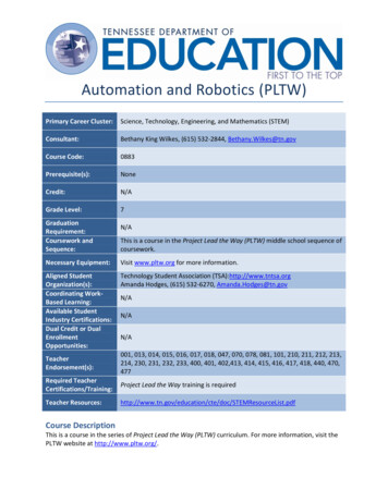
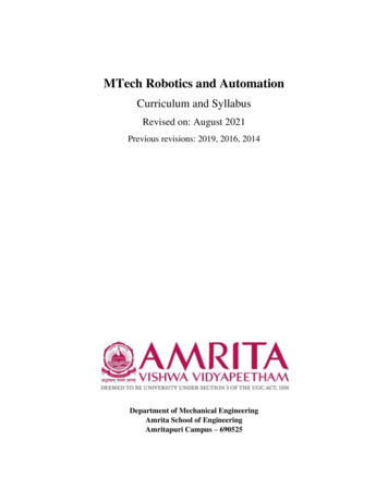
TechnologyProteomics 2006, 6, 0000–0000and character, identifying PTMs and generating proteomemaps [8]. While 2-DE is extremely useful, it suffers fromseveral technical limitations. These include the manualhandling of gels. In addition, 2-DE is time consuming. It cantake days to run and analyze a single gel [9].The quest for high-throughput formats has becomeimportant for protein characterization. One of the developments in automated production of 2-DE has been marketedby commercial entities such as NextGen Sciences and LargeScale Biology. The a2DEoptimizer by NextGen Sciencesfeatures automated gel casting, saving money for core facilities that previously bought precast gels. It can run IEF atup to 10 000 V, cast multiple gels simultaneously, and iscompletely controlled and monitored by computer. NextGenSciences claims that this improves 2-DE by increasing reproducibility and enhancing resolution of protein separations (see Fig. 3). It also has the ability to create user-definedgradient gels. Such gradients can be difficult to createmanually.Large Scale Biology, under their subsidiary, PredictiveDiagnostics, has released BAMF (Biomarker AmplificationFilter), a computer platform combining 2-DE, NMR, MS,3and biomarkers to identify individual proteins. According tothe company, BAMF successfully diagnosed 100% of ovariancancer patients using a dataset from the National CancerInstitute.There are several features that are commonly offered bymany of the newer automated gel processing systemsincluding the ability to: (i) import and export gels intostandard bit-mapped graphics formats; (ii) manipulate, preprocess, filter, and organize gel bitmaps; (iii) visualize andcompare gels; (iv) create, queue, and monitor computationalanalysis tasks; and (v) present results (e.g., peptide matchesin an excised, digested protein spot) [10].A new generation of 2-DE image analysis packages isfeaturing new flexibility and automation. These systemsinclude Progenesis by Nonlinear Dynamics, Decyder fromGE Healthcare, Delta2D from Decodon among many others.To overcome the calculation-intensive process of imageanalysis of 2-DE gels, Dowsey et al. [10] introduced Gridenabled cluster computing and the proTurbo framework inthe newly updated ProteomeGRID bioinformatics pipeline(see Fig. 4). ProteomeGRID, a high-throughput 2-DE imageanalysis computing platform now utilizes a gel-matchingFigure 3. Using the a2DEoptimizer, these images show the effectof a suitable acrylamide gradienton the resolution of proteins inthe second dimension of separation. Note that with this particular gradient, the small molecularweight proteins are betterresolved than in homogenousgel. This enables enhanced spotdetection and identificationwhen processed by MS. Reproduced, with permission, fromNextGen Sciences.Figure 4. A schematic illustration of the ProteomeGRID pipeline. ProteomeGRID is a high-throughput computingplatform designed for image analysis of 2-DE. Reproduced, with permission from [5]. 2006 WILEY-VCH Verlag GmbH & Co. KGaA, Weinheimwww.proteomics-journal.com
4G. Alterovitz et al.algorithm to overcome the bottleneck of spot matching.Researchers are currently working on implementing expression quantification as well.An important feature common to automated approachesis the use of standards for interoperability. For example,ProteomeGRID was designed to follow the HUPO Proteomics Standards Initiative General Proteomics Standards(HUPO PSI GPS) ontology for image mining [10].MS and sample preparation are undergoing automationas well. SELDI MS allows parallel processing by using several spots at once on arrays [11]. Ciphergen has manufactured a platform using SELDI technology to identify protein biomarkers. With integrated hardware, software andarrays, the ProteinChip System Series 4000 boasts fourmodes including biomarker discovery, purification, identification, and biomarker assays. Research in this area hasshown that integrating software and robotics-based hardwareinto a pipeline can yield benefits in terms of increased signalquality, reduced labor time, and lower costs [12].By using sample volumes on the order of micro- ornanoliters and capillary force, technologies such as MALDIand peptide sequencing are shrinking to the size of a compact disk (CD). These CDs can processes 96 protein samplesin parallel [13]. An example of this technology is the MALDISP1 CD Microlaboratory made by Gyrolab. The SP1 processes up to 96 protein digests at once, detecting peptideswith as low abundance as a femtomole. It offers parallelprocessing and automation at the nanoliter scale, improvingreproducibility, and reducing sample loss.Researchers at Cranfield University have developed theGenome Annotating Proteomic Pipeline (GAPP), currentlyin its beta release [14]. It is a scalable, grid-based pipelinedesigned to analyze MS data automatically. Linking users toa virtual database allows them to access GAPP’s supercomputers to do time-consuming computations over asecure connection.As automation and miniaturization develops, fully automated protein analysis may be available on a single chip [1].3Array-based proteomicsThe term protein microarray [15–18] can have many types ofinstantiations. The core, common idea is that operationsoccur in parallel and are often miniaturized. Protein arraystypically operate by using immobilized proteins on surfacessuch as glass, membranes, and beads (or other particles).The surface chemistry is designed to fix the proteins in place(see Fig. 5). Binding molecules are exposed to the array andseek out their unique target proteins. The binding of a targetprotein with a binding molecule is signified by a detectionsystem. Their advantages include speed, high specificity, andthe great amount of information derived from one test.Protein microarray technology can be broken into severalcategories including functional arrays, detection arrays, andRP arrays [19]. Functional arrays can be used to study protein 2006 WILEY-VCH Verlag GmbH & Co. KGaA, WeinheimProteomics 2006, 6, 0000–0000Figure 5. Protein array detection system – protein abundance ismeasured using labeled markers binding specifically to their substrate. This technique is the backbone of microarray technology.properties and interactions [20]. This tool can be used topredict functions based on coexpression after analyzingsmall molecule and drug-binding properties. In creatingchips, individual ligands (peptides, antibodies, etc.) are spotted onto a surface. Protein expression is then differentiallyanalyzed via biochemical interactions, providing potential,functional, and binding-based information [4, 19].Detection arrays operate in an almost opposite manner.In these, antigens or antibodies are immobilized to a surfaceand proteins are exposed to them. This technique is usefulwhen assaying antibodies and monitoring protein expression [19]. Commercial applications of antibody arrays includethe Whatman FAST Quant system. Each kit contains64 arrays of eight to ten mAb with affinities to human ormouse cytokines. It features parallel processing, performingover 500 measurements from 56 samples. Furthermore,quantitative analysis, using ArrayVision FAST can be performed minutes after scanning.The Panorama Ab Microarray Cell Signaling Kit bySigma-Aldrich features 224 different antibodies spotted onto468 glass slides (see Fig. 6). By minimizing the backgroundnoise, the kit can analyze and detect minute quantities ofprotein – as low as a few nanograms per milliliter. In lessthan 5 hr, the relative abundance of several hundred proteinsmay be determined with less than 10% variation in spotmorphology.BD Biosciences BD Lyoplate Technology allows platedesign flexibility. This is a custom service that takes userdefined specifications and prepares specialized plates thatcontain antibody and other reagents in a GMP environment.In RP arrays [21], cell lysates are immobilized to a surfaceand probed with antibodies, resulting in a multiplexed output [19]. This also allows the analysis of modified proteinsand is often used in profiling cancer [22].New developments in protein chip format and methodology has resulted in the commercially available ProteinChiptechnology from Ciphergen (it is also considered an MSwww.proteomics-journal.com
TechnologyProteomics 2006, 6, 0000–00004Figure 6. Protein expression differences within mouse tissuesusing Panorama Ab Microarray Cell Signaling Kit. One microgram of each protein extract from different tissues was labeledwith Cy3 and incubated on separate arrays. Reproduced, withpermission, from Sigma-Aldrich.automation technology and was noted in Section 2 of thisreview). ProteinChip is a parallelized approach that can provideinformation on protein structure, character, and PTMs [23]. Ithas been widely used for clinical biomarker discovery [24].Using microfluidics technology, chips can be etched withmicroscopic channels in which miniature assays are performed [2, 25]. Caliper Technologies has developed one suchdevice called the LabChip 3000 Drug Discovery System.The LabChip 3000 includes many functions and has beenmost commonly used for kinase profiling [26]. For channels10–50 mm in diameter used in the present study, less reagentwas required and fewer sample cells were necessary.The effects of protein microarrays have been seen acrossseveral fields such as medicine and pharmacology. In future,blood tests may be performed by providing fewer drops ofblood onto a chip with specific protein markers, providingvaluable diagnostic and real-time prognostic information[20]. Advantages of the protein microarray approachesinclude the ability to use small samples and built-in experimental controls for calibration.Another avenue of protein identification and screening isyeast two-hybrid (Y2H) [27]. Y2H involves the measurementof physical interactions. Using an easily differentiablereporter gene, hybrid transcription factors are made. If thetwo hybrid transcription factors react, the reporter gene istranscribed and the cell will thus phenotypically indicate theinteraction. Advantages of Y2H include its ability to be donein vivo and ease with which parallelization can be implemented to test many interaction combinations quickly.The tandem affinity purification (TAP) [28] method usesa TAP-tag to target specific proteins. The proteins are thenpurified and further analyzed via SDS-PAGE, MS, and functional assays. These interactions must be completed, for themost part, in vitro and are time consuming, requiring theaddition of many reagents. There is, however, much room foroptimization. 2006 WILEY-VCH Verlag GmbH & Co. KGaA, Weinheim5Structure and imagingThe determination of protein 3-D quaternary structures hasgreatly increased in the past few years. Analysis of the largenumber of proteins requires new high-throughput and multiplexed techniques [29]. Automated systems can helpincrease reliability, objectivity, reproducibility, and repeatability.The most common techniques for structural analysisinclude NMR, X-ray crystallography, structure predictionmethodology, and even MS to a certain degree [30]. Thesetechniques allow researchers to determine or constrain thepotential 3-D structure of proteins and protein subunits.Imaging proteomics can also detect protein–proteininteractions and protein localization. Techniques includetransfected cell arrays, green fluorescent proteinbased (GFP) labeling, and fluorescence resonance energytransfer (FRET) [31, 32]. FRET is used to detect distance-dependent interactions by using an energized fluorophore thatcan be transferred to an acceptor less than 100 Å away [33].Multiplexed surface plasmon resonance (SPR) [34, 35] isanother automated approach to the quantitative analysis ofprotein interactions. SPR is a technique that studies bioaffinity on gold and noble metal thin films. It can provide information on concentrations in a solution several hundrednanometers above the film by detecting changes in opticalproperties that result when proteins in the solution interact[36]. Advantages of SPR include its low target consumptionand freedom from radioactive labeling [37].VEGA ZZ, a molecular modeling software package, isdesigned for use by researchers analyzing protein structures.It has an extensive list of features including multiple fileformat support, atomic potential attribution, 3-D moleculareditor, and a protein–protein docking system. Figure 7 showsthe M1 muscarinic receptor–acetylcholine complex (MEPsurface and tube) as explored with VEGA [38].Despite great improvements in the automation of structural proteomics, there remain numerous challenges in thefield. For example, when analyzing proteins via X-ray diffraction, proteins must be able to crystallize. This process islargely based on trial and error – as many factors such as pHand salt concentration are involved [39]. Commercial solutions have been developed, including the Index productfrom Hampton Research, which can screen for crystallization based on factors such as pH, salt, and ionic strength.New techniques in automation and related technologies arethus not only facilitating experiments in terms of speed, butalso making a new type of large-scale proteomics researchpossible.5ConclusionProteomics is leveraging some of the automated methodologies developed for other fields such as functional genomics.However, some challenges that are new and proteomicswww.proteomics-journal.com
6G. Alterovitz et al.Proteomics 2006, 6, 0000–0000Figure 7. M1 muscarinic receptor–acetylcholine complex asexploredwithVEGA.Anenlarged view of the boxedinteraction to the right. Reproduced, with permission, fromAlessandro Pedretti.specific (such as post-transitional modifications) may benefitfrom custom-designed solutions. In addition, automation infuture may be able to take advantage of the synergisticdevelopments in other fields – by linking to that data. Thus,one area for future work involves the design of interfaces forintegration of data from many heterogeneous sources in anautomated manner. In future, automated and parallelizedpipeline solutions that integrate genomic, proteomic, andrelational information (e.g., networks) may be used in biology for discoveries that would not have been possible throughisolated, manual analysis.6References[1] Alterovitz, G., Afkhami, E., Ramoni, M., in: Liu, J. X. (Ed.), NewDevelopments in Robotics Research, Nova Science Publishers, New York, NY 2005, pp. 217–252.[2] Chapman, T., Nature 2003, 422, 665–666.[3] Kohane, I. S., Kho, A. T., Butte, A. J., Microarrays for an Integrative Genomics, MIT Press, Cambridge 2002.[4] de Hoog, C. L., Mann, M., Annu. Rev. Genomics Hum. Genet.2004, 5, 267–293.[5] Amin, A. A., Faux, N. G., Fenalti, G., Williams, G. et al., Proteins 2006, 62, 4–7.[6] Goh, C. S., Lan, N., Echols, N., Douglas, S. M. et al., NucleicAcids Res. 2003, 31, 2833–2838.[10] Dowsey, A. W., Dunn, M. J., Yang, G. Z., Proteomics 2004, 4,3800–3812.[11] Simpkins, F., Czechowicz, J. A., Liotta, L., Kohn, E. C., Pharmacogenomics 2005, 6, 647–653.[12] Alterovitz, G., Aivado, M., Spentzos, D., Libermann, T. A. etal., Proceedings of the International Conference of IEEEEngineering in Medicine and Biology, San Francisco, CA,USA 2004.[13] Gustafsson, M., Hirschberg, D., Palmberg, C., Jornvall, H.,Bergman, T., Anal. Chem. 2004, 76, 345–350.[14] Shadforth, I., Crowther, D., Bessant, C., Proteomics 2005, 5,4082–4095.[15] Dupuy, A. M., Lehmann, S., Cristol, J. P., Clin. Chem. Lab.Med. 2005, 43, 1291–1302.[16] Clarke, W., Chan, D. W., Clin. Chem. Lab. Med. 2005, 43,1279–1280.[17] Bertone, P., Snyder, M., Febs. J. 2005, 272, 5400–5411.[18] MacBeath, G., Schreiber, S. L., Science 2000, 289, 1760–1763.[19] Cretich, M., Damin, F., Pirri, G., Chiari, M., Biomol. Eng. 2005,23, 77–88.[20] Xu, Q., Lam, K. S., J. Biomed. Biotechnol. 2003, 2003, 257–266.[21] Speer, R., Wulfkuhle, J. D., Liotta, L. A., Petricoin, E. F., IIIrd,Curr. Opin. Mol. Ther. 2005, 7, 240–245.[7] Turner, E., Bolton, J., Qual. Assur. 2001, 9, 217–224.[22] Kreutzberger, J., Appl. Microbiol. Biotechnol. 2006, 70, 383–390.[8] Gerdes, S. Y., Scholle, M. D., Campbell, J. W., Balazsi, G. et al.,J. Bacteriol. 2003, 185, 5673–5684.[23] Merchant, M., Weinberger, S. R., Electrophoresis 2000, 21,1164–1177.[9] Hille, J. M., Freed, A. L., Watzig, H., Electrophoresis 2001, 22,4035–4052.[24] Issaq, H. J., Conrads, T. P., Prieto, D. A., Tirumalai, R., Veenstra, T. D., Anal. Chem. 2003, 75, 148–155. 2006 WILEY-VCH Verlag GmbH & Co. KGaA, Weinheimwww.proteomics-journal.com
Proteomics 2006, 6, 0000–0000Technology7[25] Dittrich, P. S., Manz, A., Nat. Rev. Drug Discov. 2006, 5, 210–218.[33] Phizicky, E., Bastiaens, P. I., Zhu, H., Snyder, M., Fields, S.,Nature 2003, 422, 208–215.[26] Schutkowski, M., Reineke, U., Reimer, U., Chembiochem.2005, 6, 513–521.[34] Unfricht, D. W., Colpitts, S. L., Fernandez, S. M., Lynes, M. A.,Proteomics 2005, 5, 4432–4442.[27] Uetz, P., Giot, L., Cagney, G., Mansfield, T. A. et al., Nature2000, 403, 623–627.[35] Besenicar, M., Macek, P., Lakey, J. H., Anderluh, G., Chem.Phys. Lipids 2006, DOI: 10.1016/j.chemphyslip. 2006.02.010.[28] Rigaut, G., Shevchenko, A., Rutz, B., Wilm, M. et al., Nat.Biotechnol. 1999, 17, 1030–1032.[29] Lee, H. J., Yan, Y., Marriott, G., Corn, R. M., J. Physiol. 2005,563, 61–71.[30] Stults, J. T., Arnott, D., Methods Enzymolgy 2005, 402, 245–289.[31] Nakanishi, J., Takarada, T., Yunoki, S., Kikuchi, Y., Maeda,M., Biochem. Biophys. Res. Commun. 2006, 343, 1191–1196.[32] Meyer, B. H., Martinez, K. L., Segura, J. M., Pascoal, P. et al.,FEBS Lett. 2006, 580, 1654–1658. 2006 WILEY-VCH Verlag GmbH & Co. KGaA, Weinheim[36] Alves, I. D., Park, C. K., Hruby, V. J., Curr. Protein Pept. Sci.2005, 6, 293–320.[37] Besenicar, M., Macek, P., Lakey, J. H., Anderluh, G., Chem.Phys. Lipids 2006, DOI: 10.1016/j.chemphyslip. 2006.02.010.[38] Pedretti, A., Villa, L., Vistoli, G., J. Mol. Graph Model 2002,21, 47–49.[39] Rayment, I., Structure 2002, 10, 147–151.[40] Snell, G., Cork, C., Nordmeyer, R., Cornell, E. et al., Structure2004, 12, 537–545.www.proteomics-journal.com
REVIEW Automation, parallelism, and robotics for proteomics Gil Alterovitz 1, 2, 3, Jonathan Liu4, Jijun Chow4 and Marco F. Ramoni 1 Division of Health Sciences and Technology (HST), Harvard Medical School and Massachusetts Institute of Technology, Boston, MA, USA 2 Children’s Hospital Informatics Pr