Transcription
MedDocs PublishersISSN: 2637-885XJournal of Radiology and Medical ImagingOpen Access Review ArticleAtlantoaxial rotatory subluxation in children:A review*Corresponding Author(s): NK SferopoulosAbstractDepartment of Pediatric Orthopaedics, “G. Gennimatas” Hospital, P. Papageorgiou 3, 546 35, Thessaloniki, GreeceTorticollis is a common complaint of childhood. Traumashould always be considered and carefully excluded. Allchildren with torticollis should be examined with plain radiographs to rule out a fracture or bony abnormality. Atlantoaxial rotatory subluxation has been defined as a cause oftemporary, self-resolving torticollis in children. However, onoccasion it may be a potentially severe rotational deformityof the cervical spine. Early diagnosis of the lesion, properevaluation and prompt treatment leads to a permanentresolution of the deformity, while misdiagnosis may lead tochronic deformity. In addition, fracture of the clavicle is oneof the commonest injuries in childhood. However, torticollisassociated with clavicular fracture is extremely rare. This coexistence in children should always be considered as a potential atlantoaxial rotatory subluxation. Two new cases aswell as an extensive review of the literature are presentedin this report.Fax: 0030-231-096-8265, Tel: 0030-231-096-3270Email: sferopoulos@in.gr, sferopoulos@yahoo.comReceived: Sept 02, 2018Accepted: Oct 15, 2018Published Online: Oct 19, 2018Journal: Journal of Radiology and Medical ImagingPublisher: MedDocs Publishers LLCOnline edition: http://meddocsonline.org/Copyright: Sferopoulos NK (2018).This Article is distributed under the terms of CreativeCommons Attribution 4.0 International LicenseKeywords: Atlantoaxial rotatory subluxation/displacement/fixation; Clavicular fractureReviewontoid, fracture of the first or second cervical vertebra, fractureof the first rib, atlantoaxial rotatory subluxation (or displacement) and a clavicular fracture [7-13].Torticollis is seen at all ages, from newborns to adults, andrefers to rotational deformity of the cervical spine with secondary tilting of the head. In children, it may be congenital or postnatally acquired. Acquired torticollis in children is a symptomthat is usually due to a number of benign underlying lesions.However, severe and life threatening causes have also been encountered. The latter include musculoskeletal, ophthalmologic,infectious, neurologic, and neoplastic diseases that may exhibitan early presentation, with only torticollis [1-6].Diagnosis of atlantoaxial rotatory subluxation is difficult andoften delayed. The onset is spontaneous and usually occurs following minor trauma, or may follow an upper respiratory tractinfection (Grisel syndrome) [14-18]. The child presents with torticollis and resists any attempt to move the head because ofpain. The head is tilted to one side and rotated to the oppositeside with the neck slightly flexed (a typical ‘cock robin’ positionof the head). The associated muscle spasm is noted on the sideof the ‘long’ sternocleidomastoid muscle because the muscle isattempting to correct the deformity, unlike congenital musculartorticollis, in which the spasmodic muscle causes the torticollis.Torticollis secondary to trauma is most commonly due tomuscle spasm or injury. Physical examination is essential in thediagnostic investigation of patients with acquired torticollis resulting from trauma. Rare post-traumatic causes of acquiredpainful torticollis may include fracture or dislocation of the od-Cite this article: Sferopoulos NK. Atlantoaxial rotatory subluxation in children: A review. J Radiol Med Imaging.2018; 2: 1009.1
MedDocs PublishersIn acute cases attempts to move the head cause pain. Patientsare able to increase the deformity but cannot correct it past themidline [19-26]. If the deformity becomes fixed (atlantoaxialrotatory fixation), the pain subsides but the torticollis persistsalong with diminished range of neck motion. In long-standingcases, plagiocephaly and facial asymmetry with flattening maydevelop on the side of the tilt [27-29]. Neurologic evaluationshould carefully determine any neurologic compression or vertebral artery compromise [30].by Fielding and Hawkins in 1977, is classified into four types:Type 1 is the most common form in children. It is a simple rotatory displacement without an anterior shift. Type 2 is potentiallymore dangerous. It is a rotatory displacement with an anteriorshift of 5 mm or less. Type 3 is rotatory displacement with ananterior shift greater than 5 mm. Finally, type 4 is rotatory displacement with a posterior shift. Type 3 and 4 deformities arerare, but neurological involvement or even instant death mayfollow [39,40].Conventional radiography, including anteroposterior, lateraland odontoid views, should be the first-line imaging modality.Congenital anomalies and normal variants of the immatureanatomy of the cervical spine should also be carefully definedin children suffering from torticollis after trauma [31]. The radiologist plays a key role in confirming the diagnosis [32].Diagnosis of the condition is largely clinical but may be aidedby various imaging studies, including radiographs, static or dynamic Computed Tomography (CT) scanning, three-dimensionalCT reconstructions, or magnetic resonance imaging. The radiological technique of choice for this condition is CT. Consideration should always be given to infection or other inflammatorydisease as an underlying, precipitating cause [41-50].Interpretation of radiographs is difficult. On the open mouthview there is loss of symmetry between the lateral masses ofthe atlas and the odontoid process. The lateral mass of the atlasthat is rotated anteriorly appears wider and closer to the midline(medial offset), whereas the opposite lateral mass is narrowerand away from the midline (lateral offset). The facet joints maybe obscured because of apparent overlapping. The lateral viewshows the wedge-shaped lateral mass of the atlas lying anteriorly, than the oval arch of the atlas normally lies, and the posterior arches fail to superimpose because of the head tilt. Thenormal relationship between the occiput and atlas is preserved.A lateral radiograph of the skull may show the relative positionof cervical vertebrae 1 (C1) and cervical vertebrae 2 (C2) moreclearly than a lateral radiograph of the cervical spine. Lateralflexion and extension views should be obtained to documentany atlantoaxial instability [33]. In some children, the anteriorphysiological displacement of axis on the third cervical vertebrais so pronounced that it appears pathological (pseudosubluxation) [34, 35]. Swischuk has used the posterior cervical line,drawn from the anterior cortex of the posterior arch of atlasto the anterior cortex of the posterior arch of the third cervicalvertebra, to differentiate it from pathological subluxation [36].Treatment includes observation if the complaints are mildand have been present for less than a week, short bed rest, asimple soft collar and analgesics. Most cases prove transitoryand spontaneously resolving. Whenever the stiff neck and theslightly twisted head do not resolve in a few days, more aggressive treatment should be instituted. The most important factorfor success of conservative treatment is the time from the onset of symptoms to recognition and initiation of treatment. Incases that reduction does not occur spontaneously or the rotatory subluxation is present for longer than 1 week, but less than1 month, hospitalization and cervical traction are indicated.Head-halter traction is used, but halo traction may be requiredwhen torticollis persists for longer than 1 month [51-57].Indications for operative treatment include neurologic involvement, anterior displacement, failure to achieve and maintain correction if the deformity exists for longer than 3 months,and recurrence of the deformity after an adequate trial of conservative management consisting of at least 6 weeks of immobilization [58-61]. If left untreated, persistent deformity due tothe development of secondary changes in the bony anatomy ofthe atlantoaxial joint may be evident [62].Acute post-traumatic torticollis is not necessarily the sign ofa pathologic condition of the atlantoaxial joint. It is also not necessary to obtain computed tomography scans (static or dynamic) in this group of patients at the time of presentation. However, children presenting with resistant, unresolving torticollismay suffer from atlantoaxial rotatory subluxation.Pediatric emergency physicians should have a high clinicalsuspicion for atlantoaxial rotator subluxation, particularly whena child presents with neck pain and an abnormal head posturewithout the ability to return to a neutral position [63].The diagnosis of atlantoaxial subluxation should always beconsidered in children presenting with a clavicular fracture associated with acute torticollis. It has been postulated that therotary displacement is a direct result of the traumatic injurythat produces the fracture. The head is most often laterally benttoward and rotated away from the fractured clavicle. Treatmentof the clavicle fracture is straightforward, but failure to recognize and treat the atlantoaxial rotatory subluxation promptlymay lead to a fixed deformity [64-70].Post-traumatic atlantoaxial rotatory subluxation is a rare, butpotentially severe, cause of acquired torticollis in children. Atlantoaxial rotatory subluxation represents a wide spectrum ofinjuries. Atlantoaxial rotatory fixation may occur within the normal range of rotation of the atlantoaxial joint. In these cases,the joint is neither subluxed nor dislocated. The obstruction isprobably due to capsular or synovial interposition. It may alsobe due to anterior shift of the atlas on the axis following fractures or ligamentous deficiency leading to atlantoaxial instability [37].Cases with a diagnosed atlantoaxial rotatory subluxation associated with a fractured clavicle, although very rare, have sufficiently been discussed in the world literature. However, reportson missed cases and their final outcome are not evident.The increased incidence of atlantoaxial rotatory subluxationin children, compared to adults, may be related to certain anatomical differences. The dens-facet angle of the axis is steeperin children than in adults. Meniscus-like synovial folds are foundin the first two cervical vertebrae facet joints of the spines inchildren, but not in those of adults [38].We were able to identify, from the hospital database, twocases with atlantoaxial rotatory subluxation.The former was a 10-year-old boy that presented with acutetorticollis and neck stiffness after falling on backwards a dayago. The frontal and open mouth odontoid views demonstratedThe atlantoaxial rotatory displacement, as defined initiallyJournal of Radiology and Medical Imaging2
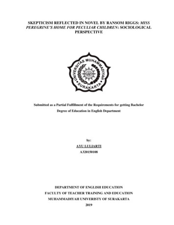
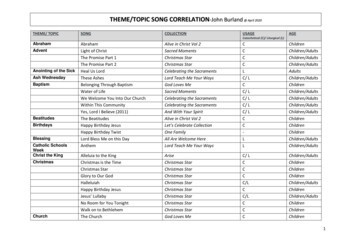
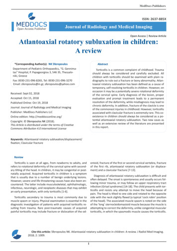
MedDocs Publishersatlantoaxial rotatory subluxation (Figure 1A). On the lateral radiograph loss of the normal lordosis was only diagnosed (Figure1B). Diagnosis was clearly secured with the CT scan findings.Coronal (Figure 1C), sagittal (Figure 1D) and axial (Figure 1E)views showed that the odontoid was lying eccentrically between the lateral masses of the atlas. A type I simple atlantoaxial rotatory displacement was diagnosed. The neck symptomswere considerably relieved immediately after gentle manipulative axial traction.Figure 2: Typical torticollis position of atlantoaxial rotatory displacement. The head is tilted to the side of the fractured clavicleand rotated to the opposite side, with slight flexion. Associatedmuscle spasm, unlike muscular torticollis, is predominantly on theside of the ‘long’ sternocleidomastoid, because the muscle is attempting to correct the deformity. Initial anteroposterior radiograph of the cervical spine, demonstrating left lateral cervical tiltdue to the midshaft clavicular fracture (A). On the lateral projection no abnormal alignment of the atlas and the axis was evidentbut there was a reversed cervical lordosis. The anterior displacement of the axis on the third cervical vertebra was not consideredpathological (B).Conflict of interest statementThe author certifies that he has no commercial associations(such as consultancies, stock ownership, equity interest, patent/licensing arrangements, etc) that might pose a conflict ofinterest in connection with the submitted article.ReferencesFigure 1: Initial anteroposterior radiograph of the cervicalspine indicated loss of symmetry between lateral masses of theatlas and odontoid process (A). Lateral cervical spine radiographshowed loss of the normal lordosis (B). Coronal CT images demonstrated narrowing of the right lateral atlantodental interval and awider left lateral atlantodental interval (C). Sagittal CT views of thecervical spine indicated no abnormal findings (D). Axial CT viewsconfirmed that the odontoid was lying eccentrically between thelateral masses of the atlas (E).The latter was an 8-year old boy that presented with torticollis associated with an injury of the left shoulder. Plain anteroposterior (Figure 2A) radiograph showed a midshaft fracture ofthe left clavicle, while the lateral view was indicative of a reversed cervical lordosis (Figure 2B). The potential severity of thecervical spine injury was overlooked. No CT scanning evaluationwas recommended. Bed rest and analgesics was the offeredtreatment. The patient reported an uneventful recovery of thetorticollis within a few days.Journal of Radiology and Medical Imaging31.White AA 3rd, Panjabi MM. The clinical biomechanics of the occipitoatlantoaxial complex. Orthop Clin North Am. 1978; 9: 867878.2.Kahn ML, Davidson R, Drummond DS. Acquired torticollis in children. Orthop Rev. 1991; 20: 667-674.3.Ballock RT, Song KM. The prevalence of non muscular causes oftorticollis in children. J Pediatr Orthop. 1996; 16: 500-504.4.Nigrovic LE, Rogers AJ, Adelgais KM, Olsen CS, Leonard JR, JaffeDM, Leonard JC; Pediatric Emergency Care Applied ResearchNetwork (PECARN) Cervical Spine Study Group. Utility of plainradiographs in detecting traumatic injuries of the cervical spinein children. Pediatr Emerg Care. 2012; 28(5): 426-432.5.Tomczak KK, Rosman NP. Torticollis. J Child Neurol. 2013; 28:365-378.6.Per H, Canpolat M, Tümtürk A, Gumuş H, Gokoglu A, YikilmazA, et al. Different etiologies of acquired torticollis in childhood.Childs Nerv Syst. 2014; 30(3): 431-440.7.Routt ML Jr, Green NE. Jefferson fracture in a 2-year-old child. JTrauma. 1989; 29: 1710-1712.8.de Beer JD, Hoffman EB, Kieck CF. Traumatic atlantoaxial subluxation in children. J Pediatr Orthop. 1990; 10: 397-400.9.Mazur JM, Loveless EA, Cummings RJ. Combined odontoid andJefferson fracture in a child: A case report. Spine. 2002; 27:E197-199.10.Papadimitriou NG, Christophoridis J, Papadimitriou A, BeslikasTA. Acute torticollis after isolated stress fracture of the first ribin a child. A case report. J Bone Joint Surg Am. 2005; 87: 25372540.11.AuYong N, Piatt J Jr. Jefferson fractures of the immature spine.Report of 3 cases. J Neurosurg Pediatr. 2009; 3: 15-19.12.Ottink KD, van Middendorp JJ, Kleinveld S, Breemans E. Traumatic atlas fracture in a child following fall on head. Ned TijdschrGeneeskd. 2009; 153: 1084-1089.13.Burkhardt M, Garcia P, Fries P, Heinzmann J, Pohlemann T, et al.Post-traumatic torticollis in a schoolchild: Fracture, congenitalanomaly or age-appropriate radiological findings of the atlas?Unfallchirurg. 2010; 113: 230-234.14.Maranich AM, Hamele M, Fairchok M. Atlanto-axial subluxation:A newly reported trampolining injury. Clin Pediatr (Phila). 2006;
MedDocs Publishers45: 468-470.and sixty children. J Bone Joint Surg Am. 1965; 47: 1295-1309.15.Sobolewski BA, Mittiga MR, Reed JL. Atlantoaxial rotary subluxation after minor trauma. Pediatr Emerg Care. 2008; 24: 852856.35.Schwarz N, Lenz M, Berzlanovich A, Smetka W. Atlanto-axial rotation and distance in small children. A postmortem study. Unfallchirurg. 2000; 103: 656-661.16.Benson M, Fixsen J, Macnicol M, Parsch K. Children’s Orthopaedics and Fractures, Springer-Verlag. 2010.36.Swischuk LE. Anterior displacement of C2 in children: Physiologic or pathologic. Radiology. 1977; 122: 759-763.17.Warner WC Jr. Pediatric cervical spine (Chapter 43). In Campbell’s Operative Orthopaedics E-Book. Azar FM, Beaty JH, CanaleST, eds. Elsevier, Philadelphia. 2017.37.Pang D. Atlantoaxial rotatory fixation. Neurosurgery. 2010; 66:161-183.18.Spiegel D, Shrestha S, Sitoula P, Rendon N, Dormans J. Atlantoaxial rotatory displacement in children. World J Orthop 2017; 8:836-845.38.Kawabe N, Hirotani H, Tanaka O. Pathomechanism of atlantoaxial rotator fixation in children. J Pediatr Orthop. 1989; 9: 569574.19.Muñiz AE, Belfer RA. Atlantoaxial rotary subluxation in children.Pediatr Emerg Care. 1999; 15: 25-29.39.Fielding JW, Hawkins RJ. Atlanto-axial rotatory fixation. (Fixedrotatory subluxation of the atlanto-axial joint). J Bone Joint SurgAm. 1977; 59: 37-44.20.Lee SC, Lui TN, Lee ST. Atlantoaxial rotatory subluxation in skeletally immature patients. Br J Neurosurg. 2002; 16: 154-157.40.21.Beier AD, Vachhrajani S, Bayerl SH, Aguilar CY, Lamberti-PasculliM, Drake JM. Rotatory subluxation: Experience from the Hospital for Sick Children. J Neurosurg Pediatr. 2012; 9: 144-148.Waegeneers S, Voet V, De Boeck H, Opdecam P. Atlantoaxial rotatory fixation. A case report and proposal of a new classification system. Acta Orthop Belg. 1997; 63: 35-39.41.22.Hussain K, Abdo MM, AlNajjar FJ, Abbo M. Not your typical torticollis: A case of atlantoaxial rotatory subluxation. BMJ Case Rep.2014; 2014.Fielding JW, Stillwell WT, Chynn KY, Spyropoulos EC. Use of computed tomography for the diagnosis of atlanto-axial rotatoryfixation. A case report. J Bone Joint Surg Am. 1978; 60: 11021104.42.Bagouri E, Deshmukh S, Lakshmanan P. Atlantoaxial rotatorysubluxation as a cause of torticollis in a 5-year-old girl. BMJ CaseRep. 2014; 2014.Kowalski HM, Cohen WA, Cooper P, Wisoff JH. Pitfalls in the CTdiagnosis of atlantoaxial rotary subluxation. AJR Am J Roentgenol. 1987; 149: 595-600.43.Murray JB, Ziervogel M. The value of computed tomography inthe diagnosis of atlanto-axial rotatory fixation. Br J Radiol. 1990;63: 894-897.44.Scapinelli R. Three-dimensional computed tomography in infantile atlantoaxial rotatory fixation. J Bone Joint Surg Br. 1994; 76:367-370.45.Ludwig K, Reisberg S. Computerized tomography in atlanto-axialrotatory fixation in childhood. Rofo. 1998; 168: 534-536.46.Hicazi A, Acaroglu E, Alanay A, Yazici M, Surat A. Atlantoaxial rotatory fixation-subluxation revisited: A computed tomographicanalysis of acute torticollis in pediatric patients. Spine (Phila Pa1976). 2002; 27(24): 2771-2775.47.McGuire KJ, Silber J, Flynn JM, Levine M, Dormans JP. Torticollisin children: Can dynamic computed tomography help determineseverity and treatment. J Pediatr Orthop. 2002; 22: 766-770.48.Alanay A, Hicazi A, Acaroglu E, Yazici M, Aksoy C, et al. Reliabilityand necessity of dynamic computerized tomography in diagnosis of atlantoaxialrotatory subluxation. J Pediatr Orthop. 2002;22: 763-765.23.24.Missori P, Marruzzo D, Peschillo S, Domenicucci M. Clinical remarks on acute post-traumatic atlanto-axial rotatory subluxation in pediatric-aged patients. World Neurosurg. 2014; 82:e645-648.25.Neal KM, Mohamed AS. Atlantoaxial rotatory subluxation in children. J Am Acad Orthop Surg. 2015; 23: 382-392.26.Powell EC, Leonard JR, Olsen CS, Jaffe DM, Anders J, et al. Atlantoaxial rotatory subluxation in children. Pediatr Emerg Care.2017; 33: 86-91.27.Wortzman G, Dewar FP. Rotary fixation of the atlantoaxial joint:Rotational atlantoaxial subluxation. Radiology. 1968; 90: 479487.28.Fielding JW, Hawkins RJ, Hensinger RN, Francis WR. Atlantoaxialrotary deformities. Orthop Clin North Am. 1978; 9: 955-967.29.Sundseth J, Berg-Johnsen J, Skaar-Holme S, Züchner M, KolstadF. Atlantoaxial rotatory fixation-a cause of torticollis. Tidsskr NorLaegeforen. 2013; 133: 519-523.30.Roach JW, Duncan D, Wenger DR, Maravilla A, Maravilla K. Atlanto-axial instability and spinal cord compression in childrendiagnosis by computerized tomography. J Bone Joint Surg Am.1984; 66: 708-714.49.Been HD, Kerkhoffs GM, Maas M. Suspected atlantoaxial rotatory fixation-subluxation: the value of multi detector computedtomography scanning under general anesthesia. Spine (Phila Pa1976). 2007; 32: E163-167.31.Samartzis D, Shen FH, Herman J, Mardjetko SM. Atlantoaxialrotatory fixation in the setting of associated congenital malformations: a modified classification system. Spine (Phila Pa 1976).2010; 35: E119-127.50.Haque S, Bilal Shafi BB, Kaleem M. Imaging of torticollis in children. Radiographics. 2012; 32: 557-571.51.Burkus JK, Deponte RJ. Chronic atlantoaxial rotatory fixation correction by cervical traction, manipulation, and bracing. J PediatrOrthop. 1986; 6: 631-635.52.Phillips WA, Hensinger RN. The management of rotatory atlanto-axial subluxation in children. J Bone Joint Surg Am. 1989; 7:664-668.53.Subach BR, McLaughlin MR, Albright AL, Pollack IF. Current management of pediatric atlantoaxial rotatory subluxation. Spine32.Roche CJ, O’Malley M, Dorgan JC, Carty HM. A pictorial review ofatlanto-axial rotatory fixation: key points for the radiologist. ClinRadiol. 2001; 56: 947-958.33.Ghanem I, El Hage S, Rachkidi R, Kharrat K, Dagher F, et al. Pediatric cervical spine instability. J Child Orthop. 2008; 2: 71-84.34.Cattell HS, Filtzer DL. Pseudosubluxation and other normal variations in the cervical spine in children. A study of one hundredJournal of Radiology and Medical Imaging4
MedDocs Publishers54.55.(Phila Pa 1976). 1998; 23: 2174-2179.64.Mihara H, Onari K, Hachiya M, Toguchi A, Yamada K. Follow-upstudy of conservative treatment for atlantoaxial rotatory displacement. J Spinal Disord. 2001; 14: 494-499.Kato T, Kanbara H, Sato S, Tanaka I. 5 cases of clavicular fracturesmisdiagnosed as congenital myogenic torticollis. Seikei Geka.1968; 19: 729-732.65.Martinez-Lage JF, Martinez Perez M, Fernandez Cornejo V, PozaM. Atlanto-axial rotatory subluxation in children: early management. Acta Neurochir (Wien). 2001; 143: 1223-1228.Goddard NJ, Stabler J, Albert JS. Atlanto-axial rotatory fixationand fracture of the clavicle. An association and a classification. JBone Joint Surg Br. 1990; 72: 72-75.66.Al-Etani H, D’Astous J, Letts M, Hahn M, Yeadon A. Masked rotatory subluxation of the atlas associated with fracture of the clavicle: A clinical and biomechanical analysis. Am J Orthop (BelleMead NJ). 1998; 27: 375-380.67.Nannapaneni R, Nath FP, Papastefanou SL. Fracture of the clavicle associated with a rotator atlantoaxial subluxation. Injury.2001; 32: 71-73.68.Bowen RE, Mah JY, Otsuka NY. Midshaft clavicle fractures associated with atlantoaxial rotatory displacement: a report of twocases. J Orthop Trauma. 2003; 17: 444-447.69.Kanik A, Sutcuoglu S, Aydinlioglu H, Erdemir A, Arun Ozer E. Bilateral clavicle fracture in two newborn infants. Iran J Pediatr.2011; 21: 553-555.70.Karski J, Matuszewski L, Jakubowski P, Karska K, Kandzierski G.Clavicle fracture associated with atlantoaxial rotator displacement, type II in an 8-year-old girl: A case report. Medicine (Baltimore). 2017; 96: e8781.56.Govender S, Kumar KP. Staged reduction and stabilisation inchronic atlantoaxial rotatory fixation. J Bone Joint Surg Br. 2002;84: 727-731.57.Weisskopf M, Naeve D, Ruf M, Harms J, Jeszenszky D. Therapeutic options and results following fixed atlantoaxial rotatory dislocations. Eur Spine J. 2005; 14: 61-68.58.Crossman JE, Thompson D, Hayward RD, Ransford AO, CrockardHA. Recurrent atlantoaxial rotatory fixation in children: a rarecomplication of a rare condition. Report of four cases. J Neurosurg. 2004; 100: 307-311.59.Crossman JE, David K, Hayward R, Crockard HA. Open reductionof pediatric atlantoaxial rotatory fixation: long-term outcomestudy with functional measurements. J Neurosurg. 2004; 100:235-240.60.Park SW, Cho KH, Shin YS, Kim SH, Ahn YH, et al. Successful reduction for a pediatric chronic atlantoaxial rotatory fixation (Grisel syndrome) with long-term halter traction: case report. Spine(Phila Pa 1976). 2005; 30: E444-449.61.Lavelle WF, Palomino K, Badve SA, Albanese SA. Chronic C1-C2rotatory subluxation reduced by C1 lateral mass screws and C2translaminar screws: A case report. J Pediatr Orthop. 2017; 37:e174-177.62.Schwarz N. The fate of missed atlanto-axial rotatory subluxationin children. Arch Orthop Trauma Surg. 1998; 117: 288-289.63.Kinon MD, Nasser R, Nakhla J, Desai R, Moreno JR, et al. Atlantoaxial rotatory subluxation: A review for the pediatric emergencyphysician. Pediatr Emerg Care. 2016; 32: 710-716.Journal of Radiology and Medical Imaging5
1B). Diagnosis was clearly secured with the CT scan findings. Coronal (Figure 1C), sagittal (Figure 1D) and axial (Figure 1E) views showed that the odontoid was lying eccentrically be-tween the lateral masses of the atlas. A type I simple atlanto-axi