Transcription
Child HealthNursingPartnering withChildren & FamiliesJane W. BallRuth C. BindlerChapter 30Alterations inGastrointestinal Function
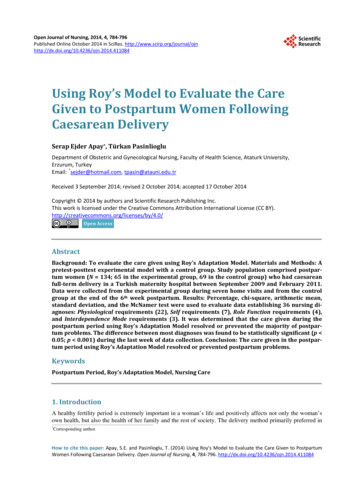
Basic GI anatomy
The internal anatomic structures of the stomach, including the pancreatic, cystic, andhepatic ducts; the pancreas; and the gallbladder.
Gastrointestinal System Digestion takes place in duodenum Enzymes aid in the digestion process
Infant vs. Children’sGastrointestinal System Liver function immature at birth Enzymes deficient until 4 to 6 months old Abdominal distention from gas commonwith infants Stomach capacity smaller Emptying time
Stomach Capacity Increase Throughout Early Childhood
Clues to GastrointestinalDisorders in Children Vomiting/ RegurgitationAbdominal painFailure to thriveStool changes– Diarrhea– Constipation Abdominal Distention
Assessment Techniques Subjective– Lifestyle and family factors– Diet– Elimination Patterns
GI assessment Objective– Observation Abdominal distentionSymmetry, bumps, bulges or massesUmbilicusPeristaltic waves– *visible rippling waves bowel obstruction– Palpation
GI Assessment– Auscultation Hyper/hyper bowel sounds– Percussion Tympany vs. dullness– Palpation Light palpationDeep palpationRebound tenderness- peritoneal inflammationMcBurney’s point
Disorders of GastrointestinalSystem Structural DefectsDisorders of MotilityIntestinal Parasitic DisordersInflammatory disordersDisorders of MalabsorptionHepatic DisordersInjuries to the Gastrointestinal System
Structural Defects Cleft lip and cleft palate– Failure of the maxillary processes to fusebetween 5 to 12 weeks gestation– Failure of the tongue to move down at thecorrect time prevents the palatine processesfrom fusing– Multifactorial causes
FIGURE 30–2A, Unilateral cleft lip. Courtesy of Dr. Elizabeth Peterson, Spokane, WA.A
FIGURE 30–2 (continued)B, Bilateral cleft lip. Courtesy of Dr. Elizabeth Peterson, Spokane, WA.B
Cleft Lip and Cleft Palate Nursing care Pre- and postoperative care Nursing diagnosis– Pre-op Imbalanced nutrition: less than body requirements Risk of aspiration Altered parenting– Post-op Risk of injury& infection Pain Altered feeding patterns
Cleft Lip& Palatecomplications Feeding p. 1100 in textSpeechChronic otitis mediaPsychological concernsDental problems
Structural Defects Esophageal atresia andtracheoesophageal fistula– Definition– Foregut fails to lengthen, separate, and fuseinto two parallel tubes (esophagus andtrachea) at 4 to 5 weeks gestation– Associated with maternal polyhydramnios
Esophageal Atresia andTracheoesophageal Fistula Nursing care– Identifying signs and symptoms of theseinfants Pre- and postoperative care– Suction is important preoperatively– Care of the gastrostomy tube postoperatively
Types of esophageal atresia
FIGURE 30–4In the most common type of esophageal atresia and tracheoesophageal fistula, the upper segment of theesophagus ends in a blind pouch connected to the trachea; the fistula connects the lower segment to the trachea.Jane W. Ball and Ruth C. BindlerChild Health Nursing: Partnering with Children & Families 2006 by Pearson Education, Inc.Upper Saddle River, New Jersey 07458All rights reserved.
TE atresia Most of these babies (about 85%) will have an upper esophagealpouch that ends blindly and a lower esophageal segment thatconnects into the trachea (Figure 1c). For 8-10%, however, there willnot be a connection between esophagus and trachea (no TEF). Thisis considered to be pure esophageal atresia (EA) with two blindesophageal ends (Figure 1a). For these babies, the distancebetween the two esophageal ends is usually longer (a long gap EA).Similarly, when the TEF is only between the upper pouch and thetrachea, the lower end of the esophagus tends to be short leaving along gap between the ends (Figure 1b).
TE Atresia Clinical symptoms– Excessive drooling– Cyanosis– Coughing– Choking Diagnosis– Pass NG tube into stomach Treatment– Surgical correction Nursing Care– IV fluids– Antibiotics– Gtube
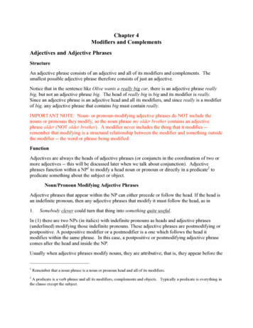
Pathophysiology of structuraldefects Pyloric stenosis– Hypertrophy of circular muscle of pyloric canal– Etiology unknown, but often affects first- bornmales.– Diagnosis by ultrasound– Stenosis occurs between stomach andduodenum
In pyloric stenosis, the hypertrophied pyloric muscle causes symptoms of projectilevomiting and visible peristalsis.
Pyloric Stenosis Clinical Manifestations:– Good eater until week 3– Projectile vomiting– Appears hungry, lethargic, fails to gain weight Nursing care Pre- and postoperative care––––––Fluid and electrolyte managementIVNG tubeAnalgesicsPrevent infectionsupport
Intussusception– Intestine invaginates into another (like a sock)– Mesentary becomes inflamed and obstructioncan occur leading to ischemia– Multifactorial causes Classic triad of Intussusception– Severe episodic pain– "Currant jelly' stool– transverse tubular abdominal mass
FIGURE 30–7syndromes.In infants, intussusception is commonly associated with measles, viral disease, and gastroenteritisJane W. Ball and Ruth C. BindlerChild Health Nursing: Partnering with Children & Families 2006 by Pearson Education, Inc.Upper Saddle River, New Jersey 07458All rights reserved.
Intussusception More common in children with cysticfibrosis, celiac disease and gastroenteritis Etiology is multifactorial Diagnosis: history, x-ray, contrast enema Nursing management:–––––Vital signsCheck for distentionFluids and electrolytesIntake and outputpain
Intussusception presentation Physical: Usually, the abdomen is soft and nontender early, but iteventually becomes distended and tender. A vertically oriented mass may be palpable in the rightupper quadrant. Currant jelly stools are observed in only 50% of cases. Most patients (75%) without obviously bloody stoolshave stools that test positive for occult blood. Fever is a late finding and is suggestive of entericsepsis.
Intussusception Emergency Department Care:– Provide rehydration and stabilization as needed.– Contrast enema is diagnostic in approximately 95% ofintussusception cases. It is therapeutic and curative inmost cases with less than 24-hour duration. Inpatient Care– Admission is indicated for all patients because up to10% of those with successful radiologic reductionhave a recurrence, usually in the first 24 hours.
Structural DefectsAbdominal Wall Defects Gastroschisis and omphalocele– Definition– Gastroschisis usually occurs to the right of theumbilicus and omphalocele occurs throughthe umbilical cord– Occurs in week 11 of gestation whenabdominal contents fail to return to theabdomen– Multifactorial causes
Structural DefectsAnorectal Malformations Anal stenosis and anal atresia– Failure of growth of urorectal septum, lateralmesoderm structures, and ectodermalstructures– Associated anomalies up to 70% of the time
Structural DefectsUmbilical Hernia Umbilical hernia– Results from imperfect closure of the umbilicalmuscle ring– Often asssociated with diastasis recti (lateralseparation of the abdominal muscles.– Etiology unknown– Around week 11 gestation the obliteratedumbilical vessels occupy the space in theumbilical ring
FIGURE 30–13The umbilical hernia of the newborn usually closes as the muscles strengthen in later infancy and childhood.From Zitelli, B., & Davis, H. (Eds.) (1994). Atlas of pediatric physical diagnosis.(2nd ed.). London: Mosby-Wolfe Publishing.
Disorders of Motility VomitingGE refluxConstipationEncopresisHirschsprung diseaseGastroenteritis
Disorders of MotilityGastroesophageal Reflux Gastroesophageal reflux– Definition– Three mechanisms allow reflux to occur Lower esophageal relaxations Incompetent lower esophageal sphincter Anatomic disruption of esophagogastric junction– Reflux acidity damages the esophagealmucosa– Causes
What is GER/ GERD? Gastroesophageal refers to the stomach andesophagus, and reflux means to flow back or return. Gastroesophageal reflux (GER) is the NORMALphysiologic passage of acidic stomach juices, or foodand fluids, back up into the esophagus. Gastroesophageal reflux disease (GERD) refers to thepathophysiologic outcomes. It is persistent orrecurrent symptoms or complications that are causedby gastric acid flowing from the stomach into theesophagus,GER is very common in infants, though it can occur atany age. It is the most common cause of vomitingduring infancy, but usually resolve by first birthday.
Clinical Manifestations of Gastroesophageal Reflux and Gastroesophageal RefluxDisease
GERD
Foods and GERD Some foods seem to affect the muscletone of the lower esophageal sphincter,allowing it to stay open longer thannormal. These include, but are not limitedto, the following: Chocolate. Peppermint. High-fat foods.
GERD Other foods increase acid production inthe stomach, including: Citrus foods. Tomatoes and tomato sauces.
Why is gastroesophageal reflux(GERD) a concern? Some infants and children who have gastroesophageal reflux maynot vomit, but may still have stomach contents move up theesophagus and spill over into the windpipe. This can cause asthma,pneumonia and possibly even SIDS (sudden infant deathsyndrome). Infants and children with GERD who vomit frequently may not gainweight and grow normally. Inflammation (esophagitis) or ulcers (sores) can form in theesophagus due to contact with stomach acid. These can be painfuland also may bleed, leading to anemia (too few red blood cells inthe bloodstream). Esophageal narrowing (stricture) and Barrett's esophagus (abnormalcells in the esophageal lining) are long-term complications frominflammation.
What are the symptoms of GERD? Belching.Refusal to eat.Stomachache.Fussiness around mealtimes.Frequent vomiting.Hiccups.Gagging.Choking.Frequent cough.Coughing fits at night.Wheezing.Frequent upper respiratory infections (colds).Rattling in the chest.Frequent sore throats in the morning.Sour taste in the mouth.
How is GERD diagnosed? Vomiting, pain associated with regurgitation, archingback and feeding refusal Difficult to differentiate between normal GER, andColic Physical Exam Chest x-ray - a diagnostic test to look for evidence ofaspiration. Upper GI (gastrointestinal) series Endoscopy Esophageal manometric studies. pH testing. Gastric emptying studies.
http://www.laparoscopy.com/pictures/lapniss.html
Nonpharmacological Treatment Watch child's food intake - limit fried and fatty foods, peppermint, chocolate,drinks with caffeine (such as colas, Mountain Dew and tea), citrus fruitand juices and tomato products.Offer smaller portions at mealtimes and include small snacks in-betweenmeals if child is hungry. Avoid letting child overeat. Allow child to let youknow when hungry or full.If child is overweight, consult your physician to set weight loss goals.Do not allow child to lie down or go to bed right after a meal. Serve theevening meal early - at least two hours before bedtime.After feedings, place infant on the stomach with the upper body elevated atleast 30 , or hold in a sitting position in your lap for 30 minutes.If bottle-feeding, keep the nipple filled with milk so infant does not swallowtoo much air while eating. Try different nipples to find one that allows yourbaby's mouth to make a good seal with the nipple during feeding.Adding rice cereal to feeding may be beneficial for some infants.Burp baby several times during bottle or breast feeding. Child may refluxmore often when burping with a full stomach
Medications. H2 receptor blockers help decrease the amount ofacid the stomach makes, which, in turn, will cut down onthe heartburn associated with reflux.– cimetidine (Tagamet )– ranitidine (Zantac ).– Famotidine (Pepcid) proton-pump inhibitors. prevent excess acid secretionin the stomach– omeprazole (Prilosec ) and– lansoprazole (Prevacid ). These medications are taken daily to. Another type of medicine helps the stomach emptyfaster. Used as adjunct with PPI on short term basis. Isused to reduce nausea post op. Side effects limit use.– (Reglan ). This medicine is usually taken three to four times aday, before meals or feedings and at bedtime.
Calorie supplements.Some infants with reflux will not be able togain weight due to frequent vomiting. Adding rice cereal to baby formula. Adding a prescribed supplement (such asPolycose or Moducal ) to formula orbreast milk to make the milk higher incalories than normal.
Tube feedings. Some babies with reflux have other conditions that makethem tired, such as congenital heart disease orprematurity. In addition to having reflux, these babiesmay not be able to drink very much without becomingsleepy. Other babies are not able to tolerate a normalamount of formula in the stomach without vomiting andwould do better if a small amount of milk was givencontinuously. In both of these cases, tube feedings maybe recommended. Formula or breast milk is giventhrough a nasogastric tube. Nasogastric tube feedingscan be given in addition to or instead of what a babytakes from a bottle.
Surgery. In severe cases of reflux, a surgical procedurecalled fundoplication may be performed if childis not gaining weight due to vomiting, hasfrequent respiratory problems or has severeirritation in the esophagus.– This procedure is usually done laparoscopically, The top portion of the stomach is wrappedaround the esophagus, creating a tight band thatgreatly decreases reflux.
Fundoplication
Pt. with GERD and sleep apnea
Gastroesophageal Reflux Nursing care– Risk for aspiration related to reflux– Fluid volume deficit related to reflux– Imbalanced nutrition, less than bodyrequirements. Important education– Feeding techniques– positioning
Motility disordersColic Colic– Paroxysmal abdominal pain of intestinal originand severe crying– Occurs between 2 weeks old and 3 monthsold– Etiology unknown
Disorders of Motilityconstipation and encopresis Constipation is a common complaint and accountsfor 25% of GI referrals– Defined by criteria of Pebble-like hard stools for a majority of bowel movements for2 weeks Firm stools more than twice per week for 2 weeks– Constipation can be caused by underlying disease,diet, or psychological factors– Three types of constipation Normal-transit constipation Defacatory disorders Slow-transit constipation– Primary vs. secondary encopresis
Constipation and Encopresis Nursing care Important education– Dietary– Medications– Fluids
Medications Osmotic Laxatives– Lactulose– Sorbitol– Milk of magnesia– Polyethylene glycol Lubricants– Mineral oil Stimulant laxatives– Dulcolax– Senna Stool softeners– Colace
Disorders of MotilityHirschsprung Disease Definition AKA congenital aganglionic magacolon Congenital absence of ganglion cells in the rectumand colon which prevents peristalsis at that portionof colon. often accompanies other genetic defects ( trisomy21, cleft palate, polydactyly, cardiac septaldefects, craniofacial anomalies Genetically acquired and occurs when there isfailure of the migration of neural crest cells inutero Colon becomes a “megacolon”
Hirschspring’s Disease Diagnosis:––––bowel pattern, history,Contrast studies ( barium enema)rectal biopsy.Rectum is small and does not contain stool Assessment:– Fluids and electrolytes– Intake and output– Abdominal distention– Stooling pattern
Disorders of Motility Gastroenteritis– Definition– Acute vs. chronic diarrhea caused by viruses,bacterial or parasites– Causes of diarrhea in children
Hirschsprung diseaseclinical manifestations Newborns––––Failure to pass meconiumRefusal to suckAbdominal distentionBile-stained emesis Older child–––––Failure to gain weightDelayed growthHistory of severe constipation alternating with diarrheaVomitingRibbon-like stool
Hirschsprung’s diseasetreatment Surgical removal of involved aganglionic bowel, andeither– End to end anastomosis to the anal canal– Temporary colostomy may be necessary for severecases with closure of the colostomy in 3-6 months. NG tube is generally inserted preop to relieve distentionand is left in place X 24 hrs. Milder cases– Dietary modification, stool softeners, and isotonic irrigations toprevent impaction.
Hirschsprung’s diseasecomplication Enterocolitis (inflammation of the intestine)– 20-60% of children after surgery– GI bleeding– Diarrhea Treatment– TPN– Lactose free diet
Hirschsprung’s diseasenursing care Monitor fluid and electrolyte balanceMaintain nutritionPreop and postop carePain reliefPromote bowel elimination
Diarrhea What is diarrhea? Diarrhea is defined either aswatery stool or increased frequency (or both)when compared to a normal amount. It is acommon problem that may last a few days anddisappear on its own. Acute (short-term, lasting less than two weeks), which isusually related to bacterial or viral infections. Chronic (long-term, lasting longer than two weeks),which is usually related to functional disorders, such asirritable bowel syndrome, or may be due to diseasessuch as ulcerative colitis, Crohn's disease, celiac sprueor Giardia.
What causes diarrhea? Diarrhea in children may be caused by anumber of conditions, including thefollowing: Bacterial infection. Viral infection. Food intolerances or allergies. Parasites. Reaction to medications.
What are the symptoms ofdiarrhea? The following are the most common symptoms fordiarrhea. However, each child may experiencesymptoms differently. Severe diarrhea may indicate aserious disease, making it important to consult yourchild's physician if any or all of the following symptomspersist: Cramping. Abdominal pain. Bloating. Nausea. Urgent need to use the restroom. Fever. Bloody stools.
Table 30–6Causes of Diarrhea in Children
Warning signs of severe diarrhea: Abdominal pain.Blood in the stool.Frequent vomiting.Loss of appetite for liquids.High fever.Dry, sticky mouth.Weight loss.Urinates less frequently (wets fewer than 6 diapers perday). Frequent diarrhea. Extreme thirst. No tears when crying.
Disorders of MotilityGastroenteritis Diarrhea Gastroenteritis is an inflammation of thestomach and intestines that may beaccompanied by vomiting and/or diarrhea. Causative agents include– Virus– Bacteria Described as– Mild– Moderate– severe
Parasitic Disorders Chart on page 1132 of text reviews majorparasites
Medications used to diarrhea Metronidazole (Flagyl)– Used to treat anaerobic bacteria and certainparasites, Giardiasis In combination for H Pylori Antidiarrea– Imodium
Inflammatory Disorders Peptic UlcerAppendicitisNecrotizing EnterocolitisMeckel’s DiverticulumRecurrent Abdominal PainInflammatory Bowel Disease
Peptic Ulcer H Pylori Review in Med surg text
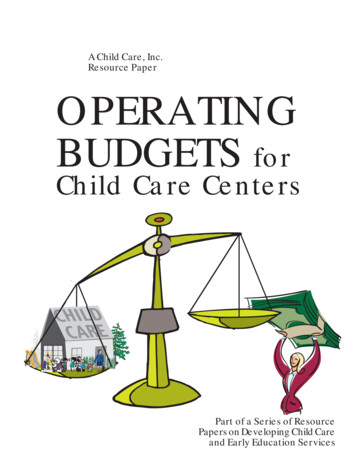
Inflammatory Disorders of GI tract Appendicitis– Definition– Caused by an obstruction of the appendiceallumen, causing edema– As edema continues, vascular supply iscompromised and bacteria invade, which canlead to a ruptured appendix
d -3315659114004055
Appendicitis Acute appendicitis is one of the mostcommon causes of abdominal pain and isthe most frequent condition that leads toemergent abdominal surgery in children
FIGURE 30–14McBurney’s point is the common location ofpain in children and adolescents with appendicitis.Jane W. Ball and Ruth C. BindlerChild Health Nursing: Partnering with Children & Families 2006 by Pearson Education, Inc.Upper Saddle River, New Jersey 07458All rights reserved.
Appendicitis Nursing care Pre- and postoperative care
Appendicitis Preop care Correction of fluid and electrolyte deficits Antibiotics
Appendicitis post op care Nursing diagnoses– Pain– Risk of infection– Anxiety
Inflammatory DisordersNecrotizing Enterocolitis Potentially life-threatening inflammatorydisease of the intestinal tract that occursprimarily in premature nfants. Most common gastrointestinal emergencyccuring during the neonatal period SIGNS OF NEC– Abdominal distention– Bilious vomiting– Bloody stools
Inflammatory Disorders Meckel’s diverticulum– Omphalomesenteric duct fails to atrophy– Outpouching of the ileum remains andcontains gastric contents causing ulceration– Bowel obstruction, perforation, or peritonitiscan occur
Meckel’s diverticulumnursing management Preop– IV for fluid and electrolyte imbalances– Test stools for occult blood Vital signs q 2 hours Post-op– Abdominal surgery– Teaching parents re: feeding, medications
Inflammatory Bowel Disease(Crohn’s Disease and UlcerativeColitis) Nursing care Important education
Inflammatory Bowel diseaseMost common chronic GI disease ofchildhood and adolescence
Inflammatory Disorders Inflammatory bowel disease (Crohn’s diseaseand ulcerative colitis)– Faulty regulation of the immune response of theintestinal mucosa– Usually genetically triggered– Crohn’s disease can cause inflammation and ulcersanywhere throughout the GI tract– Ulcerative colitis effects large intestine and rectalmucosa
Crohn’s disease Inflammation and swelling of the wall ofthe small or large intestine causes abdominal painDiarrheaFeverWeight loss Can effect anywhere in the GI tract
Crohn’s diseasecomplications Intestinal blockage– Thickening of the intestinal wall with swellingand scar tissue Fistulas– Tunnels through affected area to surroundingtissues of bladder, vagina, skin Nutritional complications– Deficiencies of proteins, calories, vitamins
Crohn’s diseasepharmacologic treatment Anti-inflammatory drugs– Sulfasalazine (po caplet)– Mesalamine (po or pr suspension) Systemic steroids– Prednisone (po)– Methylprednisolone (solumedrol) IV Immune system suppressors– Immuran– 6-mercaptopurine Antibiotics– Flagyl Probiotics– Lactobacillus (granules) 1 packet tid Antidiarrheal agents
Ulcerative Colitis Cause inflammation and ulcers in thelining of the rectum and colon Surgery cures disease– Ileostomy– Ileoanal anastomosis “pull through” 5% of ulcerative colitis develop coloncancer. Risk increases with the durationof disease
Support for children with IBD Reach out for youth with ileitis and colitis,inc.– Founded in 1979 to help children with Crohn’sdisease, ulcerative colitis and other GIdiseases.– Volunteer organization help children and theirfamilies through personal support andeducation
Table 30–7Comparison of Ulcerative Colitis and Crohn’s Disease
Malabsorption disorders Malabsorption occurs when a child isunable to digest or absorb nutrients in thediet. Disorders of malabsorption– SHORT BOWEL SYNDROME– CELIAC DISEASE– LACTOSE INTOLERANCE– CYSTIC FIBROSIS
Disorders of Malabsorptioon Short bowel syndrome– Due to shortened intestine after surgical resection ofa portion of the intestines.– Symptoms depend on which area of the bowel wasresectedIleumBile salts, fluids, and electrolyteabsorption decrease causing diarrhea.Steatorrhea, decreased absorption offat-soluble vitaminsColonFluid and electrolyte management isimpairedJejunumMostly compensated for effectively bythe remaining bowel
Celiac Diseasegluten-sensative enteropathy Celiac disease– Chronic malabsorption syndrome morecommon in white European children– Immunologic disorder characterized byintolerance for gluten found in wheat, barley,rye and oats. Affects fat absorption– ?genetic factors
Celiac Disease Clinical manifestations– Diarrhea– FTT– Abdominal pain– Large quantities of fat in stool.– Stools are greasy, foul smelling, frothy, andexcessive.– Eventually may develop protein deficiencywith wasted musculature, and abd distention.
Celiac Disease Diagnostic tests– Fecal fat content– Duodenal biopsy– Trial of gluten free diet with improvement– Serum screening for IgA Treatment– Total exclusion of gluten from diet
Gluten free dietWhat can you eatWhat is eliminatedRiceSome types of chocolate candyCornPrepared hamburgers, hotdogsTapiocaMilk preparations, malted milkBeansCanned soupMilletCatsupArrowrootvinegarDistilled productsHydrolyzed vegetable proteinPotato flourModified food starchsoyPLAYDOUGH , LIPSTICK,MEDICATIONwww.celiac.org
Disorders of MalabsorptionLactose Intolerance Inability to digest lactose due to deficiency ofenzyme Lactase, which is produced by thecells that line the small intestine. Symptoms include– Explosive, watery diarrhea,– Abdominal distention– Excessive flatus Diagnosis is made by– History– Hydrogen breath test– Stool acidity test (lactic acid buildup)
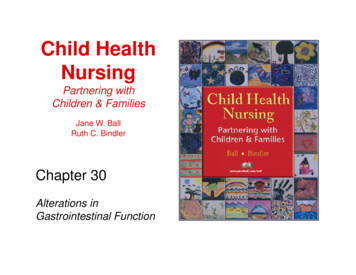
Lactose intolerancetreatment and nursing management Reduce lactose in dietSwitch to soy based formulaLactaid tablets for older childrenAssure enough calcium in dietWhich food has morecalcium?Sardines? Or Tuna?
Calcium content of common foodsFood itemgCalcium contentLactose ContentSoymilk200-300 mg/ 1 cup0Sardines 3 oz270 mg0Salmon, canned 3 oz.205 mg0Broccoli, raw 1 cup90 mg0Orange, 1 medium50 mg0Pinto beans40 mg0Tuna 3 oz.10 mg0
Hepatic disorders– Biliary atresia– Viral hepatitis– Cirrhosis
Signs of Hepatic Disorders JaundiceEasy bruising, intense itchingWhite or clay-colored stoolsTea-colored urine
Box 30–5Clinical Risk Factors for Development of Severe Hyperbilirubinemia
Table 30–8Comparison of Major Hepatitis Types
FIGURE 30–19and illness.The hepatitis virus causes degeneration and necrosis of the liver, which results in abnormal liver functionJane W. Ball and Ruth C. BindlerChild Health Nursing: Partnering with Children & Families 2006 by Pearson Education, Inc.Upper Saddle River, New Jersey 07458All rights reserved.
Box 30–6Pediatric Liver Transplantation
Injuries to the Gastrointestinalsystem Accidents and environmentalgastrointestinal disturbances– Abdominal trauma– Ingestion of foreign substances or objects– Lead poisoning
Abdominal Trauma Provide emotional support Follow care orders Prevention teaching once stabilized
Poison Ingestion Do not use ipecac Airway, hemodynamic stability, removetoxin, and support Education on prevention Most common poison?
Lead PoisoningHeavy Metal poisoning Healthy People 2010 goal: eliminatechildhood lead poisoning as a publichealth issue in the United States. From 1991 to 1994 4.4% of children ages1-6 in the US had blood lead levels abovethe limit of lead toxicity of 10ug/dL 1971 the Lead Based Point PoisoningPrevention Act was passed to ban interiorlead-based paint in residential homes.
Lead PoisoningRoutes of Exposure Mostly through lead-based paint in olderhomes– Low income areas– Antique high-end homes under renovation Paint can chip, flake and chalk into dustwhich then settles on floor or dirt. Children put more things in their mouth Child’s breathing zone is near the floorwhere dust and chips of lead may lie.
Other sources of lead exposure Water from lead pipesLead solder on canned foodLead ammunitionPool cue chalkCollectible toysJewelry
Cultural issues which contributeto lead poisoning in children Mexican– Azarcon, bright orange powder– Greta, bright yellow powder– Both are used as medicinal treatment of GI problems Tamarind– Jellied candy from Mexico packaged in ceramic jarscontaminated with lead Southeast Asia:– Paylooah remedy for rash and fever Eastern Indian– Surma, a black powder applied to the eyelids toimprove eyesight
Lead PoisoningToxicology/ absorptionThere are three major routes for absorption of leadin children’s body.LeadexposureGastrointestinalAbsorbed from GItract into bloodstreamInhalationAbsorbed intoblood streamthrough alveoliTransplacentalAbsorbed frommother into fetusthrough placenta
Lead PoisoningToxicology/ Distribution At least 99% of absorbed lead is bound toerytrocytes upon entry into the bloodstream.– 70% is stored in bone Accumulates throughout life, but can be releasedduring stress Can have a half-life of as many as 20 years.– 30% moves to major soft tissue storage sites LiverKidneyBone marrowBRAIN
Lead PoisoningPathophysiology Lead toxicity can affect any soft tissue of thebody eletal,endocrineCentral nervous systemLEAD’S MOST DETRIMENTAL PATHOPHYSIOLOGY ISNEUROTOXICITY WITHING THE PEDIATRIC CENTRALNERVOUS SYSTEM
Lead PoisoningPathophysiology/ signs & symptoms Acute lead poisoning– Nausea,– Vomiting– Anorexia– Constipation– Abdominal pain
Chronic lead poisoning Neurocognitiveeffects Developmental delay Lower IQ Speech and languageproblems Reading skills deficits Learning disabilities Lowered academicsuccess Behavioraleffects terestwithdrawal
Lead PoisoningDiagnosis 6 classesClassBlood lead levelSigns and symptomsI 9asymptomaticIIA10-14Usually asymptomatic, but can exhibitsome impairment in cognition, finemotor skills and growthIIB15-19Usually asymptomatic, but can exhibitsome impairment in cognition, finemotor skills and growthIII20-44Increased motor impairment andlethargy, possible abdominal painIV45-69Hyperirritable behavior, intermittentlethargy, decreased interest in play, N/VanorexiaV 70Encephalopathy, loss of developmentalskills, seizures
Treatment of lead poisoning Primary Prevention Secondary prevention– Chelation therapy is indicated if BLL 45ug/dL Succimer (Chemet) only oral chelating drug Ethylendiaminetetraacetic Acid (EDTA) British antilewisite (BAL) not if Fe therapy Tertiary Prevention
Foreign Object Ingestion Assessment important Prepare child and parents for x-rays andpossible removal Education on prevention
Let’s review
Potential Signs of GastrointestinalEmergencies in Infants Excessive salivation with cyanosis,coughing, and choking in newborn– Esophageal atresia and tracheoesophagealfistula A
Atlas of pediatric physical diagnosis.(2nd ed.). London: Mosby-Wolfe Publishing. Disorders of Motility Vomiting GE reflux Constipation Encopresis Hirschsprung disease Gastroenteritis. Disorders of Motility Gastroesophageal Reflux Gastroesophageal ref