Transcription
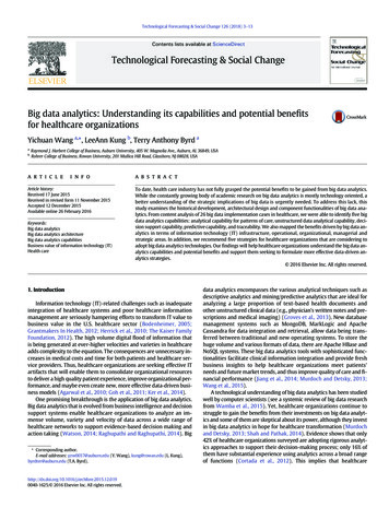
Big Self-Supervised Models Advance Medical Image ClassificationShekoofeh Azizi, Basil Mustafa, Fiona Ryan , Zachary Beaver, Jan Freyberg, Jonathan Deaton,Aaron Loh, Alan Karthikesalingam, Simon Kornblith, Ting Chen, Vivek Natarajan, Mohammad NorouziGoogle Research and Health†AbstractSelf-supervised pretraining followed by supervised finetuning has seen success in image recognition, especiallywhen labeled examples are scarce, but has received limited attention in medical image analysis. This paper studies the effectiveness of self-supervised learning as a pretraining strategy for medical image classification. We conduct experiments on two distinct tasks: dermatology condition classification from digital camera images and multilabel chest X-ray classification, and demonstrate that selfsupervised learning on ImageNet, followed by additionalself-supervised learning on unlabeled domain-specific medical images significantly improves the accuracy of medicalimage classifiers. We introduce a novel Multi-Instance Contrastive Learning (MICLe) method that uses multiple images of the underlying pathology per patient case, whenavailable, to construct more informative positive pairs forself-supervised learning. Combining our contributions, weachieve an improvement of 6.7% in top-1 accuracy andan improvement of 1.1% in mean AUC on dermatologyand chest X-ray classification respectively, outperformingstrong supervised baselines pretrained on ImageNet. In addition, we show that big self-supervised models are robustto distribution shift and can learn efficiently with a smallnumber of labeled medical images.1. IntroductionLearning from limited labeled data is a fundamentalproblem in machine learning, which is crucial for medical image analysis because annotating medical images istime-consuming and expensive. Two common pretrainingapproaches to learning from limited labeled data include:(1) supervised pretraining on a large labeled dataset such asImageNet, (2) self-supervised pretraining using contrastivelearning (e.g., [16, 8, 9]) on unlabeled data. After pretraining, supervised fine-tuning on a target labeled dataset of interest is used. While ImageNet pretraining is ubiquitous inmedical image analysis [46, 32, 31, 29, 15, 20], the use ofself-supervised approaches has received limited attention.Self-supervised approaches are attractive because they en Former intern at Google.Currently at Georgia Institute of Technology.† {shekazizi, skornblith, iamtingchen, natviv, mnorouzi}@google.com(1) Self-supervised learning on unlabeled natural images(2) Self-supervised learning on unlabeled medical imagesand Multi-Instance Contrastive Learning (MICLe) ifmultiple images of each medical condition are -rays(3) Supervised fine-tuning on labeled medical gure 1:Our approach comprises three steps: (1) Selfsupervised pretraining on unlabeled ImageNet using SimCLR [8].(2) Additional self-supervised pretraining using unlabeled medicalimages. If multiple images of each medical condition are available, a novel Multi-Instance Contrastive Learning (MICLe) is usedto construct more informative positive pairs based on different images. (3) Supervised fine-tuning on labeled medical images. Notethat unlike step (1), steps (2) and (3) are task and dataset specific.able the use of unlabeled domain-specific images duringpretraining to learn more relevant representations.This paper studies self-supervised learning for medical image analysis and conducts a fair comparison between self-supervised and supervised pretraining on twodistinct medical image classification tasks: (1) dermatology skin condition classification from digital camera images, (2) multi-label chest X-ray classification among fivepathologies based on the CheXpert dataset [23]. We observe that self-supervised pretraining outperforms supervised pretraining, even when the full ImageNet dataset(14M images and 21.8K classes) is used for supervised pretraining. We attribute this finding to the domain shift anddiscrepancy between the nature of recognition tasks in Im-3478
ised0.775CheXpert Mean AUCDermatology Top-1 600.7550.7500.745ResNet-50 (4x) ResNet-152 (2x)0.740ResNet-50 (4x) ResNet-152 (2x)Figure 2: Comparison of supervised and self-supervised pretraining, followed by supervised fine-tuning using two architectures ondermatology and chest X-ray classification. Self-supervised learning utilizes unlabeled domain-specific medical images and significantly outperforms supervised ImageNet pretraining.ageNet and medical image classification. Self-supervisedapproaches bridge this domain gap by leveraging in-domainmedical data for pretraining and they also scale gracefullyas they do not require any form of class label annotation.An important component of our self-supervised learning framework is an effective Multi-Instance ContrastiveLearning (MICLe) strategy that helps adapt contrastivelearning to multiple images of the underlying pathology perpatient case. Such multi-instance data is often available inmedical imaging datasets – e.g., frontal and lateral viewsof mammograms, retinal fundus images from each eye, etc.Given multiple images of a given patient case, we proposeto construct a positive pair for self-supervised contrastivelearning by drawing two crops from two distinct images ofthe same patient case. Such images may be taken from different viewing angles and show different body parts with thesame underlying pathology. This presents a great opportunity for self-supervised learning algorithms to learn representations that are robust to changes of viewpoint, imagingconditions, and other confounding factors in a direct way.MICLe does not require class label information and onlyrelies on different images of an underlying pathology, thetype of which may be unknown.Fig. 1 depicts the proposed self-supervised learning approach, and Fig. 2 shows the summary of results. Our keyfindings and contributions include: We investigate the use of self-supervised pretrainingon medical image classification. We find that selfsupervised pretraining on unlabeled medical images significantly outperforms standard ImageNet pretrainingand random initialization. We propose Multi-Instance Contrastive Learning (MICLe) as a generalization of existing contrastive learningapproaches to leverage multiple images per medical condition. We find that MICLe improves the performance ofself-supervised models, yielding state-of-the-art results. On dermatology condition classification, our selfsupervised approach provides a sizable gain of 6.7% intop-1 accuracy, even in a highly competitive productionsetting. On chest X-ray classification, self-supervisedlearning outperforms strong supervised baselines pretrained on ImageNet by 1.1% in mean AUC. We demonstrate that self-supervised models are robustand generalize better than baseslines, when subjected toshifted test sets, without fine-tuning. Such behavior isdesirable for deployment in a real-world clinical setting.2. Related WorkTransfer Learning for Medical Image Analysis. Despite the differences in image statistics, scale, and taskrelevant features, transfer learning from natural images iscommonly used in medical image analysis [29, 31, 32, 46],and multiple empirical studies show that this improvesperformance [1, 15, 20]. However, detailed investigations from Raghu et al. [37] of this strategy indicate thisdoes not always improve performance in medical imagingcontexts. They, however, do show that transfer learningfrom ImageNet can speed up convergence, and is particularly helpful when the medical image training data is limited. Importantly, the study used relatively small architectures, and found pronounced improvements with smallamounts of data especially when using their largest architecture of ResNet-50 (1 ) [18]. Transfer learning from indomain data can help alleviate the domain mismatch issue.For example, [7, 20, 26, 13] report performance improvements when pretraining on labeled data in the same domain. However, this approach is often infeasible for manymedical tasks in which labeled data is expensive and timeconsuming to obtain. Recent advances in self-supervisedlearning provide a promising alternative enabling the use ofunlabeled medical data that is often easier to procure.Self-supervised Learning. Initial works in self-supervisedrepresentation learning focused on the problem of learning embeddings without labels such that a low-capacity(commonly linear) classifier operating on these embeddingscould achieve high classification accuracy [12, 14, 35, 49].Contrastive self-supervised methods such as instance discrimination [45], CPC [21, 36], Deep InfoMax [22], Yeet al. [47], AMDIM [2], CMC [41], MoCo [10, 17],PIRL [33], and SimCLR [8, 9] were the first to achievelinear classification accuracy approaching that of end-toend supervised training. Recently, these methods have beenharnessed to achieve dramatic improvements in label efficiency for semi-supervised learning. Specifically, one canfirst pretrain in a task-agnostic, self-supervised fashion using all data, and then fine-tune on the labeled subset ina task-specific fashion with a standard supervised objective [8, 9, 21]. Chen et al. [9] show that this approach benefits from large (high-capacity) models for pretraining andfine-tuning, but after a large model is trained, it can be distilled to a much smaller model with little loss in accuracy.3479
nContrastive LearningChest X-rayContrastive LearningDermatology ImageMulti-Instance Contrastive LearningDermatology ImageFigure 3: An illustrations of our self-supervised pretraining for medical image analysis. When a single image of a medicalcondition is available, we use standard data augmentation to generate two augmented views of the same image. Whenmultiple images are available, we use two distinct images to directly create a positive pair of examples. We call the latterapproach Multi-Instance Contrastive Learning (MICLe).Our Multi-Instance Contrastive Learning approach is related to previous works in video processing where multipleviews naturally arising due to temporal variation [38, 42].These works have proposed to learn visual representationsfrom video by maximizing agreement between representations of adjacent frames [42] or two views of the same action [38]. We generalize this idea to representation learningfrom image datasets, when sets of images containing thesame desired class information are available and we showthat benefits of MICLe can be combined with state-of-theart self-supervised learning methods such as SimCLR.Self-supervision for Medical Image Analysis. Althoughself-supervised learning has only recently become viableon standard image classification datasets, it has alreadyseen some application within the medical domain. Whilesome works have attempted to design domain-specific pretext tasks [3, 40, 53, 52], other works concentrate on tailoring contrastive learning to medical data [5, 19, 25, 27, 51].Most closely related to our work, Sowrirajan et al. [39]explore the use of MoCo pretraining for classification ofCheXpert dataset through linear evaluation.Several recent publications investigate semi-supervisedlearning for medical imaging tasks (e.g., [11, 28, 43, 50]).These methods are complementary to ours, and we believecombining self-training and self-supervised pretraining isan interesting avenue for future research (e.g., [9]).Multi-Instance Contrastive Learning (MICLe) is used foradditional self-supervised pretraining. Finally, we performsupervised fine-tuning on labeled medical images. Figure 1shows the summary of our proposed method.3.1. A Simple Framework for Contrastive LearningTo learn visual representations effectively with unlabeledimages, we adopt SimCLR [8, 9], a recently proposed approach based on contrastive learning. SimCLR learns representations by maximizing agreement [4] between differently augmented views of the same data example via a contrastive loss in a hidden representation of neural nets.Given a randomly sampled mini-batch of images, eachimage xi is augmented twice using random crop, color distortion and Gaussian blur, creating two views of the sameexample x2k 1 and x2k . The two images are encoded viaan encoder network f (·) (a ResNet [18]) to generate representations h2k 1 and h2k . The representations are thentransformed again with a non-linear transformation networkg(·) (a MLP projection head), yielding z2k 1 and z2k thatare used for the contrastive loss.With a mini-batch of encoded examples, the contrastiveloss between a pair of positive example i, j (augmentedfrom the same image) is given as follows:\label {eq:nt xent} \ell {\mathrm {NT}\text {-}\mathrm {Xent}} {i,j} -\log \frac {\exp (\mathrm {sim}(\bm z i, \bm z j)/\tau )}{\sum {k 1} {2N} \one {k \neq i}\exp (\mathrm {sim}(\bm z i, \bm z k)/\tau )} , (1)3. Self-Supervised PretrainingOur approach comprises the following steps. First, weperform self-supervised pretraining on unlabeled imagesusing contrastive learning to learn visual representations.For contrastive learning we use a combination of unlabeledImageNet dataset and task specific medical images. Then, ifmultiple images of each medical condition are available theWhere sim(·, ·) is cosine similarity between two vectors,and τ is a temperature scalar.3.2. Multi-Instance Contrastive Learning (MICLe)In medical image analysis, it is common to utilize multiple images per patient to improve classification accuracy3480
and robustness. Such images may be taken from different viewpoints or under different lighting conditions, providing complementary information for medical diagnosis.When multiple images of a medical condition are availableas part of the training dataset, we propose to learn representations that are invariant not only to different augmentationsof the same image, but also to different images of the samemedical pathology. Accordingly, we can conduct a multiinstance contrastive learning (MICLe) stage where positivepairs are constructed by drawing two crops from the imagesof the same patient as demonstrated in Fig. 3.In MICLe, in contrast to standard SimCLR, to constructa mini-batch of 2N representation, we randomly samplea mini-batch of N bags of instances and define the contrastive prediction task on positive pairs retrieved from thebag of images instead of augmented views of the same image. Each bag, X {x1 , x2 , ., xM } contains imagesfrom a same patient (i.e., same pathology) captured fromdifferent views and we assume that M could vary for different bags. When there is two or more instances in a bag(M X 2), we construct positive pairs by drawing twocrops from two randomly selected images in the bag. In thiscase, the objective still takes the form of Eq. (1), but imagescontributing to each positive pair are distinct. Algorithm 1summarizes the proposed method.Leveraging multiple images of the same condition usingthe contrastive loss helps the model learn representationsthat are more robust to the change of viewpoint, lightingconditions, and other confounding factors. We find thatmulti-instance contrastive learning significantly improvesthe accuracy and helps us achieve the state-of-the-art resulton the dermatology condition classification task.4. Experiment Setup4.1. Tasks and datasetsWe consider two popular medical imaging tasks. Thefirst task is in the dermatology domain and involves identifying skin conditions from digital camera images. Thesecond task involves multi-label classification of chest Xrays among five pathologies. We chose these tasks as theyembody many common characteristics of medical imagingtasks like imbalanced data and pathologies of interest restricted to small local patches. At the same time, they arealso quite diverse in terms of the type of images, label spaceand task setup. For example, dermatology images are visually similar to natural images whereas the chest X-rays aregray-scale and have standardized views. This, in turn, helpsus probe the generality of our proposed methods.Dermatology. For the dermatology task, we follow theexperiment setup and dataset of [29]. The dataset was collected and de-identified by a US based tele-dermatologyservice with images of skin conditions taken using consumer grade digital cameras. The images are heteroge-Algorithm 1: Multi-Instance Contrastive Learning.Input: batch size N , constant τ , g(·), f (·), Twhile stopping criteria not met doSample mini-batch of {X}Nk 1 for k 1 to k NdoDraw augmentation functions t and t′ T ;if Xk 2 thenRandomly select xk and x′k Xk ;elsexk x′k the only element of Xk ;endx̃2k 1 t(xk ); x̃2k t′ (x′k );z2k 1 g(f (x̃2k 1 )); z2k g(f (x̃2k ));endfor i {1, . . . , 2N } and j {1, . . . , 2N } dosi,j zi zj /( zi zj );endℓ(i, j) ℓNT-Xentin Eq. (1);Pi,jN1L 2Nk 1 [ℓ(2k 1, 2k) ℓ(2k, 2k 1)];endreturn Trained encoder network f (·)neous in nature and exhibit significant variations in termsof the pose, lighting, blur, and body parts. The backgroundalso contains various noise artifacts like clothing and wallswhich adds to the challenge. The ground truth labels wereaggregated from a panel of several US-board certified dermatologists who provided differential diagnosis of skin conditions in each case.In all, the dataset has cases from a total of 12,306 uniquepatients. Each case includes between one to six images.This further split into development and test sets ensuringno patient overlap between the two. Then, cases withthe occurrence of multiple skin conditions or poor qualTrainValidationity images were filtered out. The final DDerm, DDerm,Testand DDerm include a total of 15,340 cases, 1,190 cases, and4,146 cases, respectively. There are 419 unique conditionlabels in the dataset. For the purpose of model development,we identified and use the most common 26 skin conditionsand group the rest in an additional ‘Other’ class leading toa final label space of 27 classes for the model. We refer tothis as DDerm in the subsequent sections. We also use an adExternalditional de-identified DDermset to evaluate the generalization performance of our proposed method under distributionshift. Unlike DDerm , this dataset is primarily focused on skincancers and the ground truth labels are obtained from biopsies. The distribution shift in the labels make this a particular challenging data to evaluate the zero-shot (i.e. withoutany additional fine-tuning) transfer performance of models.For SimCLR pretraining, we combine the images fromTrainDDermand additional unlabeled images from the samesource leading to a total of 454,295 images for selfUnlabeledsupervised pretraining. We refer to this as the DDerm.For MICLe pretraining, we only use images coming from3481
Trainthe 15,340 cases of the DDerm. Additional details are provided in the Appendix A.1.Chest X-rays.CheXpert [23] is a large open sourcedataset of de-identified chest radiograph (X-ray) images.The dataset consists of a set of 224,316 chest radiographscoming from 65,240 unique patients. The ground truth labels were automatically extracted from radiology reportsand correspond to a label space of 14 radiological observations. The validation set consists of 234 manually annotatedchest X-rays. Given the small size of the validation datasetand following [34, 37] suggestion, for the downstream taskevaluations we randomly re-split the training set into 67,429training images, 22,240 validation images, and 33,745 testimages. We train the model to predict the five pathologiesused by Irvin and Rajpurkar et al. [23] in a multi-label classification task setting. For SimCLR pretraining for the chestX-ray domain, we only consider images coming from thetrain set of the CheXpert dataset discarding the labels. WeUnlabeledrefer to this as the DCheXpert. In addition, we also use theNIH chest X-ray dataset, DNIH , to evaluate the zero-shottransfer performance which consist of 112,120 de-identifiedX-rays from 30,805 unique patients. Additional details onthe dataset can be found here [44] and also are provided inthe Appendix A.2.4.2. Pretraining protocolTo assess the effectiveness of self-supervised pretraining using big neural nets, as suggested in [8], we investigate ResNet-50 (1 ), ResNet-50 (4 ), and ResNet-152(2 ) architectures as our base encoder networks. Following SimCLR [8], two fully connected layers are used tomap the output of ResNets to a 128-dimensional embedding, which is used for contrastive learning. We also useLARS optimizer [48] to stabilize training during pretrainUnlabeleding. We perform SimCLR pretraining on DDermandUnlabeledDCheXpert , both with and without initialization from ImageNet self-supervised pretrained weights. We indicate pretraining initialized using self-supervised ImageNet weights,as ImageNet Derm, and ImageNet CheXpert in the following sections.Unless otherwise specified, for the dermatology pretraining task, due to similarity of dermatology images to naturalimages, we use the same data augmentation used to generatepositive pairs in SimCLR. This includes random color augmentation (strength 1.0), crops with resize, Gaussian blur,and random flips. We find that the batch size of 512 andlearning rate of 0.3 works well in this setting. Using thisprotocol, all of models were pretrained up to 150,000 stepsUnlabeledusing DDerm.For the CheXpert dataset, we pretrain with learning ratein {0.5, 1.0, 1.5}, temperature in {0.1, 0.5, 1.0}, and batchsize in {512, 1024}, and we select the model with best performance on the down-stream validation set. We also testeda range of possible augmentations and observe that the augmentations that lead to the best performance on the validation set for this task are random cropping, random colorjittering (strength 0.5), rotation (upto 45 degrees) andhorizontal flipping. Unlike the original set of proposed augmentation in SimCLR, we do not use the Gaussian blur, because we think it can make it impossible to distinguish localtexture variations and other areas of interest thereby changing the underlying disease interpretation the X-ray image.We leave comprehensive investigation of the optimal augmentations to future work. Our best model on CheXpertwas pretrained with batch size 1024, and learning rate of0.5 and we pretrain the models up to 100,000 steps.We perform MICLe pretraining only on the dermatologydataset as we did not have enough cases with the presenceof multiple views in the CheXpert dataset to allow comprehensive training and evaluation of this approach. ForMICLe pretraining we initialize our model using SimCLRpretrained weights, and then incorporate the multi-instanceprocedure as explained in Section 3.2 to further learn a morecomprehensive representation using multi-instance data ofTrainDDerm. Due to memory limits caused by stacking up to 6images per patient case, we train with a smaller batch sizeof 128 and learning rate of 0.1 for 100,000 steps to stabilizethe training. Decreasing the learning rate for smaller batchsize has been suggested in [8]. The rest of the settings, including optimizer, weight decay, and warmup step are thesame as our previous pretraining protocol.In all of our pretraining experiments, images are resizedto 224 224. We use 16 to 64 Cloud TPU cores dependingon the batch size for pretraining. With 64 TPU cores, ittakes 12 hours to pretrain a ResNet-50 (1 ) with batchsize 512 and for 100 epochs. Additional details about theselection of batch size and learning rate, and augmentationsare provided in the Appendix B.4.3. Fine-tuning protocolWe train the model end-to-end during fine-tuning usingthe weights of the pretrained network as initialization forthe downstream supervised task dataset following the approach described by Chen et al. [8, 9] for all our experiments. We trained for 30,000 steps with a batch size of256 using SGD with a momentum parameter of 0.9. Fordata augmentation during fine-tuning, we performed random color augmentation, crops with resize, blurring, rotation, and flips for the images in both tasks. We observethat this set of augmentations is critical for achieving thebest performance during fine-tuning. We resize the Dermdataset images to 448 448 pixels and CheXpert images to224 224 during this fine-tuning stage.For every combination of pretraining strategy and downstream fine-tuning task, we perform an extensive hyperparameter search. We selected the learning rate and weightdecay after a grid search of seven logarithmically spaced3482
Table 1: Performance of dermatology skin condition and Chest X-ray classification model measured by top-1 accuracy (%) and area underthe curve (AUC) across different architectures. Each model is fine-tuned using transfer learning from pretrained model on ImageNet, onlyunlabeled medical data, or pretrained using medical data initialized from ImageNet pretrained model (e.g. ImageNet Derm). Biggermodels yield better performance. pretraining on ImageNet is complementary to pretraining on unlabeled medical images.Dermatology ClassificationChest X-ray ClassifcationArchitecturePretraining DatasetTop-1 Accuracy(%)AUCPretraining DatasetMean AUCResNet-50 (1 )ImageNetDermImageNet Derm62.58 0.8463.66 0.2463.44 0.130.9480 0.00140.9490 0.00110.9511 0.0037ImageNetCheXpertImageNet CheXpert0.7630 0.00130.7647 0.00070.7670 0.0007ResNet-50 (4 )ImageNetDermImageNet Derm64.62 0.7666.93 0.9267.63 0.320.9545 0.00070.9576 0.00150.9592 0.0004ImageNetCheXpertImageNet CheXpert0.7681 0.00080.7668 0.00110.7687 0.0016ResNet-152 (2 )ImageNetDermImageNet Derm66.38 0.0366.43 0.6268.30 0.190.9573 0.00230.9558 0.00070.9620 0.0007ImageNetCheXpertImageNet CheXpert0.7671 0.00080.7683 0.00090.7689 0.0010learning rates between 10 3.5 and 10 0.5 and three logarithmically spaced values of weight decay between 10 5and 10 3 , as well as no weight decay. For training from thesupervised pretraining baseline we follow the same protocol and observe that for all fine-tuning setups, 30,000 stepsis sufficient to achieve optimal performance. For supervisedbaselines we compare against the identical publicly available ResNet models1 pretrained on ImageNet with standardcross-entropy loss. These models are trained with the samedata augmentation as self-supervised models (crops, strongcolor augmentation, and blur).4.4. Evaluation methodologyAfter identifying the best hyperparameters for finetuning a given dataset, we proceed to select the modelbased on validation set performance and evaluate the chosenmodel multiple times (10 times for chest X-ray task and 5times for the dermatology task) on the test set to report taskperformance. Our primary metrics for the dermatology taskare top-1 accuracy and Area Under the Curve (AUC) following [29]. For the chest X-ray task, given the multi-labelsetup, we report mean AUC averaged between the predictions for the five target pathologies following [23]. We alsouse the non-parametric bootstrap to estimate the variabilityaround the model performance and investigating any statistically significant improvement. Additional details are provided in Appendix B.1.1.5. Experiments & ResultsIn this section we investigate whether self-supervisedpretraining with contrastive learning translates to a betterperformance in models fine-tuned end-to-end across the selected medical image classification tasks. To this end, first,we explore the choice of the pretraining dataset for medical imaging tasks. Then, we evaluate the benefits of ourproposed multi-instance contrastive learning (MICLe) for1 gy condition classification task, and compare andcontrast the proposed method against the baselines and stateof the art methods for supervised pretraining. Finally, weexplore label efficiency and transferability (under distribution shift) of self-supervised trained models in the medicalimage classification setting.5.1. Dataset for pretrainingOne important aspect of transfer learning via selfsupervised pretraining is the choice of a proper unlabeleddataset. For this study, we use architectures of varying capacities (i.e ResNet-50 (1 ), ResNet-50 (4 ) and ResNet152 (2 ) as our base network, and carefully investigatethree possible scenario for self-supervised pretraining inthe medical context: (1) using ImageNet dataset only ,(2) using the task specific unlabeled medical dataset (i.e.Derm and CheXpert), and (3) initializing the pretrainingfrom ImageNet self-supervised model but using task specific unlabeled dataset for pretraining, here indicated as ImageNet CheXpert and ImageNet CheXpert. Table 1shows the performance of dermatology skin condition andchest X-ray classification model measured by top-1 accuracy (%) and area under the curve (AUC) across differentarchitectures and pretraining scenarios. Our results suggestthat, best performance are achieved when both ImageNetand task specific unlabeled data are used. Combining ImageNet and Derm unlabeled data for pretraining, translatesto (1.92 0.16)% increase in top-1 accuracy for dermatology classification over only using ImageNet dataset forself-supervised transfer learning. This results suggests thatpretraining on ImageNet is likely complementary to pretraining on unlabeled medical images. Moreover, we observe that larger models are able to benefit much more fromself-supervised pretraining underscoring the importance ofmodel capacity in this setting.As shown in Table 1, on CheXpert, we once again observe that self-supervised pretraining with both ImageNet3483
Table 2: Evaluation of multi instance contrastive learn
self-supervised models, yielding state-of-the-art results. On dermatology condition classification, our self-supervised approach provides a sizable gain of 6.7% in top-1 accuracy, even in a highly competitive production setting. On chest X-ray classification, self-supervi