Transcription
TEXT BOOK OF BIOMECHANICSAND EXERCISE THERAPYbyDr. C.NAGAVANI, M.P.T (Neuro)Assistant professorSusruta college of physiotherapyDilshuknagar, Hyderabad.
DEDICATED TOMy FatherC.SATYAMANDMy motherC.USHA RANI
ACKNOWLEDGEMENTFirst and foremost I want to thank Almighty and My Parentsbecause of whom I’am in this position. Thanks to my brothers Subash,Suresh and Prudhvi who are always beside me in every deed I do andhelped me in completing this book. It is my pleasure and privilege to recordmy deep sense of gratitude to Mrs. Sandhya Rani for giving me oppurtunityfor writting this book.
CONTENTSCHAPTER 1: MECHANICAL PRINCIPLES.1CHAPTER 2: GAIT.12CHAPTER 3: INTRODUCTION TO PHYSIOTHERAPY.16CHAPTER 4: INTRODUCTION TO EXERCISE THERAPY.18CHAPTER 5: STARTING POSITIONS.26CHAPTER 6: PASSIVE MOVEMENTS.31CHAPTER 7: ACTIVE MOVEMENTS.36CHAPTER 8: RELAXATION.39CHAPTER 9: JOINT MOBILITY.48CHAPTER 10: MUSCLE STRENGTH.54CHAPTER 11: STRETCHING.56CHAPTER 12: FRENKLE’S EXERCISES.59CHAPTER 13: PNF TECHNIQUES.64CHAPTER 14: HYDROTHERAPY.67CHAPTER 15: BREATHING EXERCISES.71CHAPTER 16: POSTURE.79CHAPTER 17: EXERCISE THERAPY EQUIPMENT.82CHAPTER 18: WALKING AIDS AND GAIT TRAINING.96CHAPTER 19: MASSAGE.100
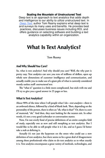
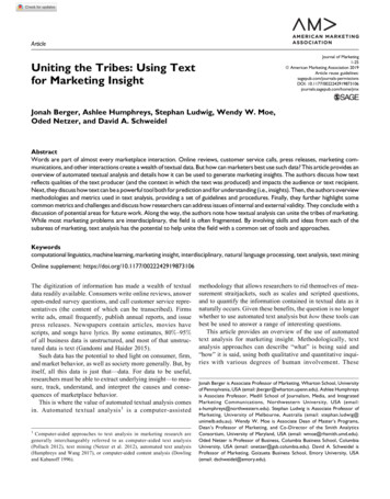
CHAPTER 1MECHANICAL PRINCIPLESDEFINITIONS:1. The study of mechanics in the human body is referred to as biomechanics.2. The study of the effects of internal and external forces on the human body inmovement and rest is called biomechanics.3. Mechanical principles applied to the study of biological functions; theapplication of mechanical laws to living structures; the study and knowledge ofbiological function from an application of mechanical principles is calledbiomechanics.Axis : It is the line about which movement takes place. An imaginary straightline around which an object rotates is called axis.Plane : It is the surface which lies at right angles to axis and in which the movement take place. These terms are used to facilitate the description of movementor direction.Types of axes : They are 3 types of axes.a. Sagittal axis: It lies parallel to sagittal suture of skull, i.e. in a anterior-posterior direction. Movement at this axes occur in a frontal plane.b. Frontal or transverse axes: It lies parallel to transverse suture of skull. It ishorizontal and at right angles to sagittal axes. Movement about frontal axes occurs in a sagittal plane.c. Vertical axes: It lies parallel to line of gravity. Movement about this axis is ina horizontal plane.SagittalFrontalVertical
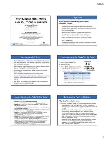
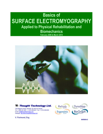
2EXERCISE THERAPYPlanes: There are three planes.1. Movement in horizontal plane (transverse plane): This plane divides thebody into upper and lower halves. Movements in transverse plane occur parallelto ground. For example in rotation of the head, the nose moves parallel to ground.Rotatory movements in a transverse plane occur around a vertical axis of motion. Movement in the horizontal plane is not affected by gravity hence it is statedas gravity free movement. Weak muscles which unable to produce movementagainst gravity can often succeed in this plane.2. Movement in frontal plane (coronal plane): the frontal plane divides thebody into front and back halves. Movements in the frontal plane occur side toside movements such as bringing the head to each of the shoulders. Rotatorymotion in the frontal plane occurs around an anterior posterior axis.3. Movement in vertical plane (sagittal plane): An anteroposterior verticalplane passing through the body from front to back, dividing it in half. It is theplane that divides the body or body segment into the right and left parts. Movements in this plane include forward and backward motions such as nodding ofthe head. Rotatory motion in the sagittal plane occurs around a coronal axis.Frontal planeSagittal planeTransverse plane
MECHANICAL PRINCIPLES3KINEMATICSKinematics is the area of biomechanics that include description of motion without regard for the forces producing it. They include.i.ii.iii.iv.Types of motion.Location of motion.Direction of motion.Magnitude of motion.(i) Types of motion: four types of motion are there.a. Rotatory motion: It is the movement of an object around a fixed axis in acurved path. Each point on the object or segment moves through the same angleat same distance. In human body the goal of most muscles appear to rotate abony lever around a fixed axis.b. Translatory motion: It is the movement of an object in a straight line. Eachpoint of an object moves through the same distance, at the same time, in parallelpaths.c. Curvilinear motion: Both rotatory and translatory motions combine to produce this motion. It is the most common form of motion produced in humanjoints Ex: thrown ball, where the ball both moves through space and rotates onits own axis.d. General plane motion: Here the object is segmented and free to move.(ii) Location of motion: Motion at a joint may occur in transverse, frontal orsagittal planes.(iii) Direction of Motion: Movement may occur either in clockwise oranticlockwise direction. Flexion and extension generally occur in sagittal planearound coronal axis. Flexion refers to rotation of one or both bony levers arounda joint axis so those ventral surfaces are being approximated. Rotation in thesame plane in the opposite direction is termed extension.
4EXERCISE THERAPYAbduction and adduction occur in frontal plane around Antero-posterior axis. Abduction is rotation of one or both segments of a joint around an axisso that the distal segment moves away from the midline of the body. Adductionin same plane but in opposite direction.Rotation occurs in horizontal plane around vertical axis. Medial rotationrefers to rotation toward the body’s midline. Lateral rotation refers to the opposite direction.(iv) Magnitude of Motion can be given either in degrees or radians.One radian 57.3010 0.01745 radiansGoniometer is most widely used measure for joint range in degrees.KINETICSKinetics means it is the area of biomechanics concerned with the forces producing motion or maintaining equilibrium. All forces are described as either Externalor Internal forces. External forces are pushes or pulls on the body arise fromsources outside the body. Ex: Gravity. Internal forces are forces act on the bodyarise from sources within human body Ex: Muscles, bones etc.Gravity: It is force by which all the bodies are attracted to earth. It is the mostconsistent force encountered by human body and behaves in a predictable manner.CENTER OF GRAVITY (COG): It is the point through which the earth’sattraction effectively acts regardless of position of body.The center of gravity (COG) of human body lies approximately at S2, anteriorto sacrum.LINE OF GRAVITY (LOG):- It is the vertical line through centre of gravity.When the human body is in fundamental standing position, the line of gravity(LOG) pass through vertex and a point between the feet, level with transversetarsal joints.
MECHANICAL PRINCIPLES5BASE OF SUPPORT: It is the area which is supported. In human body baseof support is the area bounded posteriorly by tips of heels and anteriorly by aline joining the tips of toes.EQUILIBRIUM: It results when the forces acting upon a body are balancedand the body remains at rest.Types of equilibrium are Stable equilibrium: If the forces acting upon a body at rest tend torestore it to its original position after it has been displaced, the body issaid to be in stable equilibrium. Unstable equilibrium: If a body is given an initial displacement and theforces acting upon it increase this initial displacement, the body is said tobe in unstable equilibrium. Neutral equilibrium: If, inspite of displacement of a body, the heightand position of its centre of gravity remain the same in relation to thebase, the body is said to be in neutral position.FIXATION AND STABILISATION Fixation is the state of immobility Stabilization is the state of relative immobility Active fixation of joints is obtained by co-combination of muscles. Passive fixation is by manual pressure straps, sand bags etc.FORCE: It is that which alters the state of rest of a body or its uniform motionin a straight lineThe force applies to a body is specified bya) direction of forceb) Magnitude of forceForces are classified as external force, internal force.External force: It is supplied from a source outside the body, i.e. the force ofgravity or the pressure of physiotherapist hand.Internal force: It is supplied by forces developed within the body i.e. by muscular contraction.
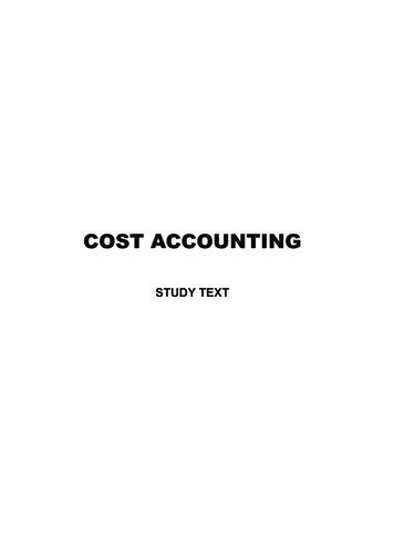
6EXERCISE THERAPYLEVER: It is a rigid bar that rotate around on axis- Forces applied to levers will produce either equilibrium or movementsuch as rotation or translation.- There are 3 orders or classes of levers.- Lever is capable of producing a movement about a fixed point calledfulcrum (F).- Work is done when a force or effort (E), applied at one point on thelever, acts upon another force or weight (W), acting at second point onlever.- The perpendicular distance from fulcrum to effort (E) is called as effortarm and from fulcrum to weight (W) is called as weight’s arm.Ist Order Lever: Here fulcrum is in between the effort and weight; it may besituated centrally, or towards either the effort or the weight, consequently theefforts and the weight arms may be equal, or may exceed the other in length.Ex: Nodding movements of head.Skull represents lever atlanto-occipital joints represents fulcrum, the weight issituated anteriorly in the face and the effort is supplied by contraction of posterior neck muscles.First order lever
MECHANICAL PRINCIPLES72nd Order Lever: The weight is in between fulcrum and effort, and the effort’sarm must therefore always exceed the weight’s arm. It helps in taking mechanical advantage, thus known as lever of power.Ex: Rising of heels to stand on toesTarsal and metatarsal bones are stabilized to form lever. Fulcrum is metatarsophalangeal joints weight of the body is transmitted to ankle joint by talus.Effort is applied by combination of calf muscles.Second order lever3rd Order Lever: Effort is in between fulcrum and weight, and weight arm musttherefore exceed the effort arm. It severs as mechanical disadvantage. It is considered as lever of velocity as it offers more velocity and less stability.Ex: When lever is forearm, fulcrum is elbow joint effort is supplied by contraction of brachialis muscle and weight is some object held in hand.Third order lever
8EXERCISE THERAPYMECHANICAL ADVANTAGE: Efficacy of force in relation to lever depends on two factors.They arei) Force exerted (W) or (E)ii) Perpendicular distance from fulcrum to the weight’s arm or efforts arm.- When both weights arm and efforts arm are of equal length no advantage is gained.- However if the length of effort arm exceeds weight arm an advantagewill be gained by the use of lever. This is known as Mechanical advantage.- Here less effort is required to lift a weight.- Mechanical advantage is obtained in 1st order lever when fulcrum isnearer to weight than to effort, and in all levers of the 2nd order. It isnever obtained in 3rd order lever.- It is the ratio of weight to effortM.A W/EPULLEYS: Pulley is a grooved wheel which rotates about a fixed axis by arope which passes round it. The axis is supported by a frame work or block.Types of pulleysi) Fixed pulleysii) Movable pulleysi) Fixed Pulleys: These are used to alter the direction of force. The pulleyblock is fixed and the rope which passes round the wheel is attached to theweight at one end and the effort is applied at the other.ii) Movable pulleys: These are used to gain mechanical advantage when liftingheavy weights. Commonly used for lifting the trunk for suspension exercises.The upper pulley is fixed to an overhead support, to which one end of rope isattached. The rope is then wound round the movable pulley, to which the weightis attached, and round the fixed pulley, the effort being applied at the free end.
MECHANICAL PRINCIPLESSingle Fixed Pulley9Two Fixed PulleySPRINGS:Spiral springs are used either to resist or to assist the force of muscular contraction, or to produce passive movement of joint, consist of a uniform coil of wirewhich is extensible.Springs in ParallelSprings in SeriesSprings used in parallel: When a spring of a specific weight is not availabletwo equal springs of half the required weight may be used in parallel to produce the same result.
10EXERCISE THERAPYSprings used in series: The weight of two equal springs arranged in series issame as that of a single spring, but the amount by which they must be extendedin order to reach the limit of extension is double that required for a single spring.ELASTICITY: It is the property of a body which regains its original shape afterthe application of force.TYPES OF MUSCLE CONTRACTION: Isometric IsotonicIsometric contraction involves the development of force by an increase inintramuscular tension without any change in length of the muscle.Isotonic contraction increases intramuscular tension accompanied by changein length of the muscle. It may either shorten or lengthen the muscle.Types of Muscle work:Work is the product of force and distance through which the force actsTypes arei)Static Muscle work: Muscles contract isometrically to balanceopposing forces and maintain stability. Therefore no work is done.ii)Concentric Muscle Work: the muscles contract isotonically inshortening to produce movement.iii)Eccentric Muscle Work: the muscles contract isotonically in lengthening. The muscle attachments are drawn apart.Range of Muscle Work: The amount of shortening or lengthening of muscleduring contraction is about 50 percent of muscles maximum length.Types of range are Inner range – muscle in its shortest position Outer range – muscle in fully extended position Middle range – muscle is neither fully shortened nor fully extended.
MECHANICAL PRINCIPLES11Group Action of Muscles: Integrated activity of many muscle groups is required for production of efficient functional movement. They arei) Agonists: Group of muscles which contract to provide the force required toproduce the movement.ii) Antagonists: These muscles oppose the action of agonists and relax progressively for permitting the movement.iii) Synergists: These groups of muscles work with agonists to provide a suitable activity and facilitates the movementsiv) Fixators: These muscles stabilize the bones of origin of the agonists andincreases their efficiency for production of movement.Limb Length Measurements: True shortening of leg is measured from the anterior superior iliac spineor upper margin of greater trochanter to lateral malleolus. Apparent shortening of leg is measured from umbilicus or xiphisternumto the level of knee joint or the tip of medial malleolus.
CHAPTER 2GAITGait may be described as a translatory progression of the body as a whole,produced by coordinated, rotatory movements of body segments.Stages of gait:I. stance phase: the stance phase begins at the instant that one extremity contacts the ground and continuous only as long as some portion of the foot is incontact with the ground.Heel strike: the beginning of the stance phase when the heel contacts the ground.Foot flat: It occurs immediately following heel strike, when sole of the footcontacts the floor.Mid stance: the point at which the body passes directly over the referenceextremity.Heel off: the point following midstance at which time the heel of the referenceextremity leaves the ground.Toe off: the point following heel off when only the toe of the reference extremityis in contact with the ground.
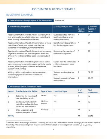
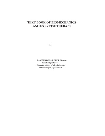
GAIT13II. Swing phase: the swing phase begins as soon as the toe of one extremityleaves the ground and ceases just before heel strike or contact of the sameextremity.Acceleration: the portion of beginning swing from the moment the toe of thereference extremity leaves the ground to the point when the reference extremityis directly under the body.Midswing: portion of the swing phase when the reference extremity passesdirectly below the body. Midswing extends from the end of acceleration to thebeginning of deceleration.Deceleration: the swing portion of the swing phase when the reference extremity is decelerating in preparation for heel strike.AccelerationMidswingDeceleration
14EXERCISE THERAPYVariables:Stance time: It is the amount of time that elapses during stance phase of oneextremity in a gait cycle.Single-support time: It is the amount of time that elapses during the periodwhen only one extremity is on supporting surface in the gait cycle.Double-support time: It is the amount of time that a person spends with boththe feet on the ground during one gait cycle.Stride length: It is the linear distance from the point of heel strike of one lowerextremity to the next heel strike of the same extremity.Step length: It is the linear distance from the point of heel strike of one lowerextremity to the next heel strike of the opposite extremity.Stride duration: It refers to amount of time it takes to accomplish one stride.Step duration: It refers to the amount of time spent during a single step.Cadence: It is the number of steps taken by a person per unit of time.Pathological gaits:1. Antalgic or painful hip gait: this is the gait of a person with a painful condition in the hip joint. To minimize the pain the person shortens the time duration ofthe stance phase on the painful side and quickly transfers the weight to the painless leg.2. Stiff hip gait: when one hip is ankylosed, it is not possible to flex at the hipjoint during walking to clear the ground in the swing phase.3. Unstable hip gait:The stability of the hip in walking is provided by the bony components of thejoint being kept in stable position by the muscles and ligaments around the joint.Any problem in these structures causes instability of hip.
GAIT15a. Trendelenberg gait: eg. Anatomical disruption on the right side Ex: nonunion fracture neck of femur. The action of gluteus medius in pulling the pelvisdownwards in the stance phase is ineffective or weak due to lack of a stablefulcrum. The pelvis drops on the opposite (i.e. left) side causing instability.b. Gluteus medius gait: when the right gluteus medius is paralyzed, it is unableto pull down the pelvis on the right due to a functional deficiency of the abductormechanism in the stance phase.4. Gluteus maximus gait: when the gluteus maximus muscle is paralyzed, thestabilizing factor is lost and the patient leans backward at the hip to passivelyextend it and keep the centre of gravity over the stance leg. This causes thebackward lurch in the gluteus maximus gait.5. Quadriceps gait: when quadriceps power is weak or paralyzed; the lockingis done by passively pushing the knee backward by the patient putting his handover the front of the lower thigh. This results in a limp and may even cause genurecurvatum.6. High stepping gait: when there is a foot drop, the foot slaps on the groundon heel strike and then drops in the swing phase. To get the foot clear the ground,the hip is flexed more and this causes the high stepping gait.7. Short leg gait: inequality of the legs is obvious when the shortening of one legis more than 1”. It leads to gait with a marked pelvic tilt downwards and anequines deformity at the foot.8. Scissoring gait: this is characteristic gait of a spastic child with marked bilateral spasm at the hips and equines spasm in the ankle.
CHAPTER 3INTRODUCTION TO PHYSIOTHERAPYPhysiotherapy (also known as physical therapy) is a health profession concerned with and the assessment, diagnosis and treatment of disease and disability through physical means. It is based upon principles of medical science, and isgenerally held to be within the sphere of conventional (rather than alternative)medicine.ROLE OF PHYSIOTHERAPY1. It provides psychological support for the patient in depression.2. It is useful in treating psychosomatic conditions.3. It helps in treating deformities and making the functionally independent.4. It plays a major role in treating neuro-muscular disorders.5. It improves the walking abilities.6. It relieves the pain, spasm etc.PRINCIPLES OF TREATMENT1. To relieve pain and spasm.2. To increase joint range of motion.3. To reduce the stiffness or contractures.4. To improve muscle power.5. To prevent deformities.6. To relieve from spasticity.7. To remove the secretions from the lungs.8. To improve the breathing capacity.9. To improve the aerobic capacity.10. Improve gait pattern etc.Methods and effects:1. Heat therapy: By heating tissues there will be rise in the temperature andincrease in the metabolitic activity. As a result of increase in the metabolism there is an increased demand for oxygen and foodstuffs, and anincreased output of waste products. There is blood flow to the part. Allthese physiological effects can be used to relieve the pain and spasm byincreasing the circulation and carrying the waste products. e.g. superficial heating modalities are wax bath, Hydrocollateral packs etc. Deepheating modalities are SWD, IFT etc.
INTRODUCTION TO PHYSIOTHERAPY172. Cryotherapy: The initial of skin to collng is vasoconstriction of bloodvessels which is useful in reducing the inflammation. Cryotherapy alsohelps in reducing the spasticity by reducing the nerve conduction velocity of muscle spindles. It also reduces pain by stimulating cold receptorswhich inhibits pain carrying fibers.3. Ultra Violet radiation thrapy is used to control skin diseases.4. Breathing exercises and postural drainage are used to reduce secretionsand improve breathing capacity.5. Passive and active exercises are used to increase joint ROM, deformities by preventing adhesion formation and maintaining circulation.6. Resisted exercises to improve strength: To improve muscle strength resisted exercises can be given with the help springs, pulleys, weights etc.7. Gait training to improve gait: after any musculoskeletal and neurologicaldeficits the patient tends to develop abnormal gait. To correct this wehave teach gait training to the patient by using assistive devices such ascrutches, frames, orthotics etc.These are some of the means of physiotherapy. There are so many otheruses which be dealt later in the text.
CHAPTER 4AN INTRODUCTION TO EXERCISE THERAPYExercise therapy is a means of accelerating the patient’s recovery from injuriesand diseases which have altered his normal way of living.The aims of exercise therapy1. To promote activity and minimize the effects of inactivity.2. To increase the normal range of motion.3. To strength the weak muscles.4. To improve the performance in daily activities.The techniques of exercise therapyMovement used in treatment may be classified as follows.I.Active movements1. Voluntary: (i) assisted(ii) Free(iii) Assisted-Resisted(iv) Resisted2. Involuntary reflexII.Passive movementsa. (i) Relaxed Passive Movements and accessory movementsb. Passive Manual Mobilization Techniques(i) Mobilizations of joints(ii) Manipulations of joints performed by(iii) Controlled sustained stretching of tightened structuresPosture: movement begins and ends in posture which is classified either asactive or passive.Active movement and posture is achieved by muscular contraction in responseto demands which are suitable to the patient’s ability.Passive movement and posture result from the application of external forceswhen the muscles are unable to contract voluntarily to permit movement or allowsupport.
INTRODUCTION TO EXERCISE THERAPY19The techniques which are most effective for obtaining the aims of treatment arethose which(i) Demand as much activity as possible.(ii) Based on patterns of movement which are the same as those used by thepatient for his normal functional activities.The approach to the patient problemThe problems arising from loss of function are different for each patient thereforetreatment must be planned according to the patient need.Assessment of the patient’s condition1. Functional tests: these are used to assess the patient’s needs and abilitieswith regard to functional activities e.g. mobility, personal care etc.2. Test of range of motion: it is measured with the help of goniometer.3. Tests for neuromuscular efficiency: these may be carried out electrically,manually or mechanically.Electrically: these may be carried out with the help of electro-myography.Manually: it can be done with manual muscle testing.Mechanically: these are done with tape measurement.Static power test: It may be recorded by means of a spring balance capable ofregistering up to 50 or 100 lbs.Dynamic power test: the maximum weight which can be lifted once through aprescribed range is called the one repetition maximum (1 R.M).4. Endurance test.5. Speed test.6. Tests for co-ordination.7. Tests of sensation.8. Measurement of vital capacity.
20EXERCISE THERAPYGONIOMETRYThe term goniometry is derived from two greek words, gonio, meaning angle,and metron, meaning measure. Therefore, goniometry refers to the measurement of angles, in particular the measurement of angles created by human joints.Types of goniometry1. Universal goniometry: These are most commonly used instrument. Thebody of a universal goniometer resembles a protractor and may from full or halfcircle. Measurement scales are located on the body (0-180 or 0-360). It consists of two arms stationary or fixed arm and movable arm. Stationary can notbe moved. Movable arm is attached to the fulcrum which is the center of thebody and it can be moved. It contain a black line extend the length of the arm formeasuring the angle.2. Gravity dependent goniometers: These are sometimes called inclinometers. They use gravity’s effect on pointers and fluid levels to measure joint position and motion.3. Electro goniometers: These are used primarily in research to obtain dynamic joint measurements. It is similar to that of universal goniometer.4. Visual estimation: Although some examiners make visual estimates of jointposition and motion but it is not a recommended position.5. Pendulum goniometer: It consists of a 360 degree protractor with a weightedpointer hanging from the center of the protractor.6. Fluid goniometer: It has fluid filled circular chamber containing an air bubble.It is similar to a carpenter’s level, but being circular, has a 360 degree scale.
INTRODUCTION TO EXERCISE THERAPY21I. UPPER LIMBShoulder joint range of motion1. FlexionRecommended testing position: supine lyingNormal ROM: 0-1800Fulcrum : acromial processMovable arm: middle line of humerusFixed arm : midaxillary line of thorax2. ExtensionRecommended testing position: prone lyingNormal ROM: 0-60Fulcrum: coracoid processMovable arm: lateral midline of the humerus.Fixed arm : midaxillary line of thorax3. Abduction and AdductionRecommended testing position: supine lyingNormal ROM: 0-180Fulcrum: acromial processMovable arm: medial midline of humerusFixed arm : parallel to the midline of the anterior aspect of the sternum.4. AdductionNormal ROM: 180-0Rest is same as abduction5. Medial rotationRecommended testing position: supine lying, with the arm placed at 90 of abductionNormal ROM: 0-70Fulcrum: olecranon processMovable arm : parallel to ulnaFixed arm : parallel or perpendicular to the floor6. Lateral rotationNormal ROM: 0-90Rest is same as medial rotation
22EXERCISE THERAPYELBOW1. Flexion and ExtensionRecommended testing position: supine lyingNormal ROM: 0-135Fulcrum: Lateral epicondyle of humerusMovable arm : lateral midline of the humerusFixed arm : midline of the humerus2. ExtensionNormal ROM: 135-0Rest is same as flexionFOREARM1. SupinationRecommended testing position: sitting with upper arm at the side of the body,elbow flexed to 90 and forearm supportedNormal ROM: 0-80Fulcrum : lateral to the ulnar styloid processMovable arm: ventral aspect of the forearm, proximal to styloid processFixed arm : anterior midline of humerus2. PronationRecommended testing position: same as supinationNormal ROM: 0-80Fulcrum: lateral to the ulnar styloid processMovable arm : dorsal aspect of the forearm, proximal to styloid process ofradiusFixed arm : anterior midline of humerusWRIST1. FlexionRecommended testing position: sitting next to a supporting surface and handfacing the ground.Normal ROM: 0-80Fulcrum: lateral aspect of the wrist over the triquetrumMovable arm : lateral midline of the fifth metacarpalFixed arm : lateral midline of the ulna
INTRODUCTION TO EXERCISE THERAPY232. ExtensionRecommended testing position: same as flexionNormal ROM: 0-70Fulcrum: at the level of capitateMovable arm : volar midline of the third metacarpalFixed arm : volar midline of the forearm3. Radial deviationRecommended testing position: same as flexionNormal ROM: 0-20Fulcrum: at the level of capitateMovable arm : dorsal midline of the third metacarpalFixed arm : dorsal midline of the humerus4. Ulnar deviation:Normal ROM: 0-30Rest is same as radial deviationII. LOWER LIMBHIP JOINT1. FlexionRecommended testing position: supine lyingNormal ROM: 0-120Fulcrum: lateral aspect of the hip jointMovable arm: lateral midline of the femurFixed arm : lateral midline of the pelvis2. Extensi
as gravity free movement. Weak muscles which unable to produce movement against gravity can often succeed in this plane. 2. Movement in frontal plane (coronal plane): the frontal plane divides the body into front and back halves. Movements in the frontal plane occur side to side movements suc