Transcription
Ob/Gyn Sonography ReviewA Q&A REVIEW FOR THE ARDMS OBSTETRICS & GYNECOLOGY EXAM
Ob/Gyn Sonography ReviewA Q&A REVIEW FOR THE ARDMS OBSTETRICS & GYNECOLOGY EXAM2nd Edition2018Kathryn A. Gill, MS, RT, RDMS, FSDMSInstitute of Ultrasound DiagnosticsSpanish Fort, AlabamawithJim Baun, BS, RDMS, RVT, FSDMSProfessional Ultrasound ServicesSan Francisco, CaliforniaTraci B. Fox, EdD, RT(R), RDMS, RVTJefferson College of Health ProfessionsThomas Jefferson UniversityDAVIESPUBLISHING
Copyright 2018 by Davies Publishing, Inc.DAVIESPUBLISHINGAll rights reserved. No part of this work may be reproduced, stored in a retrieval system, ortransmitted in any form or by any means, electronic or mechanical, including photocopying,scanning, and recording, without prior written permission from the publisher.Davies Publishing, Inc.32 South Raymond AvenuePasadena, California 91105-1961Phone 626-792-3046Facsimile 626-792-5308e-mail ichael Davies, PublisherChristina J. Moose, Editorial DirectorCharlene Locke, Production ManagerJanet Heard, Operations ManagerJim Baun, IllustrationSatori Design Group, Inc., DesignPrinted and bound in the United States of AmericaISBN 978-0-941022-26-2Library of Congress Cataloging-in-Publication DataNames: Gill, Kathryn A., author. Baun, Jim, author. Fox, Traci B., author.Title: OB/GYN sonography review : a Q&A review for the ARDMS obstetrics &gynecology exam / Kathryn A. Gill with Jim Baun, Traci B. Fox.Description: 2nd edition. Pasadena, California : Davies Publishing, Inc.,2017. Preceded by OB/GYN sonography review : a review for the ARDMSobstetrics & gynecology exam, 2003/2004 / Kathryn A. Gill, Misty H.Sliman, Peter W. Callen. c2003. Includes bibliographical references andindex.Identifiers: LCCN 2016052047 ISBN 9780941022262 (alk. paper)Subjects: MESH: Obstetrics Genital Diseases, Female--ultrasonography Pregnancy Complications--ultrasonography Examination QuestionsClassification: LCC RG527.5.U48 NLM WQ 18.2 DDC 618.1/07543--dc23 LC record availableat https://lccn.loc.gov/2016052047
ReviewersMarianna Desmond, EdM, RDMS, RT(R)Lead SonographerMidwest Center for Women’s HealthKenosha, WisconsinAnnmarie Lobdell, MHS, RDMS, RDCS, RVTClinical Technology ConsultantMindray ZONAREMahwah, New JerseyJulia A. Drose, BA, RDMS, RDCS, RVTManager, Divisions of Ultrasound andPrenatal Diagnosis and GeneticsUniversity of Colorado HospitalAssociate Professor of RadiologyUniversity of Colorado School of MedicineDepartment of RadiologyAurora, ColoradoDarla J. Matthew, BAS, RT(R)(S), RDMSProgram DirectorDiagnostic Medical SonographyDoña Ana Community CollegeLas Cruces, New MexicoJanice Hickey Scharf, BSc, RDMS, CRGS,FSCClinical Applications SpecialistUltrasound, Philips Healthcare CanadaMarkham, Ontario, CanadaDiane M. Kawamura, PhD, RT(R), RDMS,FAIUM, FSDMSBrady Presidential Distinguished ProfessorDepartment of Radiologic SciencesWeber State UniversityOgden, UtahFrederick W. Kremkau, PhD, FACR,FAIMBE, FAIUM, FASAProfessor of Radiologic SciencesDirector, Program for Medical UltrasoundCenter for Applied LearningWake Forest University School of MedicineWinston-Salem, North CarolinaDebra L. Krukowski, BS, RDMS, RTProgram Coordinator/FacultySchool of Health CareersTriton CollegeRiver Grove, IllinoisSusan Nager, BS, RDMSDiagnostic Medical Sonography InstructorAmerican College of Medical CareersOrlando, FloridaRegina K. Swearengin, BS, RDMSDepartment Chair, SonographyAustin Community CollegeAustin, TexasMark G. Torchia, MSc, PhDExecutive Director, Centre for theAdvancement of Teaching and LearningAssociate Professor of SurgeryUniversity of ManitobaWinnipeg, Manitoba, CanadaClaudia von Zamory, BA, RDMS, RVTUltrasound Program DirectorGurnick Academy of Medical ArtsSan Mateo, CaliforniaKerry E. Weinberg, PhD, MA, MPA, RT(R),RDMS, RDCS, FSDMSDirector and Associate ProfessorDiagnostic Medical Sonography ProgramSchool of Health ProfessionsLong Island UniversityBrooklyn, New Yorkv
Preface to the 2nd EditionT IS MOCK EXAM is Step 2 in Davies’ 1-2-3 Step Ultrasound Education & Test PreparationHsystem. Together, the three publications in this system form a targeted, integrated,silver-bullet method that prepares sonographers to take, and pass, the Ob/Gyn specialtyexamination given by the American Registry for Diagnostic Medical Sonography (ARDMS). Youcan learn more about the companion volumes—Step 1, Ob/Gyn Sonography: An IllustratedReview, and Step 3, ScoreCards for Ob/Gyn Sonography—by visiting Davies’ website atwww.daviespublishing.com.The second edition of Ob/Gyn Sonography Review has been revised, expanded, and fully updatedto cover all the topics identified on the ARDMS exam content outline—and more. We have addednearly 200 questions and updated more, for a total of 700 registry-like items, and the mock examnow contains 230 images, schematics, and anatomic illustrations.The actual ARDMS exam contains approximately 170 questions. Mastery of these 700 registrylike items and their underlying concepts—as explained in the companion Step 1 review textby Jim Baun, Ob/Gyn Sonography: An Illustrated Review—will thoroughly arm you to pass thisARDMS specialty exam to obtain the RDMS credential or simply to cross-train for added expertise.Designed as an adjunct to your regular study, this mock exam will help you precisely determineyour strengths and weaknesses and gives you clear explanations and page-specific referencesto Davies’ Step 1 text so that you can study most effectively. The Step 3 flashcard component,ScoreCards for Ob/Gyn Sonography, ensures your mastery of basic facts, values, and principles—and also your ability to think on your feet—with numerous image-based questions. We also highlyrecommend Kathryn Gill’s masterful Ultrasound in Obstetrics and Gynecology: A Practitioner’sGuide, which, with its particular strengths in clinical protocols, tips, and pitfalls, forms a valuablecomplement to the Step 1 review text. All these study aids come with SDMS-approved CME creditand are resources that sonographers will want to keep in their reference libraries.Facts about Ob/Gyn Sonography Review:4 This mock exam covers the material on the ARDMS exam content outline in effect as of2018. Readers are advised to check the ARDMS website, www.ardms.org, for the latestupdates. This mock exam itself is continuously updated and revised as necessary.4 This mock exam focuses exclusively on the Obstetrics and Gynecology specialty exam toensure thorough coverage of even the smallest subtopic and task on the exam. (For thosepreparing for the Sonography Principles and Instrumentation exam, see Davies’ UltrasoundPhysics Review: SPI Edition, available at www.DaviesPublishing.com.)4 In preparing this mock exam, we have referred to the last best ARDMS content outlineas a guideline for coverage. At the same time, we have organized the content using acomprehensive, subject-driven approach to ensure that all important topics are fullyaddressed. The ARDMS exam content outline provides a generalized categorical overviewtogether with very specific clinical tasks, but it can omit and assume the mastery of keyintermediate topics you must know to pass the examination. Hence our hybrid approachgives you the best of both worlds.4 This second edition of Ob/Gyn Sonography Review contains 700 registry-like questions—37%more than the first edition. Twice as many questions are now illustrated, with over 230images and schematics.vii
viii Ob/Gyn Sonography Review, 2nd Edition4 While otherwise in ARDMS exam format, this Davies mock exam makes deliberate andjudicious use of answer-choice formats that occasionally depart from those on the ARDMSexam. We use multiple-choice items with five, not four, possible choices—thereby increasingboth the difficulty of each question and the time needed to answer it. We also sprinkle in afew answer-choice variants (such as “A and B” and “not/except” items). The point? To giveyou not only practice for the registry exam but also an educational tool that will exercisethose neural pathways in more than one direction. Registry candidates who stretch beyondthe standard format and master these items at an average rate of 1 minute apiece will beexceptionally well prepared for the actual exam.4 The answer key located in Part 5 contains not only the answers but also conciseexplanations that are clear and authoritatively referenced for fact checking and furtherstudy. We recommend that you have a copy of the Davies Step 1 review text, Ob/GynSonography: An Illustrated Review by Jim Baun, for extended and well-illustrated coverageof your weak areas, as well as Kathryn Gill’s Ultrasound in Obstetrics and Gynecology: APractitioner’s Guide. The answer key provides page-specific references to these integratedtexts along with other authorities. No more searching for sources and pages!4 This mock examination has been approved by the Society of Diagnostic Medical Sonography(SDMS) as a CME activity. A CME application form, quiz, and full submission instructionsare included in Part 6. Passing this quiz will qualify the applicant for 12 CME credits. Amodest administrative processing fee applies at the time of submission, and more than onesonographer may submit this activity for CME credit. These credits are accepted by ARDMS,the Alliance for Physician Certification and Advancement (APCA), the American Registryof Radiologic Technologists (ARRT), and other organizations toward meeting their CMErequirements. Some credentials carry stipulations regarding specialty areas in which CMEcredits may be earned. Always check with the organization that governs your credential(s).All the credits in this activity may be applied to maintain the RDMS credential.4 A bibliography of current authoritative text references appears In Part 7. It includes theall-important Step 1 review text by Jim Baun, the Step 3 ScoreCards for Ob/Gyn Sonographyby Traci B. Fox, and the complementary Ultrasound in Obstetrics and Gynecology byKathryn A. Gill.4 Finally, Part 8 functions as an index to the ARDMS exam content outline. Here you will findthe full outline, complete with all tasks, as published on the ARDMS website. Each task onthe exam outline is cross-referenced to any and all questions in this Ob/Gyn mock examthat pertain to that task, for your convenience in targeting your study on specific tasks.We also encourage you to consult the ARDMS website at www.ardms.org for the latestinformation and updates on the Ob/Gyn specialty examination.ARDMS Advanced Item Type (AIT) QuestionsAll the ARDMS exams now include Advanced Item Type (AIT) questions that assess practicalsonography instrumentation skills. For the Ob/Gyn specialty exam, these AIT questions includewhat ARDMS calls “Hotspot” questions. Hotspot items display an image and question, requiringyou to indicate the correct answer by marking directly on the image using your cursor. This typeof question is considered advanced because it requires a higher level of clinical thinking andprocessing than required when you are answering a conventional multiple-choice question. InDavies’ mock exam, similar questions are identified as AIT—Hotspot questions. These items askyou to identify what an arrow in the image is pointing at or to indicate the label on an image thatcorresponds to the correct answer.Another type of AIT question, the AIT—SIC (Semi-Interactive Console) item, requires the examineeto use a semi-interactive console to correct a problem with the image presented. These itemsare currently limited to the Sonography Principles and Instrumentation (SPI) examination, but as abonus feature we have identified such items for your reference.
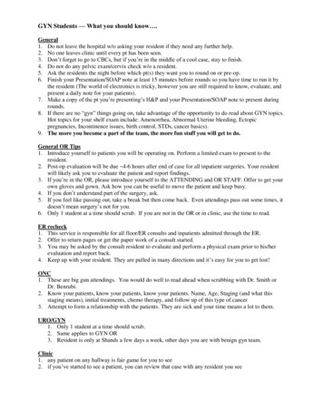
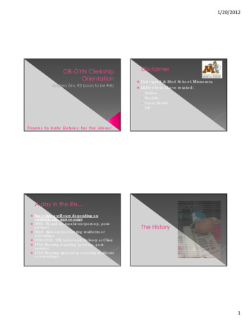
Ob/Gyn Sonography Review, 2nd Edition ixFinally, PACSim items—case-based Picture Archive and Communication Simulation questions—are not included in this Ob/Gyn sonography mock exam due to the multifaceted interactive natureof these questions. Currently there are approximately two PACSim questions on the Ob/Gynexam. These items are designed to simulate a reading workstation experience, a picture archiveand communication system. Each item presents a brief case description, or clinical history.Candidates must read the case/clinical history of the patient, evaluate existing image(s), andcomplete the ultrasound report by selecting options from a drop-down menu. PACSim questionsmove the examinations closer to clinical practice, improving the level of certainty that the testtaker has demonstrated the basic levels of skill, knowledge, and abilities when the ARDMS awardsa credential. Davies recommends that those taking the Ob/Gyn specialty exam visit the ARDMSwebsite at http://www.ardms.org/Pages/PACS.aspx to access the PACSim User Guide, read FAQs,see sample questions, and watch a video tutorial.How to Use This Mock ExamOb/Gyn Sonography Review effectively simulates the content of the ARDMS Ob/Gyn specialtyexam. Current ARDMS standards call for approximately 170 multiple-choice questions to beanswered during a three-hour period. That is, you will have an average time of just over 1 minuteto answer each question. Timing your practice sessions according to the number of questions youneed to finish will help you prepare for the pressure experienced by Ob/Gyn candidates takingthis exam. It also helps to ensure that your practice scores accurately reflect your strengths andweaknesses so that you can study more efficiently and with greater purpose in the limited timeyou are able to devote to preparation—especially if you use Davies’ integrated 1-2-3 Step testprep products.IMPORTANT NOTE: Although many of our customers remark on similarities between ourquestions and those of the actual exam, do not be misled into thinking you should memorizethese questions and answers. They are here to give you practice, to teach you things you may notknow, and to reveal your strengths and weaknesses so that you know where to put your energyas you prepare for the exam.ARDMS test results are reported as a “scaled” score that ranges from a minimum of 300 to amaximum of 700. A scaled score of 555 is the passing score—the “passpoint” or “cutoff score”for all ARDMS examinations. The scaled score is simply a conversion of the number of correctanswers that also, in part, takes into account the difficulty of a particular question. You cansearch on the Internet for “Angoff scoring method” if you want to learn more about scaledscoring. Suffice it to say that it helps to ensure the fairness of the exams.We include below and strongly recommend that you read “Taking and Passing Your Exam,” byDon Ridgway, RVT, who offers useful tips and practical strategies for taking and passing theARDMS examinations.Finally, you have not only our best wishes for success but also our admiration for taking this bigand important step in your career.Kathy GillJim BaunTraci B. FoxKathryn A. Gill, MS, RT, RDMS, FSDMSJim Baun, BS, RDMS, RVT, FSDMSTraci B. Fox, EdD, RT(R), RDMS, RVT
Taking and Passing Your Examby Don Ridgway, RVT*Preparing for Your Exam . . .Study. And then study some more. Knowing your stuff is the most important factor in yoursuccess. Start early, set a regular study schedule, and stick to it. Make your schedule specificso you know exactly what to study on a particular day. Write it down. Establish realistic goalsso that you don’t build a mountain you can’t climb.As to what you study, don’t just read aimlessly. Focus your efforts on what you need to know.Rely on a core group of dependable references, referring to others as necessary to firm up yourunderstanding of specific topics. Let the ARDMS exam outline guide you. And use different butcomplementary study methods—texts, flashcards, and mock exams—to exercise those neuralpathways.Ease down on studying the week before. Wind down, reduce stress, build confidence, andrest up. Don’t cram! And no studying the night before. You had your chance. Watch a movie, relax,go to bed early, and sleep well.Organize your things the night before. Lay out comfortable clothes (including a sweater orsweatshirt in case the testing center is cold), pencils, your ARDMS test-admission papers, car andhouse keys, glasses, prescriptions, directions to the test center, and any other personal items youmight need. Be prepared!The Day of Your Exam . . .Eat lightly. You do not want to fall asleep during the exam. Go easy on the coffee or tea so yourbladder doesn’t distract you halfway through the exam.Arrive early. Plan to arrive at the test center early, especially if you haven’t been there before.Take directions, including the telephone number of the testing center in case you have to makecontact en route. You don’t need a wrong-offramp adventure.Be confident. As you wait for the exam to begin, smile, lift both hands, wave them towardyourself, and say, “Bring it on.”During the Exam . . .Read each question twice before answering. Guess how easy it is to get one word wrong andmisunderstand the whole question!Try to answer the question before looking at the choices. Formulating an answer beforepeeking at the possibilities minimizes the distractibility of the incorrect answer choices, which inthe test-making business are called—guess what?—distractors.*Don Ridgway is the author of Introduction to Vascular Scanning: A Guide for the Complete Beginner and editor of VascularTechnology Review. He is Professor Emeritus at Grossmont College in El Cajon, California.x
Ob/Gyn Sonography Review, 2nd Edition xiKnock off the easy ones first. First answer the questions you feel good about. Then go backfor the more difficult items. Next, attack the really tough ones. Taking notes on long or trickyquestions often can jog your memory or put the question in new light. For questions you justcannot answer with certainty, eliminate the obviously wrong answer choices and then guess.Guessing. Passing the exam depends on the number of correct answers you make. Becauseunanswered questions are counted as incorrect, it makes sense to guess when all else fails. TheARDMS itself advises that it is to the candidate’s advantage to answer all possible questions.Guessing alone improves your chances of scoring a point from 0 (for an unanswered question) to25% (for randomly picking one of four possible answers). Eliminating answer choices you know orsuspect are wrong further improves your odds of success. By using your knowledge and skill toeliminate two of the four answer choices before guessing, for example, you increase your oddsof scoring a point to 50%.Pace yourself; watch the time. Work methodically and quickly to answer those you know, andmake your best guesses at the gnarly ones. Leave no question unanswered.Don’t despair 50 minutes into the exam. At some point you may feel that things just aren’tgoing well. Take 10 seconds to breathe deeply—in for a count of five, out for a count of five. Relax.Recall that you need only about three out of four correct answers to pass. If you’ve preparedreasonably well, a passing score is attainable even if you feel sweat running down your back.Taking the Exam on Computer . . .Some candidates express concern about taking the registry exam on a computer. Most folksfind this to be pretty easy; some find it offputting, at least in prospect. But the computerizedexams are quite convenient: You can take the exam at your convenience (a far cry from thedays of one exam per year), you know whether or not you passed before you leave the testingcenter (compare that to waiting weeks and even months, as used to be the case), and you canreschedule the exam after 90 days if you happen not to pass the first time (rather than waitinganother six months to a year). Another good point: The illustrations are said to be clearer oncomputer than in the booklets at a Scantron-type exam.Taking the test by computer is not complicated. The center even gives you a tutorial to be sureyou know what you need to do. You sit in a carrel with a computer and answer the multiplechoice questions by pointing and clicking with a mouse. There is a clock on the display lettingyou know how much time is left. Use it to pace yourself. Scratch paper is available; make liberaluse of it.You can mark questions for answering later. A display shows which questions have not beenanswered so you can return to them. When you have finished, you click on “DONE,” and you findout immediately whether you passed.It’s nothing to be afraid of. The principles are the same as those for any exam. Be methodical andkeep breathing.
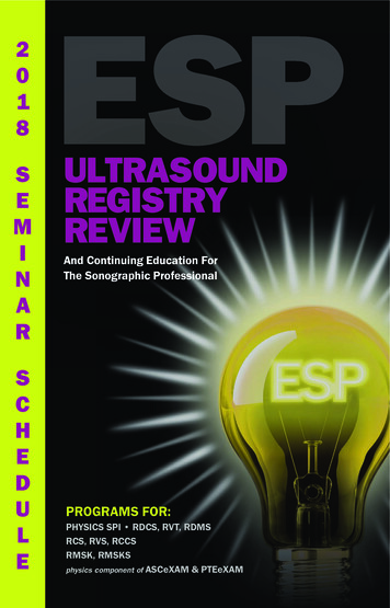
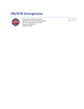
xii Ob/Gyn Sonography Review, 2nd EditionSummary . . .Preparing for the exam:4 Study.4 Use flashcards.4 Join a study group.4 Wind down a week before.4 Don’t cram.4 Relax!The day of your exam:4 Eat lightly, avoid coffee.4 Arrive early.4 Take a sweater.4 Be confident!During the exam:4 Read each question twice.4 Answer the question before looking at the answer choices.4 Answer the easy ones first.4 Guess when necessary.4 Pace yourself.4 Don’t despair.Taking the exam on computer:4 Just point and click.4 Take notes.4 Mark and return to the hard questions.4 Use the on-screen clock to pace yourself.4 Be methodical.4 Breathe!
ContentsReviewers vPreface to the 2nd Edition viiTaking and Passing Your Exam xColor Plates xviiPART 1Obstetrics 1First trimesterGestational ageGestational sacYolk sacEmbryonic developmentOvaries/corpus luteumCul-de-sacEctopic pregnancyPregnancy failureSecond and third minalGastrointestinalGenitourinaryExtremitiesFetal branesUmbilical cordAbruptionPreviaMasses and lesionsMaturity/gradingxiii
xiv Ob/Gyn Sonography Review, 2nd EditionDopplerPhysiologyAccretaAssessment of gestational ageGestational sacEmbryonic size/crown-rump lengthBiparietal diameterFemur lengthAbdominal circumferenceHead circumferenceTranscerebellar measurementsBinocular measurementsCephalic indicesFetal lung maturityOtherComplicationsIntrauterine growth restrictionBiophysical profile/nonstress testMultiple gestationsMaternal illnessAntepartumFetal therapyPostpartumAmniotic fluidAssessmentPolyhydramniosOligohydramniosFetal pulmonic maturity studiesGenetic studiesMaternal serum testingAmniotic fluid testingChorionic villus samplingDominant/recessive risk occurrenceFetal demiseFetal abnormalitiesCranialFacialNeckNeural tube
Ob/Gyn Sonography Review, 2nd Edition Abdominal ardiacSyndromesOtherCoexisting disordersLeiomyomaCystic massesTrophoblastic diseaseSolid/mixed massesMyometrial contractionOtherPART 2Gynecology 117Normal pelvic anatomyUterusOvariesFallopian tubesSupporting structuresCul-de-sacVasculatureDoppler flowGynecology-related studiesPhysiologyMenstrual cyclePregnancy testsHuman chorionic gonadotropin (hCG)FertilizationPediatricPrecocious pubertyHematometra/hematocolposSexual onCausesxv
xvi Ob/Gyn Sonography Review, 2nd EditionMedications and treatmentOvulation induction (follicular monitoring)Assisted reproductive technology (ART)—GIFT, IVF, yPelvic pathologyCongenital uterine malformationUterine massesOvarian massesEndometriosisPolycystic ovarian diseaseInflammatory diseaseDoppler flow studiesGynecology-related studiesOtherExtrapelvic pathology associated with gynecologyAscitesLiver metastasisHydronephrosisOtherPART 3Patient Care 171PART 4Physical Principles and Instrumentation 177PART 5Answers, Explanations, and References 183PART 6Application for CME Credit 327PART 7Bibliography 353PART 8 ARDMS Exam Content Outline Tasks Cross-Referenced to Mock ExamQuestions 355
PART 1ObstetricsFirst trimesterSecond and third trimestersPlacentaAssessment of gestational ageComplicationsAmniotic fluidGenetic studiesFetal demiseFetal abnormalitiesCoexisting disorders1
2 Ob/Gyn Sonography Review, 2nd EditionFirst TrimesterGestational ageGestational sacYolk sacEmbryonic developmentOvaries/corpus luteumCul-de-sacEctopic pregnancyPregnancy failure1. When sonographers use the term gestational age, they mean:A. Weeks or days since conceptionB. Weeks or days since fertilizationC. Weeks or days since implantationD. Weeks or days since the first day of the last menstrual periodE. Weeks or days since the last day of the last menstrual period2. Up to 10 weeks’ gestational (menstrual) age, the mean diameter of the normal gestationalsac should grow approximately:A. 0.5 mm per dayB. 1.0 mm per dayC. 2.0 mm per dayD. 3.0 mm per dayE. 4.0 mm per day3. A successful pregnancy outcome is associated with a yolk sac that:A. Occupies more than 30% of gestational sac volumeB. Has a round shapeC. Measures less than 2 mm in diameter at 8–10 weeksD. Measures greater than 6 mm in diameterE. Has irregular echogenic borders4. Which sonographic finding is associated with an abnormal pregnancy?A. Spherical gestational sac within the uterusB. Double sac sign within an intrauterine gestational sacC. Oval gestational sac within the uterusD. Defined double decidual ring around the intrauterine gestational sacE. Embryo with a calcified yolk sac
Ob/Gyn Sonography Review, 2nd Edition 35. An incomplete abortion is defined as:A. Spontaneous abortion with retained products of conceptionB. Anembryonic abortionC. Heterotopic pregnancyD. Subchorionic hemorrhageE. Ectopic pregnancy without bleeding6. When does the trilaminar embryonic disc form?A. During organogenesisB. During implantationC. During transit through the fallopian tubeD. During neurulationE. During gastrulation7. A patient presents with a positive pregnancy test and bright red spotting. By dates she is8–9 weeks. What does this transverse image demonstrate?A.B.C.D.E.Second gestational sacAn anembryonic pregnancyNormal amnionSubchorionic hemorrhagePlacental abruption8. What is being measured in the image in question 7?A. Gestational sacB. Biparietal diameterC. Crown-rump lengthD. Embryonic discE. Abdominal circumferenceAIT—Hotspot.**Advanced Item Type (AIT) Hotspot items marked here are similar to Hotspot items on the ARDMS exam. On the exam, theseHotspot items require you to indicate the correct answer by marking directly on the image using your cursor. In Davies’ Ob/Gynmock exam, similar questions are identified as AIT—Hotspot and ask you to identify what an arrow in the image is pointing at or toindicate the label on an image that corresponds to the correct answer. Another type of AIT question, the AIT—SIC (Semi-InteractiveConsole) item, requires you to use a semi-interactive console to correct a problem with the image presented. These items are currently limited to the Sonography Principles and Instrumentation (SPI) examination, but as a bonus feature we have also identifiedthese items in this mock exam.
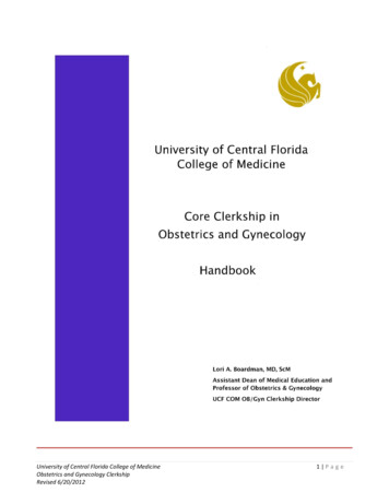
4 Ob/Gyn Sonography Review, 2nd Edition9. In the image in question 7, what is the arrow pointing to?A. Amniotic cystB. Fetal headC. Gestational sacD. Yolk sacE. Umbilical cordAIT—Hotspot.10. Your patient relates a history of amenorrhea for 7 weeks. Her home pregnancy test wasnegative, but her serum beta-hCG exceeds 4000 mIU/ml. What does the image belowdemonstrate?A.B.C.D.E.Pseudocyesis with an endometrial cystFluid contained within the endometrial cavityNormal early intrauterine pregnancyDegenerating submucosal fibroidNormal empty uterus with periovulatory endometrium11. This patient presented with bleeding and cramping. Her pregnancy test is weakly positive.Three days prior, a living IUP was documented in the uterus. This transvaginal image suggests:A. Normal intrauterine pregnancyB. Incomplete abortion
Ob/Gyn Sonography Review, 2nd Edition 5C. Ectopic pregnancyD. PseudocyesisE. Placental abruption12. The primitive hindbrain can be seen as a cystic structure within the embryonic head. Whatis the name of this structure?A. ProsencephalonB. MesencephalonC. EncephaloceleD. DiencephalonE. Rhombencephalon13. What is a heterotopic pregnancy?A. A cervical ectopic pregnancyB. A fertility-assisted pregnancyC. An abdominal ectopic pregnancyD. Coexisting intrauterine and ectopic pregnanciesE. A twin ectopic pregnancy14. In a ruptured ectopic pregnancy, which section of the fallopian tube is potentially the mostlife-threatening?A. LigamentousB. AmpullaC. InterstitialD. FimbrialE. Isthmic15. Which statement about ectopic pregnancies is NOT true?A. Interstitial ectopics are more serious than those located in the ampulla.B. The ovary is the second most common site for ectopic pregnancy.C. The increased incidence of ectopic pregnancies is mostly attributable to sexuallytransmitted diseases.D. The most common clinical symptom is pain.E. If a patient has had a previous ectopic pregnancy, she is at increased risk for a recurrentectopic pregnancy.16. A longitudinal image shows an intrauterine gestational sac that occupies one-half of theendometrial canal. The sac size indicates that the gestational age of the pregnancy shouldbe:A. 4 weeksB. 6 weeksC. 8 weeksD. 10 weeksE. 12 weeks
6 Ob/Gyn Sonography Review, 2nd Edition17. A patient presents with a positive pregnancy test, bleeding, and cramping. The sonogramreveals an intrauterine gestational sac containing an echogenic structure but no heartbeat.Measurement of the structure shows it to be 11 mm in length. The most likely diagnosis is:A. Anembryonic pregnancyB. Missed abortionC. Placenta previaD. Partial moleE. Early pregnancy failure18. What is the arrow pointing to in this image?A.B.C.D.E.AmnionYolk sacSeptumChorionSynechiaeAIT—Hotspot.19. Which of the following is NOT an indication of ectopic pregnancy?A. Fluid in the cul-de-sacB. Adnexal massC. Double decidual ringD. Fluid within the endometrial cavityE. Fluid in the right upper quadrant
Ob/Gyn Sonography Review, 2nd Edition 20. This sagittal transvaginal image demonstrates a normal-appearing intrauterine gestationalsac. The hypoechoic structure indicated by the calipers most l
Ob/Gyn Sonography Review, 2nd Edition ix Finally, PACSim items—case-based Picture Archive and Communication Simulation questions— are not included in this Ob/Gyn sonography mock exam due to the multifaceted interactive nature of these questions. Currently there are approximately two PACSim questi