Transcription
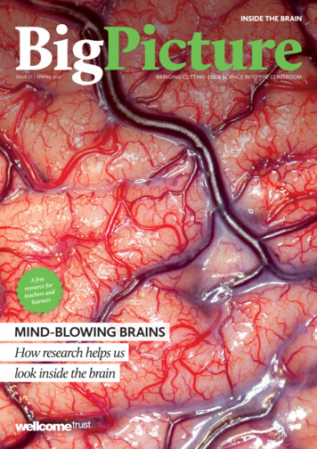
INSIDE THE BRAINISSUE 17 SPRING 2013bringing CUTTING-EDGE SCIENCE INto THE CLASSROOMMIND-BLOWING BRAINSHow research helps uslook inside the brain
The vital stats of brains and brain imaging0.5 l1.13 l1.26 lSource: e: orIonisingRadiationSource: www.ncbi.nlm.nih.gov/pmc/articles/PMC3386878/?tool pubmedBrain size and massSpeed of nerve transmission4HumanStructural imagingHow researchers and clinicianscan see inside our brains.6real voicesThree people talk about therole of brains in their lives.Chimp0.42 kg85–35 m/s80–120 m/s223.5 m/s1.4 kg4.8 kgFrom top: unmyelinated fibre, thin myelinated fibre, thick myelinated fibre, cruising commercial airliner.Sources: faculty.washington.edu/chudler/cv.html, forums.x-plane.org1050Relative brain size andshape viewed from topTotal number of synapses in human neocortexGo to www.wellcome.ac.uk/bigpicture/brain for more teaching resources, includingextra articles, videos, image galleries,curriculum links, lesson ideas, a poster andan animation of the action potential. You canalso download the PDF of this magazine orsubscribe to the Big Picture series.2 BIG PICTURE 17: inside the brain302043.131.6100JapanSources: faculty.washington.edu/chudler/facts.html, 1.00029/fullONLINEPutting this diagram together, wefound that different sources gavedifferent numbers for the same thing.Why don’t they match?401214FINDING DATAMRI units (scanners) per million peopleMRI unitsKnow your mindLooking at some social, ethicaland legal issues to do with the brain.80–120 m/s 179–268 mph0.5–2 m/sWhy the brain?Exploring the basics of brainstructure and function.MythbustingPutting to rest some commonbrainy misconceptions.86bnThere is roughly the same number of glia(supporting cells).ElephantFunctional imagingWays to study what is happeningin the brain.100 mSvLevel at whichchanges in bloodcells can be readilyobserved7.8 mSvAverage annual radondose in CornwallArea relates to dose in millisieverts.2Neurons by numbersA numerical tour of brainsand brain imaging.0.07 mSvTransatlantic flight2.7 mSvAverage annual radiationdose in the UK0.0 lINSIDE1.4 mSvCT scan of the head0.005 mSv135g bag of Brazil nuts1.5 l1.0 l0.02 mSvChest X-ray10 mSvCT scan of thewhole bodyAverage human brain volumeNumber of neurons in thehuman brainUSA22.65.9GreeceUK2.0MexicoSource: www.oecd.org (2010 data)(0.15 quadrillion)150 000 000 000 000Well, data can be interpreted indifferent ways, and estimates can bemade using different methods and/orbaseline data. Definitions matter, too –different sources might define ‘volume’or ‘mass’ differently.Which should you choose? The sourceitself is important – is it reliable? Arethe figures recent? How might anorganisation’s ‘agenda’ affect how itcalculates and presents data?The neocortex is a six-layered part of the cerebral cortex found in mammals.Source: postcog.ucd.ie/files/Pakkenberg%202003.pdfSPRING 2013 3Cover image: The surface of the human brain during an operation. Robert Ludlow/Wellcome Images. Top and bottom row: Neuron illustration. VLADGRIN/iStockphoto.The brain is one of ourmost fascinating organs.Developments in technologyand medicine mean thatdoctors and scientists canexamine our brains in moreways and more detail thanever before, all withouthaving to open up the body.Join us as we find out moreabout how imaging researchhas changed the way we canlook inside the human brain.Neurons by numbersEquivalent radiation doses
Why the brain?Finding your way aroundThe brain has mystified and inspired philosophers, doctorsand scientists for centuries, and our fascination with ittoday only seems to be growing. So what exactly does this1.4 kg of tissue do for us?In the systemLudovic Collin/Wellcome ImagesYou and your brainThe autonomicnervous system(divided into thesympathetic andparasympatheticsystems) is anotherpart of the peripheralnervous system. It controlsinvoluntary (autonomic) functions suchas heart rate, breathing and digestion.The brain is incredibly complex andcan be affected by many diseases, but westill know only a fraction of what thereis to know about how it works. A bettercomprehension of how the brain workswill enable scientists to understandmental processes and behaviour betterand, eventually, to prevent or treat anyproblems that occur.White and greyInside the brain, only white and grey matterThe brain is made of grey and whitematter. Grey matter contains the cellbodies of neurons (nerve cells) and theirlocal connections to each other. Whitematter contains bundles of nerve fibresthat connect distant brain regions toone another; it gets its name from thefatty myelin that insulates the axons,which makes the tissue look white tothe naked eye.The brain contains 86 billionneurons. They are specialised toproduce electrical signalscalled action potentials. Whenresting, neurons produce a fewaction potentials (or ‘fire’) everysecond, but this increases when theybecome active. Neurons form intricatenetworks and communicate with eachother across junctions called synapses.Action potentials cause the neuronon one side of the synapse to release aneurotransmitter that travels acrossthe synapse to the neuron on the otherside. Receptor proteins detect theneurotransmitter and generate newaction potentials in response.FAST FACTThe world’s largest brain bank is at Harvard, where there are over 7000 brains.Source: www.youtube.com/watch?v cVWb5OCgOr8Wellcome ImagesYour brain is your body’s control centreYour brain underpins who you are. Itstores your knowledge and memories,gives you the capacity for thoughtand emotion, and enables you tocontrol your body. The brain is justone part of the nervous system.Together with the spinal cord, it makesup the central nervous system.The brain is the control centre,which sends and receives information toand from the body via the spinal cord.This involves the peripheral nervoussystem, which contains mixed nervesmade up of sensory and motor nervefibres. Sensory fibres carry informationabout what the body is sensing into thespinal cord, and motor fibres transmitsignals from the central nervoussystem to the muscles and glands.Below is an annotated guide to the brain. Remember, many simple and complex psychologicalfunctions are mediated by multiple brain regions and – at the same time – a single brain areamay control many psychological functions.Exploring the nervous systemCerebral hemispheres: The two halves of thebrain, each of which controls and receivesinformation from the opposite side of the body.Cortex: The thin, folded structure onthe outside surface of the brain.A cluster of neurons in culture.Like other systems in the body, the nervoussystem consists of tissue that is made ofcollections of cells, each of which containsthousands of different molecules. The cellsof the nervous system can be broadly dividedinto two types – neurons and glia.Neurons are specialised to produceelectrical signals called action potentials.They form networks and communicate witheach other by transmitting chemical signalsacross tiny gaps called synapses.Different projections from the cell bodyconnect one neuron with others. An axoncarries the action potential away from the cellbody, while many dendrites carry impulsesfrom other neurons towards the cell body.Neurons have distinctive shapes that areclosely related to their function. Primarysensory neurons, for example, carryinformation from the body into the spinalcord. They have a single fibre that splitsinto two. One branch goes out towardsthe body and the other goes into the spinalcord. Motor neurons in the spinal cord havedendrites that receive signals from other cells,and a long axon that extends to the musclesthrough peripheral nerves. Interneuronsconnect sensory and motor neurons toeach other and have short dendrites and abranched axon.The other cells of the nervous systemare called glia. They include Schwann cells,which form the myelin sheaths that wraparound the axons of sensory and motorneurons, principally in the peripheralnervous system (the nerves and nerve cellsoutside of the brain and spinal cord). Themyelin sheath is not continuous but isinterrupted by gaps called nodes of Ranvier.Although the myelin sheath is an electricalinsulator, the gaps actually speed up nerveconduction because they permit saltatoryconduction, where action potentials ‘jump’from one node to the next, and this increasestheir conduction speed.Pituitary gland: The ‘master gland’ of the body,which releases hormones that control growth,blood pressure, the stress response and thefunction of the sex organs.Cerebellum: The ‘little brain’ that controlsbalance and coordinates movements. It’s normallyrequired for learning motor skills, such as riding abike, and is involved in thought processes.Substantia nigra: The ‘black substance’contains cells that produce theneurotransmitter dopamine and the pigmentmelatonin, giving it a black appearance.Hypothalamus: The interface between thebrain and pituitary gland. It controls theproduction and release of hormones.FrontallobeSpinal cord: A large bundle of millions of nervefibres, which carries information back and forthbetween the brain and the body.Medulla oblongata: Controls vital involuntaryfunctions such as breathing and heart rate.ParietallobeTemporallobeCranial nerve nuclei: Clusters of neurons in thebrain stem. Their axons form the cranial nerves.OccipitallobeFor even more on the brain(including the limbic system)and neurons, synapses andneurotransmitters, order ordownload our poster at www.wellcome.ac.uk/bigpicture/brainMORE ONLINE: www.wellcome.ac.uk/bigpicture/brain4 BIG PICTURE 17: inside the brainSPRING 2013 5
Magnetic resonance imagingA technique that slices you up?In your headhasa/ShutterstockWe can image, or ‘scan’, the brain to examine its structure and function inliving people and other animals. This can be done using various methods, suchas computerised tomography (CT), magnetic resonance imaging (MRI) andpositron emission tomography (PET), alone or in combination. Researchers usethese methods to try to understand how the healthy brain develops, performs itsfunctions and changes as we get older, as well as to study the changes that occur inneurological conditions such as Alzheimer’s disease and stroke.In the earliest stages of Alzheimer’s disease, for example, the hippocampusbegins to shrink, eventually leading to problems with memory. In stroke, damage tothe grey matter and the white matter connecting different parts of the brain oftenaffects a person’s ability to speak and move. Doctors can image the brain to observethese changes to diagnose diseases more accurately. Brain imaging can also be usedto monitor the progression of a disease, as well as the effects of therapy.CT angiography of an aneurysm.Sved Attila Oliver/ShutterstockCT scans of the human brain.DISADVANTAGESADVANTAGESSpot the differenceCT- It is non-invasive- I t is very useful for producing imagesof soft tissues, such as the brain,eyes, ligaments and cartilage- I t can produce images in any plane(e.g. horizontal or vertical)- It is non-invasive- It is very useful for imaging hardtissues such as bone- It is quick – a scan can take as littleas five minutes- It produces highly detailed images- It can be used on patients withmetallic implants- I t is expensive- I t cannot be used on patients withartificial pacemakers or metallicimplants- I t is not portableComputerised tomography (CT), orcomputerised axial tomography (CAT), usesX-rays and a computer to produce detailedimages of horizontal slices through thebody and brain. It is not used for researchpurposes very often but is widely used inhospitals to diagnose and monitor a varietyof medical conditions, including arthritisand pneumonia. CT can also be used to lookfor brain tumours, haemorrhages and thedamage caused by strokes, and to examineinjuries to the bones and internal organs. Itis often used to examine abnormalities seenin normal X-rays in more detail.Some companies are offering people‘health MOTs’: paid-for CT and MRI scansand other body imaging. The NuffieldCouncil on Bioethics has published a reporton the potential risks and benefits of this‘direct-to-consumer body imaging’ and hascalled for private whole-body CT imagingto be banned /personalisedhealthcare-body-imaging).Magnetic resonance imaging (MRI)is well suited to visualising softtissues such as the brain. It relies onthe magnetic properties of atoms toproduce images. An MRI scanner is alarge cylinder containing an extremelypowerful magnet. When a patientlies inside the scanner, the magneticfield it produces causes the protonsin atomic nuclei to align themselveswith it. The scanner then transmitsradio waves through the body, makingthe protons alter their alignment.This causes the nuclei to produce tinyrotating magnetic fields that can bedetected and recorded by the scanner toconstruct the image.Researchers use MRI to look at thestructure of the brains (both livingand dead) of humans and animals. In2007, neuroscientists used it to scanthe brains of two stroke patients whodied about 150 years ago. More recently,researchers transplanted human cellsinto the brains of rats to help themrecover from stroke and used MRI todetect the structural changes caused bythe transplanted cells.Doctors use MRI to visualise thechanges that occur in a wide varietyof neurological diseases, includingAlzheimer’s disease, epilepsy andmultiple sclerosis. In Alzheimer’sdisease, certain parts of the brain beginto shrink many years before symptomsappear. MRI scans can detect thisshrinkage, which is important becauseearly detection can lead to earliertreatment. In addition, the method isused to diagnose brain tumours andto determine exactly where they are sothey can be surgically removed.- It is not portable- Some patients are allergic to thecontrast dyes that are injected forsome CT scans- It exposes the patient to radiationFAST FACTAll mammals have a wrinkled cortex,but the human cortex is much larger inarea (in proportion to the overall brain)than any other animal’s. Source: www.dana.org/news/brainhealth/detail.aspx?id 10060Neuroscientists see the brain asconsisting of hundreds of specialisedareas organised into multipleinterconnected networks. Theyuse imaging to visualise the whitematter tracts that form connectionswithin and between networks andexamine how the connections break indiseases such as stroke. One methodthat is increasingly being used forthis is diffusion tensor imaging(DTI) tractography, a form of MRIthat detects the diffusion of watermolecules along axons.DTI tractography reveals thebrain’s largest pathways. Last year,Harvard researchers used it to imagelarge fibres throughout the entirebrain for the first time. Researchersat Cardiff University are developinga more advanced method, called DTItractometry, to image the microscopicstructure of the pathways and providedetails such as the density anddiameter of the axons in the tracts.Some of this work is part of a largerproject called the Human ConnectomeProject, which some researchers sayis too ambitious. If it is eventuallypossible to produce a complete map ofthe connections in the brain, even thatwill not tell us everything about howthe brain works.MRI scan of a human brain after a stroke (dead tissue in red) and 3D MRI of a dog’s head.Check the volumeVolume changes in the brain can tell us about disease and ageingMRINeuroscientists want tounderstand how our brainpathways link upAn imaging technique where protons get in a spinIt used to be the case that to see the brain, you would needto open up the skull to expose it. Now, many non-invasivescanning technologies mean we can see inside the headwithout touching a scalpel.Imaging the living brain is a valuable thing to doWell connected(L) Zephyr/Science Photo Library. (R) Thierry Berrod,Mona Lisa Production/Science Photo LibraryStructural imagingComputerisedtomographyVoxel-based morphometry (VBM) is a type of analysis applied to MRI images thatis used to measure the volume of specific brain structures. By comparing healthyand diseased brains, researchers can detect the subtle structural changes thatoccur in neurological and psychiatric conditions. It can detect the reduction ofhippocampus volume that occurs in Alzheimer’s disease and depression, as wellas the thinning of the cerebral cortex that occurs as a normal part of ageing.Memory researchers in London have been investigating the changes thatoccur in taxi drivers’ brains as a result of learning. To qualify for the job,London’s taxi drivers have to learn ‘the Knowledge’ (every street name, landmarkand direction of traffic flow in a six-mile radius of Charing Cross) so they cannavigate the city effectively. Using VBM, the researchers found that those whoqualified had an increased volume of grey matter in the hippocampus, a partof the brain known to be involved in generating maps and forming spatialmemories. They have also found that the longer a person has been driving a taxi,the larger their hippocampus.Looking atthe pastHow did people explore the brainwithout imaging it?Before brain imaging was invented, doctorswould observe the behaviour of people withbrain damage and then examine their brainsafter they died. This enabled them to linkthe changes in behaviour to specific partsof the brain. One early example is PhineasGage, who had a metre-long iron rodpropelled by an explosion through his skull.His experience led doctors to link the frontallobes to social behaviour. Read about Gageand other historical case studies at www.wellcome.ac.uk/bigpicture/brain.MORE ONLINE:ONLINE: www.wellcome.ac.uk/bigpicture/brainMOREFossilised bones show humans were butchering meat 3.5 million years ago. Read more at www.wellcome.ac.uk/bigpicture/food6 BIG PICTURE 17: inside the brainSPRING 2013 7
Functional imagingImaging can be used to study neurotransmitters tooFrom reconstructing YouTube clips from brain scans to ‘watching’ aperson’s brain as they slip under an anaesthetic, functional brain imagingis producing fascinating insights into how our brains work.EEG and MEGImaging techniques that detect electrical activityBOLD thinkingWhat does fMRI really measure?The most common form of fMRI isblood oxygenation level dependent(BOLD) fMRI. It is based on the ideathat neurons require more energy whenthey fire and that increased blood flowto active parts of the brain supplies theoxygen required. This form of MRImeasures changes in the concentrationof oxygen in red blood cells.The main limitation of fMRI is that itmeasures brain activity indirectly, usingblood flow as an indication that neuronsare active. In 2009, researchers in theUSA published important work showingthat parts of the brain that receive moreoxygenated blood do not necessarilybecome more active. The researchersused fMRI to scan the brains of monkeyswhile they looked at pictures, and theyfound that the brain anticipates whichof its parts will be activated over thenext few seconds and pre-empts itself bysending more blood to them. Most areasthat receive more blood are more active,but some areas that receive more blooddo not become more active.Pasieka/Science Photo Librarythe brain waves that occur during sleep.Researchers used EEG to compare thevisual cortex activity of people who areborn blind with those who are not blind,and they found that the visual cortexof blind people was active.EEG can also be used to detect electricalsignals associated with specific sensorystimuli, thought processes or movements.Detecting and measuring these ‘eventrelated potentials’ can be done at a singleelectrode, but in practice, many are used.They are spread across the scalp, to helpresearchers pinpoint where in the brain theneurons responsible for the potentials are.Functional MRIAn imaging technique to show brain activation during tasksFunctional magnetic resonance imaging(fMRI) is used to image the parts of thebrain that become active during differentmental processes. In medicine, fMRI canbe used to map brain functions in patientswho are about to undergo neurosurgeryto remove a brain tumour or abnormaltissue that causes epileptic seizures. Thishelps neurosurgeons to minimise the riskof accidentally removing or damaging theparts of the brain involved in functions suchas language and memory.fMRI studies show that different brainareas are specialised for certain functions,often supporting data from previousneurological and psychological studies. Forexample, the frontal lobe contains areasthat plan and control voluntary movements.The frontal lobe also contains areasspecialised for complex functions such asmaking decisions and judgements, whichare important for social interactions. fMRIstudies show that activity in these areas andtheir detailed structure is altered in peoplewith autism, but further research is neededto confirm this.fMRI can also be used to ‘decode’ brainactivity. In 2011, researchers in Californiascanned participants’ brains while theywatched different YouTube clips; theythen recorded activity in the primaryvisual cortex, which contains cells thatdetect contrast and the orientation ofmoving edges. Researchers then showedthe participants different film clips whilescanning their brains again and were ableto construct low-quality copies of what theparticipants saw by decoding primary visualcortex activity. Our thoughts are muchmore complex than this simple mappingof visual images, so we are a long way fromdecoding the content of minds.Imaging can be used to measurethe levels of a neurotransmitter, itsreceptors and its transporters (whichremove neurotransmitters from thesynapse after release). This is veryuseful because many neurologicalconditions involve the death ofneurons that produce a specificneurotransmitter, or alterations in theactivity or distribution of receptors ortransporters within certain parts ofthe brain. The relationship betweendiseases and neurotransmittersis complex. For example, otherneurotransmitters than those discussedbelow could also be involved in theconditions presented.In Parkinson’s disease, dopamineproducing neurons in the midbrain die.Alzheimer’s disease, depression andschizophrenia involve alterations in thetransporter for the neurotransmitterserotonin. Attention deficithyperactivity disorder (ADHD) hasbeen associated with malfunctionsof dopamine receptors and dopadecarboxylase, one of the enzymesneeded for making dopamine, in thefrontal cortex. ADHD, depressionand schizophrenia can all involvedisturbances in the function ofreceptors and transporters for theneurotransmitter noradrenaline.The imaging methods used mostoften to measure neurotransmitterlevels are positron emission tomography(PET) and single-photon emissioncomputed tomography (SPECT). Bothinvolve injecting a radioactive ‘tracer’substance that binds to the moleculebeing studied. The scan reveals wherethe tracer binds, and the intensity ofthe radioactive signal in each brain areais related to the level of the moleculebeing studied.Neurotransmitter receptors come inmultiple forms, or subtypes, and eachsubtype has multiple variations. Sometracers bind to one particular subtypeor more specifically to one variation ofa subtype. To date, we know of at leastfive subtypes of receptor for dopamine– D1 to D5. Genetic mutations in theD4 subtype of the dopamine receptor(DRD4), for example, are linked toADHD, Parkinson’s disease andschizophrenia, as well as drug use andpersonality traits such as aggression andimpulsiveness. PET and SPECT can beused to examine exactly how alterationsin the DRD4 receptor are linked tothese conditions and behaviours.Other ways to imageMany different imaging methods are in use and developmentYouTubeElectro-encephalography (EEG) andmagneto-encephalography (MEG) arefunctional imaging methods used tomeasure brain activity directly and noninvasively (from outside the head). EEGdetects synchronised electrical activityof large groups of neurons, whereas MEGdetects the tiny changes in magnetic fieldsthat this electrical activity is associatedwith. The images produced by EEG andMEG are not very localised, but they canmonitor how electrical activity changeswith time very precisely.EEG requires electrodes to be attachedto the scalp. It can be used to detect generalpatterns of electrical activity, such asUniversity of Durham, Simon Fraser/Science Photo LibraryKnow your neurotransmittersOne imaging technique that is nowwidely used by researchers is calciumimaging. When neurons fire, theconcentration of calcium ions insidethem increases. This can be detectedin slices of brain tissue or in liveanimals, using fluorescent dyes thatare sensitive to the concentrationincreases combined with two-photonmicroscopy (a type of microscopy usedparticularly to image living cells).This technique requires an operationto expose the brain and is not usedin humans.Near-infrared spectroscopy (NIRS)is a method that detects changes inblood flow around the brain. It worksby transmitting light with a wavelengthof 700–900 nanometres throughthe head; the light passes throughskin, bones and brain tissue but isabsorbed differently by oxygenated anddeoxygenated blood. The person beingscanned wears a cap containing lasersor light-emitting diodes; this allowspeople to do more things while theirbrain is examined because they are notenclosed in a scanner. NIRS is cheaperthan fMRI and is particularly useful insmall babies, whose skulls are thinnerthan those of children or adults andtransmit the light used by NIRS better.Other techniques includefunctional electrical impedancetomography by evoke response(fEITER). This uses electrodes attachedto the head, which transmit electricalcurrents that are interrupted by thebrain’s electrical activity. Researchersrecently used fEITER to image howbrain activity changes when people loseconsciousness under anaesthesia.Hands-on researchResearchers can alter your brain functionSome techniques enable researchers to alteractivity in the living human brain. One ofthese is transcranial magnetic stimulation (seeabove), which uses a figure-of-eight-shapedcoil placed on the scalp to deliver magneticpulses to parts of the brain directly under thecoil. This can be used in the lab to interruptbrain activity, to see how different brainregions contribute to certain functions. In theclinic, transcranial magnetic stimulation canalso be used to assess brain damage in peoplewho have suffered a stroke and to aid theirrehabilitation.Deep brain stimulation is an invasivesurgical procedure in which thin wireelectrodes are implanted into the brain. Bypassing current through the wires from acontrol box outside the head, you can alterthe activity of the region where the wiresare implanted. Deep brain stimulation canhelp alleviate some symptoms of Parkinson’sdisease, and so far it has been used to treatabout 90 000 patients with the condition. Itis also being used to treat other conditions,such as depressionand obsessive–compulsivedisorder, as well asdrug addiction.FAST FACTA 2011 study suggests that the increase ingeneral intelligence seen during adolescencecan be attributed to increases in mentalspeed. Source: Coyle TR et al. Psychol Sci2011;22(10):1265–9.MORE ONLINE: www.wellcome.ac.uk/bigpicture/brain8 BIG PICTURE 17: inside the brainSPRING 2013 9
Is it true that Neuroscientists believe that learningand memory change the physicalstructure of the brain. Animalresearch shows that forming a newmemory involves the strengtheningof synapses in a network ofneurons. The same is probably trueof humans, although we still do nothave the techniques to observe thesechanges directly in the human brain.Learning can cause other changes in thebrain, some of which can be seen in the humanbrain using various imaging techniques. In 2004, for example, researchersused MRI to show that learning to juggle for three months increases thedensity of grey matter in parts of the visual cortex specialised for processingcomplex visual motion. More recently, another group of researchersshowed that learning to juggle also leads to changes in the brain’s whitematter tracts. Using diffusion tensor imaging, they found that it increasesthe density of white matter underneath the intraparietal sulcus, which isinvolved in several mental functions, including memory for where thingsare in space and perceptual–motor skills (such as hand–eye coordination).Learning to read as an adult also causes significant changes in brainstructure. In a unique study, researchers in London and Spain examined thebrains of former Colombian guerrillas who learned to read Spanish afterreturning to mainstream society. Using MRI, they detected increases in thevolume of grey matter in the left frontal and temporal lobes, both of whichcontain areas specialised for processing language. we only use 10 per centof our brains?Many people think that we only use 10per cent of our brains and that we canharness the rest to boost our mentalabilities. This is the most popularmyth about the brain, and there is noscientific evidence for it – we almostcertainly use all of it. The brain is a veryhungry organ, consuming nearlyone-quarter of the body’s energy,despite accounting for just 2 per centof body mass. From the point of viewof evolution, it just doesn’t make senseto have a large organ that consumes somuch energy if only 10 per cent of it isactually needed.Eric Boucher/Shutterstock learning can physicallyalter the brain?The makers of Brain Gym claim thatparticular physical activities enablestudents to access parts of the brain thatthey normally do not use, and that pushing“Brain Buttons” on parts of the body, orperforming certain repetitive movements,can improve blood flow to certain partsof the brain or improve certain brainfunctions.www.braingym.org/faq states: “Ourprimary evid
also download the PDF of this magazine or . itself is important – is it reliable? Are the figures recent? How might an organisation’s ‘agenda’ affect how it . Doctors can image the brain to observe these changes to diagnose diseases more ac