Transcription
www.nature.com/scientificreportsOPENReceived: 28 August 2018Accepted: 7 December 2018Published: xx xx xxxxThree-dimensional organization oftranszonal projections and othercytoplasmic extensions in themouse ovarian follicleValentina Baena& Mark TerasakiEach mammalian oocyte is nurtured by its own multi-cellular structure, the ovarian follicle. We usednew methods for serial section electron microscopy to examine entire cumulus and mural granulosacells and their projections in mouse antral ovarian follicles. Transzonal projections (TZPs) are thincytoplasmic projections that connect cumulus cells to the oocyte and are crucial for normal oocytedevelopment. We studied these projections in detail and found that most TZPs do not reach the oocyte,and that they often branch and make gap junctions with each other. Furthermore, the TZPs thatconnect to the oocyte are usually contacted on their shaft by oocyte microvilli. Mural granulosa cellswere found to possess randomly oriented cytoplasmic projections that are strikingly similar to the freeended TZPs. We propose that granulosa cells use cytoplasmic projections to search for the oocyte, andcumulus cell differentiation results from a contact-mediated paracrine interaction with the oocyte.The mammalian ovarian follicle is a complex tissue structure that nurtures the growth of the oocyte and alsoserves as the endocrine organ which supplies the female hormones estrogen and progesterone1. In the large antralstage, a basal lamina encloses about 1000 granulosa cells, which form multiple layers around the oocyte. The 2–3layers of cells adjacent to the oocyte are known as cumulus cells (or cumulus granulosa cells), while the cells in theouter layers of the follicle are known as mural granulosa cells.The follicle begins development as a small oocyte surrounded by a single layer of thin somatic cells (“primordial follicle”) and grows to full size over the course of 3–4 estrus cycles2 (each cycle is 4 days). Follicledevelopment involves multiple paracrine interactions3,4. For instance, growth-differentiation factor-9 (GDF9) issynthesized by the oocyte and is required for the follicle to develop past the single layer stage5.Early follicle growth is autonomous but later, the follicle becomes responsive to follicle stimulating hormone(FSH) from the pituitary. This hormone stimulates the differentiation of cumulus cells and outer mural granulosacells, as well as the final stages of growth. The mural granulosa cells synthesize estrogen, and the hypothalamusmonitors the number of mural granulosa cells by sensing the estrogen present in the blood. When this reaches athreshold level, the hypothalamus signals to the pituitary to release a pulse of luteinizing hormone6 (LH). LH actson the follicle to start the ovulation process: the mural granulosa cells are reprogrammed to synthesize progesterone, the oocyte resumes meiosis, and the cumulus cells reorganize (cumulus expansion) to be expelled from thefollicle along with the oocyte.Gap junctions connect all cells in the follicle and have a critical role in follicle development and function7. Gapjunctions transmit nutrients taken up by the granulosa cells to the oocyte8. Furthermore, they transmit the LHsignal throughout the follicle. The LH receptors are present only on the outer mural granulosa cells9. LH bindingcauses a reduction of cGMP in these cells, which in turn lowers the cGMP levels in other granulosa cells and inthe oocyte by diffusing through the gap junctions10. Elevated cGMP levels in the oocyte maintain it arrested inmeiotic prophase, and the reduction of cGMP caused by LH reinitiates meiosis in preparation for fertilization11.A parallel pathway involving EGF also lowers cGMP12.The gap junctions between cumulus cells and the oocyte are present on remarkable structures called transzonalprojections13,14 (TZPs). These are thin cytoplasmic projections that originate from the cumulus cells and traversethe 3–5 micron-thick extracellular matrix of the oocyte (zona pellucida). They contact the oocyte surface atDepartment of Cell Biology, UConn Health, Farmington, CT, USA. Correspondence and requests for materials shouldbe addressed to M.T. (email: terasaki@uchc.edu)Scientific Reports (2019) 9:1262 https://doi.org/10.1038/s41598-018-37766-21
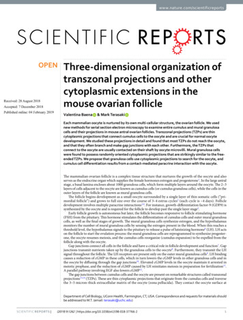
www.nature.com/scientificreports/Figure 1. Cumulus cells send numerous projections through the zona pellucida. (A) Schematic of a mouse antralfollicle. Black square represents area magnified in (A’). (A’) Schematic of several cumulus cells that send transzonalprojections (TZPs) through the zona pellucida to connect to the oocyte. Cumulus cell bodies can be adjacent toor displaced from the zona pellucida. (B) SEM image of a cross-section through cumulus cells, zona pellucida(ZP, outlined by dashed lines), and oocyte of an antral follicle. Video 1 shows 202 serial sections of this area andhighlights a cumulus cell with all of its projections. Scale bar, 5 μm. (B’) High-magnification of a region in the zonapellucida showing several TZPs in cross-section imaged by SEM. Scale bar, 1 μm. (C) Reconstruction of every TZPsent by three neighboring cumulus cells, each shown in a different color. TZPs were segmented from 405 serialelectron micrographs, encompassing a volume of 22.5 6.7 18.2 μm (x, y, z). ZP, zona pellucida.adherens junctions15 and gap junctions16. Because TZPs provide the site of gap junctional communication andpossibly of other interactions between the oocyte and cumulus cells, they are crucial structural elements of thefollicle. However, due to their density and complexity, the three-dimensional organization of oocyte components,TZPs, and their junctions is poorly understood.Automation and new computer capabilities have improved serial section electron microscopy so that it ismuch more feasible to produce large, three-dimensional fields of view at high resolution17. We used the ATUM(automated tape-collecting ultramicrotome) method with scanning electron microscopy18 (SEM) to examineentire cells and their relationships to neighboring cells in mouse antral ovarian follicles. The images provide newinformation on follicle structure, particularly the inter-relationships of the TZPs and the oocyte cell surface.They also reveal the presence of cytoplasmic projections among granulosa cells within the follicle. As in othertissues and systems, these projections may be essential for cell communication during normal development andfunction19–21.ResultsCumulus cells extend connected and free-ended transzonal projections (TZPs).The observations to be described below were obtained from antral follicles from prepubertal mice without exogenous hormonal stimulation. At this stage, the oocyte is surrounded by a several micron-thick extracellular matrix, thezona pellucida. Cumulus cells on the other side of the zona pellucida contact the oocyte by sending cytoplasmicprojections through the zona pellucida (TZPs) (Fig. 1A).To visualize TZPs in three dimensions, we collected 600 serial sections of 40–45 nm thickness (total depth27 μm) and imaged volumes of 43 43 μm (x, y) at a 3.5–5 nm per pixel resolution. The imaged volumes werecentered to contain the zona pellucida, oocyte surface, and cumulus cell bodies (Fig. 1B). We were able to traceevery TZP sent by individual cells (Video 1 and Fig. 1C), and record their interactions with other TZPs and withthe oocyte surface (described below). As can be seen in video 2, TZPs arise from cumulus cells located at varyingdistances from the oocyte.We found that the majority of TZPs do not reach the surface of the oocyte (Fig. 2A,C,D). On average, eachcumulus cell had 31 5 (n 8) TZPs that do not reach the surface of the oocyte, and 9 2 (n 8) TZPs that reachthe oocyte and make a junction with it (Fig. 2B–D) (data shown as mean standard error of the mean, unlessspecified otherwise). We will refer to these as free-ended and connected TZPs, respectively.We measured the lengths of connected and free-ended TZPs in serial electron micrographs. The lengths ofconnected TZPs had a bell curve-type distribution with an average length of 7.9 1.9 μm (n 70) (mean standard deviation). Free-ended TZPs had a different kind of distribution, in which the number of projectionsdecreased with length (Fig. 2E). The average length of free-ended TZPs was 2.9 2.3 μm (n 248).Connected TZPs were significantly longer than the average thickness of the zona pellucida, which was4.1 0.3 μm (n 3). The thickness of the zona pellucida was measured between the cumulus cell boundary andScientific Reports (2019) 9:1262 https://doi.org/10.1038/s41598-018-37766-22
www.nature.com/scientificreports/Figure 2. TZPs can be free-ended or connect to the oocyte. (A) Serial section SEM images of a free-endedTZP. The TZP (arrow) ends on section 4 without making contact with the oocyte or another TZP. Scale bar,500 nm. (B) A series of SEM images showing every other section of a TZP that connects to the oocyte. Blacktriangles indicate the TZP-oocyte site of contact. Scale bar, 500 nm. (C) Treelines representing every TZP sentby one cumulus cell. Free-ended TZPs are shown in blue, and connected TZPs are shown in red. (D) Averagenumber of TZPs per cumulus cell. TZP types were divided into free-ended (blue) and connected (red). 8cumulus cells from 2 different follicles were analyzed. Total number of projections was 316. Average is shown asmean standard error of the mean. (E) Histogram of the length of free-ended (blue) and connected (red) TZPsfrom the data in D. An example of a remarkably long connected TZP can be seen in Video 3.the oocyte surface at five locations in sections containing the widest diameter of the oocyte. Connected TZPsoften extended along the surface of the oocyte making long junctions with it, thereby increasing their averagelength. Unexpectedly, some connected TZPs looped back to the zona pellucida after making a long junction withthe oocyte surface (Video 3). Additionally, many free-ended TZPs were longer than the thickness of the zonapellucida (Fig. 2E), but these did not reach the oocyte surface because they traveled obliquely through the zonapellucida (Fig. 2C).Connected TZPs were slightly thicker than free-ended TZPs (75 2 nm (n 28), and 68 2 nm (n 30),respectively), and organelles were more often seen in connected TZPs rather than free-ended TZPs. During ouranalysis, we encountered two cumulus cells in the process of mitosis in two of the follicles analyzed. One cell wasin prophase and one in prometaphase. Interestingly, both mitotic cumulus cells had free-ended and connectedTZPs (Supplementary Fig. 1), suggesting that cumulus cells maintain their connection with the oocyte duringmitosis.TZPs often contact each other and make gap junctions. Our data revealed novel characteristics ofTZPs throughout the zona pellucida. For instance, we found that 12% of the TZPs analyzed branched into one ormore projections (Supplementary Fig. 2, and see Supplementary Fig. 3B for an additional example). Our data alsorevealed numerous examples of side-to-side and end-to-end contacts between TZPs (Video 4 and SupplementaryFig. 3A). Video 4 shows three examples of TZP contact sites, all found within just 21 sections (a thickness of 1μm), providing an example of how common these interactions are within the zona pellucida. Individual contactslike these would be difficult to resolve by light microscopy. TZP-TZP contacts were small, spanning through 1–440 nm sections. Most occurred in the upper half of the zona pellucida, closer to the cumulus cells than to theoocyte. We traced interacting TZPs back to their cell of origin to determine if they were derived from differentcells (Supplementary Fig. 3B). From 82 TZP-TZP contacts analyzed, 78% were between TZPs from different cellsand 22% were between TZPs from the same cell. The presence of same cell TZP-TZP contacts suggests that TZPsform contacts indiscriminately, in other words, two TZPs about to make a contact are not restricted whether theyare derived from the same cell or from two different cells.Gap junctions within the zona pellucida have previously been observed by immunofluorescence22. However,due to the limited resolution of SEM ( 3 nm for these studies), it was not possible to identify small gap junctionsScientific Reports (2019) 9:1262 https://doi.org/10.1038/s41598-018-37766-23
www.nature.com/scientificreports/in our data. To investigate if the TZP-TZP contacts consisted of gap junctions, we collected sections from thesame follicles that had been previously analyzed, and used transmission electron microscopy (TEM) to image thezona pellucidae. We found that most, but not all, contacts in the upper half of the zona pellucida were gap junctions (Supplementary Fig. 3C). Supporting the idea that TZPs can form gap junctions at their endings, we foundthat some TZPs (18 of 325) ended in an invaginated annular junction within a cumulus cell body (SupplementaryFig. 4). Based on our previous immunogold studies23, these are likely to be invaginated gap junctions.Connected TZPs and oocyte microvilli make contacts with each other. As described previously13,24,TZPs end in junctions at the oocyte surface that contain adherens junctions, as identified by an electron-densedeposit25 (Fig. 3A,B). We reconstructed 12 of these junctions and found that they range from 0.39 μm to 3.59 μmin length, and that most lie along the oocyte surface (Fig. 3C), while a few form invaginations into the oocytecytoplasm.Mammalian oocytes have a dense network of microvilli on their surface26. In our analysis, oocyte microvilli were generally uniformly distributed along the oocyte surface, and were 1.06 0.09 μm (n 45) long.Interestingly, we often noticed areas in the zona pellucida where microvilli appeared “clumped” (Fig. 3D). Serialsection analysis revealed that these clumped areas consisted of one or two TZPs connected to the oocyte, whichwere closely associated with 3–6 oocyte microvilli (Fig. 3E and Video 5). Video 5 shows an example of a connected TZP that is almost continuously coupled with a long microvillus and then becomes surrounded by 5–6short microvilli as it gets close to the oocyte surface. In some cases, microvilli were seen alongside the TZP for adistance of up to 6 μm (Fig. 3F). Although most TZPs that reached the oocyte surface were contacted by microvilliin this manner, some were not. Only one example of a microvillus contacting a free-ended TZP was seen.To test whether TZP-microvilli contacts were gap junctions, we inspected thin sections by TEM as describedin the previous section. The oocyte cytoplasm and microvilli can usually be distinguished from TZPs by a difference in electron density (see example in Video 3) or by the presence of precipitate that often forms in theoocyte cytoplasm and microvilli, but not on TZPs (see example in Video 5). This allowed us to identify possibleTZP-microvilli contacts in the TEM images. In contrast to contacts seen in the upper half of zona pellucida, thesedid not appear to be gap junctions (data not shown).Cytoplasmic projections are also found outside of the zona pellucida (non-TZPs).During ouranalysis of TZPs, we found that many cumulus cells had some projections directed away from the oocyte towardother cells, and that mural granulosa cells had similar projections. The number and directionality of the cytoplasmic projections had a striking dependence on cell location within the follicle. Projections not found in the zonapellucida will be referred to as non-TZP cytoplasmic projections.Cumulus cells were identified as any cell that was connected to the oocyte by means of TZPs. As describedbefore, these cells differed based on whether the cell body was located adjacent to the zona pellucida or displacedaway from it (Fig. 4A,B). Cumulus cells that were directly adjacent to the zona pellucida extended most projections toward the oocyte as TZPs, and only a few away from it (Fig. 4C). These cells had an average of 51 4TZPs and 10 3 non-TZP cytoplasmic projections (n 5) (connected and free-ended TZPs were pooled togetherfor these studies). Displaced cumulus cells, which were located 1–2 cell diameters away from the oocyte, had adecreased number of TZPs, and an increased number of non-TZP cytoplasmic projections. These were generally oriented toward other cells and sometimes invaginated into neighboring cell bodies (Fig. 4D and Video 1).Video 1 highlights one of these cells and every cytoplasmic projection derived from it, including TZPs. Thesecumulus cells had 22 9 TZPs and 27 3 non-TZP cytoplasmic projections (n 3) (see Fig. 7A). In summary,cumulus cells that are further displaced from the oocyte have fewer TZPs and more non-TZP cytoplasmic projections compared to cumulus cells adjacent to the oocyte (see Video 2).To analyze the projections of mural granulosa cells, we imaged volumes of 71 71 27 μm (x, y, z) at a resolution of 5–6 nm per pixel, centered on mural granulosa cells of the follicles that were previously used to studyTZPs. Cells chosen for analysis were selected if they were centrally located within the field-of-view and the cellbody was completely within the volume.Inner mural granulosa cells, which are not connected to the oocyte or to the basal lamina (Fig. 5A), had anaverage of 23 2 projections per cell (n 14). These projections were oriented in many directions with no consistent bias (Fig. 5B,C and Videos 6 and 7). Some of these projections were remarkably long (up to 13.7 μm) andfrequently invaginated into neighboring cells. Video 6 shows several examples of such invaginations; in particular, one cell (colored in blue) showed 7 projections, all of which invaginated into its neighboring cell (located toits upper right). We have not detected fused membranes between the invaginated projections and the cells intowhich they invaginate (See video 10 for an example of an invaginated projection).Outer mural granulosa cells were identified as those that had a visible connection with the basal lamina(Fig. 6A). Many outer mural granulosa cells had their cell body located 2–3 cell layers away from the basal lamina,but connected to it through a thick long cytoplasmic process27 (Fig. 6B,C). Numerous thin cytoplasmic projections, similar to those found in the other cell groups, were seen originating from these elongated cells (Fig. 6C andVideo 8), with an average of 28 5 projections per cell (n 4). Strikingly, outer mural granulosa cells that werelocated directly adjacent to the basal lamina (Fig. 6A,D and Video 8) had a significant reduction in the number ofprojections, having 7 1 (n 3) per cell. In summary, mural granulosa cells that are not connected to the oocyteor to the basal lamina have numerous projections that are oriented randomly, while cells that are connected to thebasal lamina differ based on the location of their cell body: cells that are further displaced from the basal laminahave a larger number of projections compared to cells directly adjacent to the basal lamina (Video 9 and Fig. 7A).We then characterized the endings of non-TZP cytoplasmic projections from cumulus, inner mural, and outermural granulosa cells (analysis of 427 projections from 20 different cells). Most projections (38%) ended onScientific Reports (2019) 9:1262 https://doi.org/10.1038/s41598-018-37766-24
www.nature.com/scientificreports/Figure 3. Contacts between TZPs and oocyte components. (A) TEM image of TZPs that make adherens junctionswith the oocyte surface. Adherens junctions are identified by an electron-dense region at the site of TZP-oocytecontact (black triangles). Mv, oocyte microvilli. Scale bar 500 nm. (B) Serial section SEM images of a TZP (blackarrow) that makes an adherens junction with the oocyte surface. Oo, oocyte. Scale bar, 300 nm. (C) Reconstruction ofan adherens junction made by the TZP shown in B. Reconstruction is 2.0 1.1 1.7 μm (x, y, z), sp
Department of cell Biology, Uconn Health, farmington, ct, USA. correspondence and requests for materials should be addressed to M. t. (email: terasaki@uchc.edu) Received: 28 August