Transcription
RAPIDJOURNALOFNEUROPHYSIOLOGYVol. 76, No. 3, September1996. Printedin U.S.A.PUBLICATIONEye-Centered, Head-Centered, and Intermed iate Cod ing ofRemembered Sound Locations in Area LIPBRIGITTESTRICANNE,RICHARDA. ANDERSEN,AND PIETROCentre de Recherche Cerveau et Cogniscience, Universite Paul Sabatier,Toulouse, France; Division of Biology, California Institute of Technology,Presbyterian Medical Center, Department of Psychiatry, New York, NewSUMMARYANDCONCLUSIONS1. The lateral intraparietal area (LIP) of the posterior parietalcortex lies within the dorsal cortical stream for spatial vision andprocesses visual information to plan saccadic eye movements. Weinvestigated how LIP neurons respond when a monkey makes saccades to the remembered location of sound sources in the absenceof visual stimulation.2. Forty-three (36%) of the 118 neurons sampled showed significant auditory triggered activity during the memory period. Thisfigure is similar to the proportion of cells showing visually triggered memory activity.3. Of the cells showing auditory memory activity, 44% discharged in an eye-centered manner, similar to the way in whichLIP cells discharge for visually initiated saccades. Another 33%responded in head-centered coordinates, and the remaining 23%had responses intermediate between the two reference frames.4. For a substantial number of cells in all three categories, themagnitude of the response was modulated by eye position. Similarorbital ‘ ‘gain fields’ ’ had been shown previously for visual saccades.5. We propose that area LIP is either at the origin of, or participates in, the transformationof auditory signals for oculomotorpurposes, and that orbital gains on the discharge are part of thisprocess.6. Finally, we suggest that, by the level of area LIP, cells areconcerned with the abstract quality of where a stimulus is in space,independent of the exact nature of the stimulus.INTRODUCTIONMAZZONIFaculte de Medecine de Rangueil, 31 000Pasadena, California 91125; and ColumbiaYork 10032-2603such as V2, V3, V4, V3a, and PO and these areas in turnreceive input from primary visual cortex. The visual activityof LIP cells has been found to be represented in a retinalreference frame (i.e., attached to the moving eye), but monotonically modulated by both eye and head position (Andersen et al. 1990; Brotchie et al. 1995) . The nature of thesemodulations is consistent with the area containing an implicitrepresentation of space distributed over the neural population. This distributed coding has the attractive feature thateye-, head-, or body-centered coordinates can be extractedfrom the neural population. The question addressedin thispaper is how are auditory signals interface with this distributed representation of visual space in area LIP.Visually triggered memory activity has been described inarea LIP (Barash et al. 1991b; Gnadt and Andersen 1988).Special tasks that dissociate the direction of intended eyemovements from sensory stimuli have shown that, for asubstantial fraction of LIP cells, this memory activity codesthe movement the animal intends to make rather than thestimulus per se. Other experiments have also shown thatauditory stimuli will evoke memory activity when the animalplans an eye movement to the remembered sound location(Mazzoni et al. 1993). Jay and Sparks ( 1984, 1987; superiorcolliculus) and Russo and Bruce ( 1994; frontal eye fields)have shown that visual and auditory stimuli are brought intoa common eye-centered coordinate frame that is used bythese areas to code the motor error required to foveate atarget. It is conceivable that area LIP might demonstrate asimilar common coordinate frame becauseit projects to thesetwo oculomotor areas and is involved intimately in processing sacades(Blatt et al. 1990). In addition, LIP alreadyreceives eye position signals necessary to match visual andauditory locations ( Andersen et al. 1990). In the currentstudy, we tested the effect of different eye positions on theresponsesof LIP neurons to the remembered locations ofsounds. Our goal was to determine the coordinate frame ofthis auditory-triggered activity. We also wished to determineif the auditory memory activity was modulated by eye position, similar to the modulation found for visual memoryactivity. Such results would show the state of sensory integration in the posterior parietal cortex and possibly indicatethe mechanismsused.Early researchersconsidered the posterior parietal cortexto be a classical association area, important for combininginformation from the various sensory modalities to form amultimodal representation of space( Critchley 1953; Hyvarinen 1982; Mountcastle et al. 1975). Later investigationsgenerally have focused on the coding of only a single sensorymodality, usually vision or touch, within this area. It is currently not well understood how information from the different modalities, which are coded in different coordinateframes, are associated in the posterior parietal cortex. Thelateral intraparietal area (LIP) has been postulated to playa role in processing saccadic eye movements. This proposalwas based on the findings that the area projected to motorstructures involved in processing of saccadesand that itscells discharged prior to saccades.Further anatomic experiments (Blatt et al. 1990) showed that area LIP is a classic METHODSextrastriate visual area based on its anatomic connections.Recordings were made from 118 area LIP neurons while a rhesusIt receives most of its inputs from other extrastriate areas monkey performed a memory saccade task. In this task, dia0022-3077/96 5.00 Copyright0 1996 The AmericanPhysiologicalSociety207 1
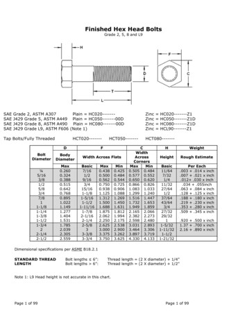
2072B. STRICANNE,auditorydelayedsaccadesfromR. ANDERSEN,various0(0) iif@fixation3 fixation(olANDpointspoints(ol5 speakersBsen soryresp onsefixationlightSACmemoryresponselight b FFN0II750msII500msII500P. MAZZONImsgrammed in Fig. 1, the animal made saccades to the rememberedlocations of sound sources from various initial eye positions, withthe head fixed and in complete darkness (see figure legend fordetails). We chose to arrange the speakers only horizontallybecause horizontal discriminationof sounds is much more accuratethan vertical discriminationand memorized saccades are alreadyless accurate than saccades to visible targets (Gnadt et al. 1991) .A 12-deg separation between the speakers appeared to be the optimum for the monkey’s sound localization ability ( -80% correctfor an accuracy of 5 deg around the target). This, however, limitedsaccades to 12 and 24 deg because 36-deg saccades are very unnatural, potentially uncomfortable, and thus likely to decrease substantially the monkey’s performance.We monitored the monkey’s eye movements using the scleralsearch coil technique (Robinson1963 ) . The animal receiveda reward of apple juice each time he accomplishedthe tasksuccessfully. Single-cell recordings were made with either glasscoated or insulated tungsten microelectrodes.Each condition ofthe experimentalparadigm was presented between 8 and 12times, and all conditions were interleaved using a random blockdesign.We analyzed the cell responses during the delay period, usingthe mean firing rate during the last 400 ms of this 500-ms timesegment. We used a general linear model (a more robust versionof the analysis of variance) to test the significance (for P values 0.05) of the effect of two discrete variables on cell responses:the horizontal component of the motor error of the saccade (ME)and the head-centeredlocation of the auditory target (HL). AP value 0.05 for the variable HL but not for ME means thatthe response field is stable relative to the head, whereas a significant P value only for the variable ME means that the responsefield moves with the eyes. If, for a given cell, both variableshave a P value 0.05, it consequently means that this cell hasan intermediateresponse field, which could be fixed relativelyto the head for a certain range of stimuli and move with theeyes for another range.We also tested whether initial orbital position influences theactivity through a second general linear model with two discretevariables: the one to which the cell responds selectively (as revealed in the previous test) and starting eye position (EP). Whenboth ME and HL were significant in the first test, we tested theeffect of orbital position with each factor separately; conservatively, the P value for EP had to be significant in both cases(P 0.05) for the effect to be acknowledged.IIFIG. 1.Schematicrepresentationof the experimentalparadigm.Stimulationapparatus (A) consists of 5 speakersdisplacedhorizontally,centeredstraight ahead from themonkeyand separated by 12 deg. A round fixationlight(0.5 deg; 45 cd/m2) is back projectedon a translucentscreen 10 deg above either of 3 central speakers. Auditorystimulus is a white noise burst of 20-20,000Hz and 7080 dB. B : timing in task: monkeyfixates for a total of1,750 ms; after 1 st 750 ms of fixation, a 500-ms noise burstis emitted from 1 speaker; finally, after a delay of 500 ms,the fixationspot extinguishes,and the monkeymakes asaccade to the rememberedsound location.iDFLREsu LT sDuring the delay period between offset of the sound andthe cue signal to make the eye movement, 43 cells (36%)showed a significant modulation (P 0.05) for one or bothof the variables of target location or movement vector. Theother cells were either unresponsive during the delay periodor, occasionally, were responsive but nonselective for eithertarget location or movement vector. About 75% of thesecells had a strong contralateral preference.Three general cell types were recognized, based on theplots of their response fields; some cells responded in eyecentered coordinates, others responded in head-centered coordinates, and others were intermediate between these twocoordinate frames. Figure 2A shows an example of a cellcoding in eye-centered coordinates. The left plot presentsthe cell response to different sound source locations. Thethree curves obtained from three initial eye positions indicatethat the cell’s response field shifts with different eye positions. The plot on the right shows the same cell’s responsebut now plotted against the motor error of the saccade; thealignment of the curves shows that the shift was equal tothe difference in eye position and that this neuron respondsmost when the animal was planning a saccadeto a location12 deg to the right, independent of starting eye position.For some neurons, the response to a given sound sourcedoes not change when the saccade metrics are changed.These cells code auditory targets invariantly relative to thehead. Such a neuron is shown in Fig. 2B. The alignmentsof the three curves in the left plot but not in the right plotshow that the cell’s auditory receptive field does not changefor different eye positions, remaining aligned on the cell’s‘ ‘preferred’ ’ sound location (here straight ahead).A final group of cells appearedto be intermediate betweeneye- and head-centered coordinate frames. Typically thesecells showed only a partial shift of the receptive field witheye position or a shift for two eye positions but not thethird. Figure 2C shows one particular example of such anintermediate cell; here the three curves are not aligned oneither plot.
ENCODINGOF AUDITORYSPACEIN LIP207324A 24firingrate(H Z 12.0-24-12d122416B 16firingrate(H Z )Tuning curves of 3 representativeLIP neuFIG. 2.rons (A-C). Mean response (averagedover the last400 ms of delay period, mean SE) is plotted againsthead-centeredlocation of sound (I@) or horizontalcomponentof motor error (right).Three lines in eachplot correspondto neural response obtained from agiven starting eye position:12 deg right fixation,-;central fixation,- - - ; 12 deg left fixation,A : neuron with an eye-centeredresponse field:curves obtained for 3 differentfixationsare alignedon right, but not on kft, showing a strongest responsewhen animal was planning a saccade to a location 12deg to the right for all 3 fixations (P values: 0.9279for head-centeredlocation; 0.001 for eye-centeredlocation).B: neuron with a head-centeredresponsefield: reverse situation of neuron A, aligned curves onleft but not right, show strongest response for thespeaker straight ahead (P values 0.0468 for head-centered location;0.3776 for eye-centeredlocation).C:cell with an intermediateresponse field: here curvesare aligned completelyon neither plot, but still partially aligned on both (P values: 0.0018 for headcentered location;0.0 138 for eye-centeredlocation).l.880I-24-12d1224c 12-24-1201224-24-120122412firingrate(H Z 6/\0’I-24-12head centered01224location (deg.)eye centeredFor all three types of cells, initial eye position can inducea. gain on the neural discharge. Figure 3 shows a neuronwith an oculocentric response field centered on downwardsaccades, as indicated by the alignment of the three curvesinthe motor error plot. However, different starting eye positions significantly modulated the amplitude of the cell’s response, resulting in a vertical shift of the curves. A similargain effect for eye position was seen for cells with responsefields in head-centered coordinates and for intermediatecells. The gains were either increasing in the direction ofthe selectivity (8/ 16), or in the opposite direction (8/ 16).To obtain a more quantitative assessment of the relativedistributions of the three types of activity described above,we analyzed the data statistically (see METHODS).Figure 4location(deg.)shows the distribution of these 43 cells into different categories based on this analysis. Forty-four percent ( 19/43) ofthe selective cells respond in oculocentric coordinates (P 0.05 for motor error but not for target location), another33% (14/43) have head centered response fields (P 0.05for target location only), and the remaining 23% ( 10/43)are intermediate cells (significant P values for both variables) (see figure legend).A second statistical test analyzed the effect of initial eyeposition on the cell response (see METHODS).For six of the19 eye-centered cells, 2 of the 14 head-centered cells, and8 of 10 intermediate cells, the magnitude of the activity wasaffected by eye position. None of these cells were significantfor eye position only, that is, simply coding the position of-
2074B. STRICANNE,1616R. ANDERSEN,AND88.-24-12head centered01224location (deg.)-24-12eye centeredd2015-co"25 1050eyecentered4. Populationsummary,obtained from statistical analysis (generalmodel with speaker locationand motor error as discrete variables;see METHODS),showing distributioninto 3 categoriesdescribedin text.Because this analysis collapsed data across all 3 eye positions, it tended toaverage out effects of eye position. A second statistical analysis used startingeye position and factor relevant from 1st analysis as discrete variables.Ifcell was of intermediateclass, then 2 models ( 1 with motor error and eyeposition and the other with target location and eye position)were requiredto be significantfor both factors to qualify as a gain effect. Unshaded partof each column shows number of cells in each category for which initialeye position modulates the strength of response.FIG.linearFIG. 3.Neuron with an oculocentricresponse field modulated by initial eye position (P values: 0.2959 for head-centered location;0.0001 for eye-centeredlocation).Changinginitial eye position modulates amplitudeof neural response.Indeed, 3 curves are aligned on right only, with strongestresponse for downwardsaccades for all 3 fixation positions,but intensity of the response is strongest for fixation on right(P values in second model: 0.0001 for motor error and 0.0050for starting eye position).24location (deg.)the eyes in the orbits. The relative proportions of cells withthese effects are represented in Fig. 4 by the unshaded partof the bars.Finally, because monkeys can orient their pinnae towardthe direction of gaze, different pinnae positions could modifythe properties of a given sound entering the ear canals (Bruceet al. 1988). We controlled for pinnae effects by recordingwhile the monkey’s ears were taped down tightly. Out ofeight cells recorded in this condition, we found one witha head-centered receptive field and three with oculocentricresponse fields, one of which was also modulated by orbitalposition. The remaining four cells were not selective duringthe delay period. The results from this control made it veryunlikely that the animal’s pinnae orientation would accountfor the transformation of auditorv inputs outlined above.-EzP. MAZZONIDISCUSSIONThirty-six percent of the cells in our sample showed memory activity after presentation of the auditory target. Thisproportion is similar to the number of cells in LIP showingactivity in visual memory saccade tasks (Barash et al. 199 1a;Gnadt and Andersen 1988). In this group, we observed bothshifts of response fields and modulations of response amplitude with different fixation positions. Amplitude modulations, usually called gain fields, also commonly occur in thecoding of visual saccades in LIP (Andersen et al. 1990).We also found selective responses that occur during thepresentation of the auditory stimulus. These responses aresimilarly distributed between the three categories of eye-,head-centered, and intermediate coordinates. However, it isnot yet possible to resolve whether this activity is a sensoryresponse or an early elaboration of an activity related to amemory or a plan.An explicit head-centered coding at the single neuron levelin the posterior parietal cortex would appear, at first glance,to be surprising. Numerous studies have shown that visualreceptive fields in this region generally code the retinal position of stimuli; an exception has been the report of a fewPO neurons, which code in head-centered coordinates (Galletti et al. 1993 ). The present result is less surprising whenconsidering that the activity is derived from auditory stimuli,which generally are coded in head-centered coordinates. Infact, it is the oculocentric representation of auditory information that is more interesting, because this coding requires atransformation from head- to eye-centered coordinates. Wefound in LIP both types of representations, plus cells withintermediate coding between the two. This observation suggests that area LIP is either at the origin of, or participatesin, the transformationof auditory signals for oculomotorpurposes and that this is the functional significance of theco-occurrence of the different cell types.LIP has very wide response fields that do not appear tobe arranged in a strict topography. In addition, eye positioninfluences the response amplitude of cells in each category.The somewhat disordered representation in LIP might be acomputational advantage allowing neighboring interactionsbetween cells gated by eye position. These interactions couldresult in the appropriate transformationsfrom head- to eye-
ENCODINGOF AUDITORYcentered coordinates through local circuits. This transformation function is suggested by the output of LIP, which projects strongly to both the superior colliculus (SC) and thefrontal eye fields (FEF) (Blatt et al. 1990)) two areas essential in the programming of saccades (Schiller et al. 1980).Both the SC and the FEF are active in the context of auditoryguided saccades (Jay and Sparks 1984, 1987; Russo andBruce 1994)) and both areas have auditory responses in eyecentered coordinates.Microstimulationsof the posterior parietal cortex showedthat fixation position influences both the amplitude and direction of the saccades evoked (Kurylo and Skavenski 1991;Thier and Andersen 1991, 1996). Looking at the systematiceffect of gaze angle, these studies revealed sites from whichthe stimulations would evoke saccades of a constant vectorand other sites which would bring the eyes to a zone fixedrelative to the head. Neural network models of LIP havesuggested that the combination of visual information in eyecentered coordinates
Eye-Centered, Head-Centered, and Intermed iate Cod . Presbyterian Medical Center, Department of Psychiatry, New York, New York 10032-2603 . (Blatt et al. 1990) showed that area LIP is a classic extrastriate visual area based on its anatomic connections. It rece