Transcription
Moon and Park Cell Communication and 1(2020) 18:109RESEARCHOpen AccessPrion peptide-mediated calcium levelalteration governs neuronal cell damagethrough AMPK-autophagy fluxJi-Hong Moon and Sang-Youel Park*AbstractBackground: The distinctive molecular structure of the prion protein, PrPsc, is established only in mammals withinfectious prion diseases. Prion protein characterizes either the transmissible pathogen itself or a primaryconstituent of the disease. Our report suggested that prion protein-mediated neuronal cell death is triggered bythe autophagy flux. However, the alteration of intracellular calcium levels, AMPK activity in prion models has notbeen described. This study is focused on the effect of the changes in intracellular calcium levels on AMPK/autophagy flux pathway and PrP (106–126)-induced neurotoxicity.Methods: Western blot and Immunocytochemistry was used to detect AMPK and autophagy-related proteinexpression. Flow cytometry and a TdT-mediated biotin-16-dUTP nick-end labeling (TUNEL) assay were used todetect the percentage of apoptotic cells. Calcium measurement was employed using fluo-4 by confocalmicroscope.Results: We examined the effect of calcium homeostasis alterations induced by human prion peptide on theautophagy flux in neuronal cells. Treatment with human prion peptide increased the intracellular calciumconcentration and induced cell death in primary neurons as well as in a neuronal cell line. Using pharmacologicalinhibitors, we showed that the L-type calcium channel is involved in the cellular entry of calcium ions. Inhibition ofcalcium uptake prevented autophagic cell death and reduction in AMP-activated protein kinase (AMPK) activityinduced by human prion peptide.Conclusion: Our data demonstrated that prion peptide-mediated calcium inflow plays a pivotal role in prionpeptide-induced autophagic cell death, and reduction in AMPK activity in neurons. Altogether, our results suggestthat calcium influx might play a critical role in neurodegenerative diseases, including prion diseases.Keywords: Calcium, Prion, Autophagy flux, Neurodegeneration, AMPKBackgroundProtein accumulation in the brain is related to thepathogenesis of various neurodegenerative diseases, suchas Parkinson’s disease, Alzheimer’s disease, as well asprion diseases [1]. Although prion disease is intermittent, it is lethal due to the non-availability of medical* Correspondence: sypark@jbnu.ac.krBiosafety Research Institute, College of Veterinary Medicine, Jeonbuk NationalUniversity, Gobong ro, Iksan, Jeonbuk 54596, South Koreatreatments to stop or interrupt the disease progression.Furthermore, prion transmission is a dangerous menaceto community health due to the high resistance of prionproteins to standard disinfection methods [2]. Thenormal cellular prion protein (PrPc) is converted toscrapie-associated prion protein (PrPsc) through progressive changes involving the conversion of its α-helicaldomains to β-sheets [3]. These diseases share a generalmechanism that includes structural modification of the The Author(s). 2020 Open Access This article is licensed under a Creative Commons Attribution 4.0 International License,which permits use, sharing, adaptation, distribution and reproduction in any medium or format, as long as you giveappropriate credit to the original author(s) and the source, provide a link to the Creative Commons licence, and indicate ifchanges were made. The images or other third party material in this article are included in the article's Creative Commonslicence, unless indicated otherwise in a credit line to the material. If material is not included in the article's Creative Commonslicence and your intended use is not permitted by statutory regulation or exceeds the permitted use, you will need to obtainpermission directly from the copyright holder. To view a copy of this licence, visit http://creativecommons.org/licenses/by/4.0/.The Creative Commons Public Domain Dedication waiver ) applies to thedata made available in this article, unless otherwise stated in a credit line to the data.
Moon and Park Cell Communication and Signaling(2020) 18:109disease-causing protein, leading to the generation of selfreplicating particles and subsequent pathogenesis withinthe central nervous system (CNS) [4]. However, it remains unclear whether scrapie pathogenesis associatedneurotoxicity is through PrPc or other mechanisms [5].Human prion peptide corresponding to amino acid residues 106–126, can form fibrils in vitro and is noxious tohippocampal neurons [6]. It might be possible that thetoxic form of PrP is created directly from PrPc or as apredecessor to pathological PrP [7].Prion diseases are correlated with the deregulation ofautophagy flux, as evidenced by the deposition ofmassive autophagic vacuoles in a hamster model ofscrapie [8]. These autophagic vacuoles often enlarge insize and increase in number as neurons age, ultimatelyoccupying the whole neurites [9]. Autophagic cell death,also known as programmed cell death II, is involved inthe intracellular process that results in the dilapidationof cytosolic constituents inside the lysosomes [10]. Thefunction of the lysosome is well-characterized in programmed cell death, although its exact role remainsblurred [11]. Recent studies investigating the degradationof PrPsc revealed that the lysosome performs an imperative role in prion protein degradation and autophagy fluxmay be the main mechanism of PrPsc transport to thelysosomes [12–14]. Based on these observations, theauthors suggested that autophagy flux might be beneficial in various diseases. Xu et al. advocated that themacro-autophagic system is stimulated in the scrapieinfected animals and human prion diseases [15]. ULK1,an autophagy protein, is necessary for cell survivalduring nutrient deficiency and is phosphorylated byAMPK [16].AMP-activated protein kinase (AMPK) has beenconsidered as a therapeutic target for several neurodegenerative diseases [17]. AMPK plays an essential role inpreserving energy balance by phosphorylating andsubsequently inactivating key enzymes involved invarious biosynthetic pathways [18–20]. AMPK functionsas a protein kinase that senses metabolic stress due toATP depletion [21]. AMPK can be activated throughphosphorylation by a tumor suppressor kinase, LKB1,under energy-deficient conditions and by CaMKK-β inresponse to depletion of intracellular calcium [22–24].CaMKK activates AMPK in an AMP-independentmanner and is itself activated by an increase in theintracellular calcium levels [25]. Thus, calcium mightplay an essential role in the activation of AMPK.Vitamin D3 improves the concentration of cytosoliccalcium ions and generates autophagy flux [26, 27].Maria et al. proposed an increase in the concentration ofintracellular calcium induces autophagy flux, which isdependent on the autophagy-related genes (Atg) andCaMKK- β [28].Page 2 of 12Calcium ions (Ca2 ) are secondary messengers thatplay important roles in various signal transductionprocesses [29]. In healthy neurons, Ca2 -related cascadescontrol numerous cellular functions, including genetranscription, exocytosis, intracellular respiration, andmembrane trafficking [30]. Perturbation of calciumregulating mechanisms results in extreme intracellularCa2 , thereby triggering cell death [31]. Several reportssuggest that PrP (106–126) alters calcium homeostasisthrough L-type calcium channels impairment [32, 33].However, the molecular mechanisms regulatingapoptosis during prion-mediated calcium influx remainslargely unexplored.Our previous study showed that prion proteinmediated neuronal cell death is triggered by theautophagy flux [34]. However, the relationshipbetween intracellular calcium levels, AMPK activity,and autophagy flux has not been described yet. Thisstudy is focused on the effect of the changes inintracellular calcium levels on AMPK/autophagy fluxpathway and PrP (106–126)-induced neurotoxicity.Our results suggest that preventing the calciumentry into the neurons affect AMPK/autophagy fluxpathway and attenuates prion protein-mediatedneurotoxicity. These outcomes suggest that regulating intracellular calcium levels, AMPK activity, andautophagy flux may be a practical therapeuticapproach for neurodegenerative disorders includingprion disease.MethodsCell cultureThe primary murine cortex neuronal cells wereprepared from embryonic 18-day ICR mice. Briefly,the brain was dissected in Hanks Buffered SalineSolution without Mg2 and Ca2 (HBSS: GIBCO,Grand Island, NY, USA), and digested in 0.25%trypsin containing DNAse I (2000 Units/mg) (GIBCO,Carlsbad, CA, USA) for 30 min at 37 C. The obtainedcell was diluted in fetal bovine serum (FBS), andcultured with Neuro basal media containing B-27 intissue culture flasks coated with 50 μg/ml poly-Dlysine at a density of 3–4 105 cells/cm2. Humanneuroblastoma cell line SK-N-SH was obtained fromthe American Type Culture Collection (ATCC, Rockville, MD, USA). SK-N-SH cells were cultured inMinimum Essential Medium (Hyclone Laboratories,Logan, UT, USA) containing 10% FBS (GIBCO, GrandIsland, NY, USA) and gentamycin (0.1 mg/mL) in ahumidified incubator at 37 C with 5% CO2. SK-N-SH(human neuroblastoma cell line, passage no.14) wasacquired from the American Type Culture Collection(ATCC, Rockville, MD, USA).
Moon and Park Cell Communication and Signaling(2020) 18:109Page 3 of 12PrP (106–126) treatmentConfocal microscopySynthetic prion peptides PrP (106–126) (sequence, -Gly-Ala-Val-Val-Gly-Gly-Leu-Gly) were synthesized byPeptron (Seoul, Korea). The peptides were dissolved insterile dimethyl sulfoxide (DMSO) at a concentration of10 mM (stock) and stored at 20 C.For confocal microscopy, the cells were cultured oncoverslips. After treatment, the cells were fixed with 4%PFA (in PBS) for 20 min at room temperature (RT) andpermeabilized using 0.3% Triton X-100 (in PBS) with 5%horse serum for 10 min. Subsequent incubations wereperformed in permeabilization buffer. The cells were incubated with primary antibodies for 60 min at RT,washed four times with PBS and incubated with AlexaFluor 488, Alexa Fluor 568, and Alexa Fluor 647 conjugated secondary antibodies at a concentration of 0.3 μg/ml each for 60 min at RT. The coverslips were placed inmounting medium and imaged on a Zeiss LSM710microscope equipped with a standard set of lasersthrough a 63 oil objective. Excitation wavelengths were488, 543, and 633 nm. Bandpass filters were set at 500–550 (Alexa Fluor 488), 560–615 nm (Cy3, Alexa Fluor568) and 650–750 nm (Alexa Fluor 647). Image acquisition was accomplished at the 12-bit rate. Settings wereoptimized to ensure suitable dynamic range, lowbackground, and appropriate signal/noise ratio.Annexin V assayCells in the logarithmic phase were collected andcultured in 24-well plate at 4 104 cells/well. Cellsurvival was evaluated using an annexin V Assay kit(Santa Cruz Biotechnology, CA, USA) following to themanufacturer’s procedure. The fluorescence was determined at 488-nm excitation and 525/30 emission usinga Guava EasyCyte HT System (Millipore, Bedford, MA,USA).Lactate dehydrogenase assayCytotoxicity was measured by evaluating the levels oflactate dehydrogenase in the cell culture supernatantusing the lactate dehydrogenase (LDH) detection kit(Takara Bio, Inc., Tokyo, Japan) following the manufacturer’s protocol. LDH activity was detected by measuringabsorbance at 490 nm using a microplate reader (SpectraMax M2, Molecular Devices, Sunnyvale, CA, USA).BacMam transductionGFP-tagged wild-type or mutant LC3B was overexpressed in neuronal cells via viral transduction usingPremo Autophagy Sensor LC3B-GFP (BacMam 2.0) kit(Life Technologies, P36235). Briefly, LC3B-FP or LC3B(G120A)-FP viral particles (MOI 30) were added to thegrowth medium and autophagosomes dynamics werevisualized using fluorescence microscopy. The mutantLC3B (G120A)-FP was used as negative control.ImmunocytochemistryCells in the logarithmic phase were collected andcultured in 1% gelatin-coated cover-slit (12 mm; NalgeNunc International, Naperville, IL) in 24-well plate at4 104 cells/well. After treatment, fixed with 4% PFA inPhosphate Buffered Saline for 20 min at roomtemperature (RT). Cells were washed in sterilized TrisBuffered Saline with 0.1% Tween 20 (TBST) for 10 min,then blocked for 15 min in TBST with 5% FBS, and thenincubated for 3 h at RT with the primary antibodies(anti-phospho-AMPKα diluted 1:100; #2535, CellSignaling Technology) diluted with in TBST with 5%FBS. Alexa Fluor 488-labeled donkey anti-rabbit IgGantibody (Molecular Probes, A21206) diluted 1:1000 wasemployed to visualize channel expression using fluorescence microscopy (Nikon Eclipse 80i). Image was evaluated using the NIS-Elements F ver4.60 Imaging software.Measurement of Ca2 ionsNeuronal cells cultured on collagen-coated confocaldishes were incubated with 5 μM Fluo-4 AM (Invitrogen,Thermo Fisher Scientific) in culture media containing1% FBS at 37 C for 40 min. The cells were washed threetimes with HBSS (Hank’s Balanced Salt Solution). Intracellular calcium dynamics were visualized using a confocal microscope (Zeiss) with 488 nm excitation and 530nm emission. For [Ca2 ]i calculation, the method ofTsien et al. [35] was employed with the followingequation: [Ca2 ]i Kd(F Fmin) / (Fmax F), where Kdis 345 nM for Fluo-4, and F is the observed fluorescencelevel. Each tracing was calibrated for the maximal(Fmax) by adding ionomycin (2 μM) and for the minimum intensity (Fmin) by adding EGTA (5 mM) at theend of each measurement.Western blot analysisCells in the logarithmic phase were collected andcultured in 6-well plate at 3 105 cells/well. After treatments, cells were washed with PBS and lysed in lysisbuffer [25 mM HEPES (4-(2-hydroxyethyl)-1-piperazineethanesulfonic acid), pH 7.4, 100 mM NaCl, 1 mMethylene diamine tetra acetic acid (EDTA), 5 mM MgCl2,0.1 mM dithiothreitol (DTT), and a protease inhibitormixture]. Equal quantities of cellular proteins (morethan 15 μg/μl) were electrophoretically resolved on a10% sodium dodecyl sulfate (SDS) poly-acrylamide geland transferred to a nitrocellulose membrane. Immunoreactivity was detected through consecutive incubationwith blocking solution using 5% skim milk and primaryantibodies, followed by the corresponding horseradish
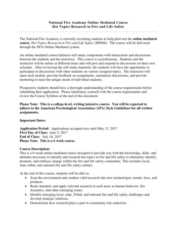
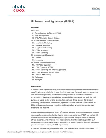
Moon and Park Cell Communication and Signaling(2020) 18:109Page 4 of 12Fig. 1 PrP (106–126) increased the intracellular Ca2 levels and induced apoptosis in neurons. a Neuroblastoma cells (SK-N-SH) were exposed tofluo-4 AM and the changes in the intracellular Ca2 levels were evaluated using confocal microscopy. The time point of PrP (106–126) orscrambled-PrP (106–126) treatments (100 μM) is shown by arrows. [Ca2 ]i was measured at 200 s after the treatment in three independentexperiments, indicate that, average kinetics of Ca2 in the PrP groups more than the sc-PrP groups. Data are represented as mean SEM. b Greenfluorescence (fluo-4) intensity that represents intracellular calcium concentration, changes time-dependently in SK-N-SH cells. c Primary neuronsand SK-N-SH cells were exposed to different doses PrP (106–126) for 24 h. Cell viability was determined by Annexin V assay using FITC-annexin V,which binds to phosphatidylserine of the plasma membrane during the apoptotic process. d The bar graph represents the average number ofannexin V negative cells. e LDH (lactate dehydrogenase) assay was performed to measure the LDH released into the culture medium. The resultsrepresent at least three independent experiments. Data are expressed as the mean SEM. *** p 0.001, compared to control (one-way ANOVA).PrP, Prion peptide (106–126); sc-PrP, scrambled Prion peptideperoxidase-conjugated secondary antibodies, and finallydeveloped using enhanced chemi-luminescence substances i.e. west save gold detection kit (LF-QC0103,AbFrontier Inc.). The primary antibodies that antiphospho-AMPKα diluted 1:1000 (#2535, Cell SignalingTechnology), anti-AMPKα diluted 1:1000 (#2532, CellSignaling Technology), anti-LC3B diluted 1:1000 (#4108,Cell Signaling Technology), anti-P62 diluted 1:1000(#5114, Cell Signaling Technology) and anti-β-actindiluted 1:5000 (A5441, Sigma Aldrich) using antibodysolution (1% skim milk in TBST) were used forimmunoblotting. Images were inspected using a Fusion
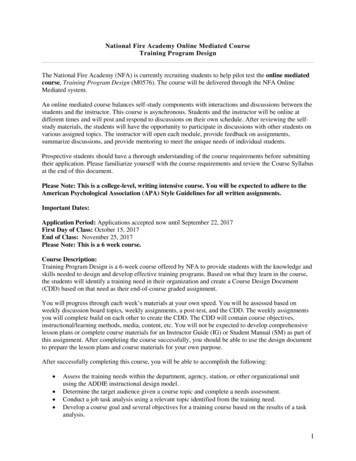
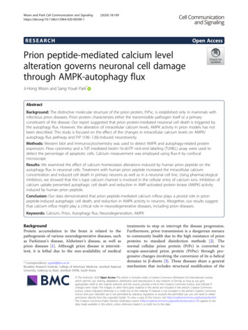
Moon and Park Cell Communication and Signaling(2020) 18:109Page 5 of 12Fig. 2 PrP (106–126)-mediated increase in the intracellular Ca2 induced neuronal apoptosis. a SK-N-SH cells were treated with fluo-4 AM and thechange in Ca2 levels was measured using confocal microscopy. The time point of EGTA treatment (2 mM) is shown by the ① arrow and PrP(106–126) treatment (100 μM) is shown by the ② arrow. Data are represented as mean SEM. [Ca2 ]i was measured at 200 s after the treatmentin three independent experiments. b SK-N-SH cells were incubated with EGTA (a calcium chelator) for 1 h and then treated with 100 μM PrP(106–126) for 24 h. Cell viability was evaluated using FITC-annexin V, indicate that EGTA decreased PrP-mediated neurotoxicity. c The bar graphrepresents the average number of annexin V negative cells. The results represent at least three independent experiments. Data are expressed asthe mean SEM. * p 0.05, ** p 0.01, compared to the PrP-treated group (one-way ANOVA). PrP, Prion peptide (106–126); EGTA, Egtazic acidFX7 imaging system (Vilber Lourmat, Torcy Z.I. Sud,France). Densitometry of the signal bands was evaluatedusing the Bio-1D software (Vilber Lourmat, Marne LaVallee, France).TEM (transmission electron microscopy) analysisCells were fixed in 2% glutaraldehyde (Electron Microscopy Sciences, Hatfield, PA, USA) and 2% paraformaldehyde (EMS, USA) in 0.05 M sodium cacodylate (pH 7.2;Electron Microscopy Sciences) for 2 h at 4 C. After, thesamples were fixed in 1% osmium tetroxide (ElectronMicroscopy Sciences) for 1 h at 4 C, dehydrated by incubating in alcohol solutions of increasing concentration(25, 50, 70, 90 and 100%) for 5 min each and entrenchedin epoxy resin (Embed 812; Electron MicroscopySciences) for 48 h at 60 C following the manufacturers’instructions. Ultrathin sections (60 nm) were cut usingthe LKB-III ultratome (Leica Microsystems GmbH, Wetzlar, Germany) and stained using 0.5% uranyl acetate(Electron Microscopy Sciences) for 20 min and 0.1% leadcitrate (Electron Microscopy Sciences) for 7 min at roomtemperature. Images were captured on a Hitachi H7650
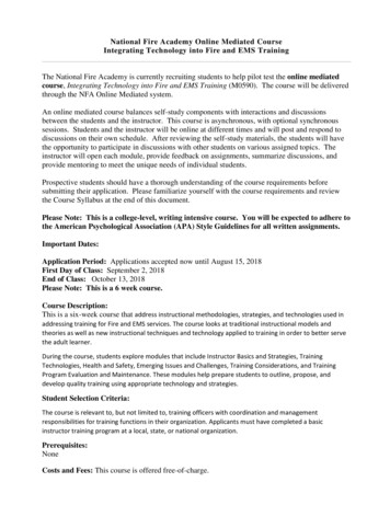
Moon and Park Cell Communication and Signaling(2020) 18:109Page 6 of 12Fig. 3 PrP (106–126)-mediated increase in intracellular Ca2 occurs via L-type calcium channel. a SK-N-SH cells were exposed to fluo-4 AM andthe alterations in Ca2 levels were evaluated using confocal microscopy. The time point of isradipine (10 μM), L651,582 (20 μM) (L-type calciumchannel blocker), ω-conotoxin GVIA (20 nM) (T-type calcium channel blocker) or ML218 (2 μM) (N-type calcium channel blocker) treatments isdesignated by the ① arrow and 100 μM of PrP (106–126) treatment is indicated by the ② arrow. Data are represented as mean SEM. [Ca2 ]iwas measured at 200 s after the treatment in three independent experiments. b The bar graph represents the average of the peak value ofcalcium levels. c Green fluorescence (fluo-4) intensity represents intracellular calcium concentration in SK-N-SH cells using confocal microscopy,indicate that L-type calcium channel blockers decreased PrP-mediated Ca2 influx. d SK-N-SH cells were incubated with isradipine or L651,582 for1 h and then exposed to PrP (106–126) (100 μM) for 24 h. Cell viability was assessed by annexin V assay using FITC-annexin V, indicate thatisradipine and L651,582 decreased PrP-mediated neurotoxicity. e The bar graph represents the average number of annexin V negative cells. f LDHassay was performed to measure LDH released into the medium. Annexin V assay an
a Guava EasyCyte HT System (Millipore, Bedford, MA, USA). Lactate dehydrogenase assay Cytotoxicity was measured by evaluating the levels of lactate dehydrogenase in the cell culture supernatant using the