Transcription
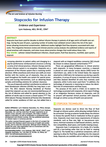
The Art and Science of Infusion NursingStopcocks for Infusion TherapyEvidence and ExperienceLynn Hadaway, MEd, RN-BC, CRNI ABSTRACTStopcocks have been used for decades to deliver infusion therapy in patients of all ages and in all health care settings. During the past 20 years, a growing number of studies have validated concern about the risk of the openlumen allowing intraluminal contamination. Additional studies highlight fluid flow dynamics associated with stopcocks. This integrative literature review and clinician practice survey analyzes the published evidence and reports ofactual practices with stopcocks, and raises issues about practice changes that could reduce these risks.Key words: catheter-related bloodstream infection, closed system, fluid flow dynamics, manifold, open system,stopcockIncreasing attention to patient safety and changing to apay-for-performance reimbursement structure is drivingscrutiny of all clinical practices. Infusion therapy and theentire infusion system is no exception. Stopcocks are acomponent of the infusion system that is receiving carefulattention. While anesthesia and critical care staffs are mostfamiliar with the routine use of stopcocks, they are alsoused throughout the health care system for special procedures and nursing interventions, such as obtaining bloodsamples and instilling solutions for catheter clearance.The use of stopcocks and their contribution to catheter-related bloodstream infection (CR-BSI) has long been a concern. The 2011 Infusion Nursing Standards of Practicestated that stopcock use was not recommended because ofthe risk of infection and called for special attention to theaddition of sterile caps to ensure a closed system.1(pS31) The2016 Infusion Therapy Standards of Practice (the Standards)called for similar avoidance of their use, but added that astopcock with an integral needleless connector (NC) shouldbe chosen to reduce stopcock contamination.2(pS72)There are geographical differences in clinical practicewith stopcocks. In many European countries, a stopcockis placed on each vascular access device (VAD) lumen forintermittent use, while in the United States the stopcockattached to a VAD hub is for temporary use during a specificprocedure. A survey of nurses in neonatal units in Australiaand New Zealand identified 10 units that routinely usedstopcocks, while 3 units never used them.3 In addition, thetechnology of stopcocks is improving to reduce the identified risks of contamination and flow issues.The purpose of this work is 3-fold: (1) to explore thetechnology associated with stopcocks; (2) to report findingsof an integrative literature review on stopcocks; and (3)to present self-reported clinical practices with stopcocksthrough an online survey of health care personnel.STOPCOCK TECHNOLOGYAuthor Affiliation: Lynn Hadaway Associates, Inc, Milner, Georgia.Lynn Hadaway, MEd, RN-BC, CRNI , is president of Lynn HadawayAssociates, Inc, and has more than 40 years’ experience as aninfusion nurse, educator, and consultant. She holds a master’sdegree in education, as well as certification in professional staffdevelopment and infusion nursing. She has published extensivelyon infusion topics in numerous journals.The author received financial support in the form of a project feeto conduct this literature search and survey of health care professionals, and to write this manuscript. Although Elcam Medical, Inc,funded the work, that organization had no involvement with theeditorial content of the article.Corresponding Author: Lynn Hadaway, MEd, RN-BC, CRNI , LynnHadaway Associates, Inc, 126 Main St, PO Box 10, Milner, GA30257 (lynn@hadawayassociates.com).DOI: 10.1097/NAN.000000000000025824 Copyright 2018 Infusion Nurses Society Stopcocks are devices used to direct the flow of fluidthrough an infusion system, while allowing multiple fluids tobe connected. Turning an exterior handle allows the clinicianto choose the specific fluid or medication to flow at a giventime. Stopcocks must meet many requirements of industrystandards, such as flow rate, high- or low-pressure resistance, chemical resistance to lipid-based solutions, sterilization compatibility, dead space, and handle-rotating torque.Stopcocks have 3 components: (1) a body, (2) a handle,and (3) a luer-locking collar that can be fixed or rotating(Figure 1). The handle has a cylindrical component, with anopening to allow for fluid flow, extending into the stopcockbody. Stopcocks are available with or without a luer-lockingJournal of Infusion NursingCopyright 2018 Infusion Nurses Society. Unauthorized reproduction of this article is prohibited.
Figure 1 Standard stopcock components. Reproduced and used withpermission from Elcam Medical.collar on the male port; however, the national standard ofpractice is to use a luer-locking mechanism on all connections in the infusion administration system.2 Stopcocks havea male port that luer locks to the hub of the VAD, or thefemale end of an administration set. The opposite end andthe side ports have a female port design to accept the maleluer-locking connection of an administration set or syringe.Stopcocks are also offered in a manifold design, asingle device with several stopcocks assembled togetherin a straight line. This device is used most often by anesthesiologists and in critical care settings. Each stopcockhas the same internal design, although the handles maybe different colors. The different colors can be used toindicate different medications in each stopcock, althoughreliance on color coding as the sole indicator for choosingthe lumen for medication administration can lead to errorsand is not recommended.2 Because fluids flow through thesame channel, a manifold system may not be appropriatefor infusing incompatible solutions.Stopcocks are available as a standard design, in additionto high-flow and high-pressure designs. The size of theopening determines the maximum flow rate. By rotatingthe handle, the hole is positioned toward one of the flowpaths, creating a multidirectional rotating valve (Figure 2).Stopcocks can be designed with 1, 2, 3, or 4 fluid flow paths,all controlled by the handle. High-flow stopcocks have thesame design with a larger opening. High-pressure stopcocksare designed with more robust connectors and handles towithstand higher pressures for interventional cardiologyand radiology procedures (Table 1).The internal diameter of intravenous (IV) administrationsets includes standard bore, macrobore, and microbore.There are no standard measurements for each of thesegroups, and each manufacturer must provide sets that arecompatible. Also uniformity in the internal diameter is notrequired.4 A standard bore set may be approximately 3 mm,while a blood administration set is approximately 4 mm, anda microbore set is usually less than 1.5 mm.5 The opening of astandard stopcock is 2.33 mm, slightly less than the 3 mm of astandard administration set. However, the stopcock will allowa maximum flow rate of about 500 mL/min and usually doesnot have a negative effect on flow rates in clinical practice.Dead space is an issue with stopcocks. The lumen of theunused port will initially contain air when the stopcock is firstadded to the infusion system. After infusion through the sideport, fluid or medication remains inside the space (Figure 3).A syringe attached to the side port of the stopcock is used toaspirate the air or flush residual fluid or medication into the primary flow pathway. A new stopcock design that has eliminatedthis problem allows fluid from the primary flow pathway toflow into and out of this space in a U-shaped pattern, flushingout trapped air bubbles or residual medication (eg, UltraportZer0, B. Braun Medical, Bethlehem, PA; Marvelous Stopcock,Elcam Medical, Hackensack, NJ) (Figure 4). Maximum flow ratein this new design is 220 mL/min with a maximum pressuretolerance of 29 pounds per square inch (psi).Stopcocks are an open system, requiring some type of closure to reduce the entry of microorganisms into the lumen. Asterile dead-end cap is attached to the lumen when the extraports are not connected to an administration set or syringe.Uncapped, open stopcock lumens pose a significant risk forcontamination leading to CR-BSI. Stopcocks currently areavailable with the integration of a luer-activated NC on thestopcock body, creating a closed system.Unique stopcock features correspond to cultural differences around the globe. For example, in the United States,stopcocks include an “off” handle to indicate the directionof flow that is closed, while European health care professionals prefer “on” so that the handle indicates the openflow directions.LITERATURE REVIEW RESULTSFigure 2 Handle with fluid pathway hole in a standard stopcock.Reproduced and used with permission from Elcam Medical.VOLUME 41 NUMBER 1 JANUARY/FEBRUARY 2018The search of published literature was planned to addressthe following questions:1. In which clinical settings and among patients of whatage are stopcocks used for the delivery of infusiontherapy through any type of VAD?2. What are the reported clinical outcomes of stopcockuse for infusion therapy?The process chosen was an integrative literature review,a method that uses empirical and theoretical publications toachieve a greater understanding of a problem or issue. ThisCopyright 2018 Infusion Nurses Society 25Copyright 2018 Infusion Nurses Society. Unauthorized reproduction of this article is prohibited.
TABLE 1Stopcock CharacteristicsStandardHigh FlowHigh PressureOpening size7 Fr, 2.33 mm9 Fr, 3 mm2.25 mmMaximum flow rate 500 mL/min 750 mL/min 510 mL/minMaximum pressure43 psi29 psi1200 psi0.2-0.4 mL0.32-0.57 mL0.56 mLPriming volumeAbbreviations: Fr, French; min, minute; mL, milliliter; mm, millimeter; psi, pounds per square inch.method is specifically important for evidence-based practicebecause it is not limited to experimental research methodsalone.6 All types of evidence, as outlined in the Standards,were included.2(pS10) Criteria for inclusion were (1) any articlediscussing stopcock use in specific procedures and the associated clinical setting (ie, Level V2[pS10]) and (2) any clinicalor laboratory research study using a stopcock on any typeof VAD. All quasi-experimental and experimental researchdesigns were included (ie, Levels I, II, III, and IV2[pS10]).Search terms included stopcock, stopcock catheter intravenous, and stopcock catheter infusion. The search wasconducted using PubMed from the US National Library ofMedicine of the National Institutes of Health, and GoogleScholar to retrieve publications not listed on PubMed.The original search yielded almost 5000 articles. Furtherrefinement by reading article abstracts eliminated all articles related to the use of stopcocks in other specialties,such as intraoperative procedures, along with management of endotracheal intubation/ventilation, nasogastrictubes, intracranial pressure monitoring, and many others.Publications from January 2000 through June 2016 wereincluded. One hundred thirty-three articles were found andexamined closely; 12 identified clinical stopcock use, and 28were clinical or laboratory research studies.Available publications were divided into 2 groups: (1)those that presented procedure details involving stopcockuse for specific infusion-related procedures, and (2) clinicalor laboratory research studies examining stopcocks for anytype of infusion therapy or VAD management.Figure 3 Trapped fluid or air in side port. Reproduced and used withpermission from Elcam Medical.Figure 4 Constant flushing of the side port. Reproduced and usedwith permission from Elcam Medical.26 Copyright 2018 Infusion Nurses Society Published Clinical Stopcock UsesPublications involving specific procedures with stopcocksincluded intra-arterial blood pressure monitoring, bloodsampling from central vascular access devices (CVADs),CVAD clearance procedures, and contrast-enhanced ultrasound for CVAD tip location identification.Intra-arterial blood pressure monitoring proceduresrequire frequent assessment to ensure complete stopcockclosure and that a minimum number of stopcocks areused in the system. Air in the infusion system, use of longand flexible tubing, and multiple stopcocks can result in aserious overestimation of systolic blood pressure.7-9 To prevent arterial backflow of blood into some types of infusiontubing, 1 report recommended the use of a stopcock onJournal of Infusion NursingCopyright 2018 Infusion Nurses Society. Unauthorized reproduction of this article is prohibited.
both ends of the pressure monitoring line.10 Stopcocks alsoare used within the system to monitor for central venouspressure, through both peripherally inserted and centrallyinserted central venous catheters.11Blood sampling from CVADs and arterial catheters mayemploy stopcocks. A study designed to evaluate the reliabilityand validity of using a bundle checklist for obtaining bloodsamples from pediatric oncology patients included stopcocksin its procedure. The routine use of stopcocks was determined by the opinion of 3 experts in the facility. The checklistincluded the use of a “closed stopcock method”; however,there was no further explanation of procedure details or thebenefits of using a stopcock on the CVAD hub.12 Concern fordrug adsorption or adherence to the intraluminal catheterwalls raises concern for obtaining accurate laboratory datawhen the blood sample is taken from the same lumen usedfor drug infusion. A study testing the “mixing” method, alsoknown as the push-pull method, for drawing blood samplesfrom a single-lumen CVAD used a stopcock in the protocol.However, there was no discussion of why the stopcock wasneeded.13 On another note, hospital-acquired anemia is asignificant problem associated with blood sampling. The literature was searched, and there was no mention of the use ofstopcocks for obtaining blood samples from any type of VAD.Procedures for clearing material obstructing a CVAD lumenfrequently involve the use of stopcocks. The primary reasonfor lumen occlusion is thrombus, but drug precipitate andlipid accumulation may also cause the occlusion. The occluding material may be anywhere along the CVAD lumen, andthere may be fluid between the occlusion and the hub. Thegoal is to remove the fluid and create negative pressure insidethe catheter lumen. When the negative pressure is released,the solution in the attached syringe will be pulled into thelumen without using force on the syringe plunger. This reduces the possibility that overpressurization could cause catheterdamage, and it allows the solution to reach the occludingmaterial more readily. Stopcocks have been described for thisprocedure in medical-surgical nursing unit,14 home care,15special procedures unit,16 and oncology settings.16Although postprocedure chest x-rays and electrocardiograms are the most common methods for identifying CVADtip location, stopcocks now have a role. They are beingused for CVAD tip location with ultrasound. The stopcockis used to create agitated saline, usually a mixture of 90%sodium chloride and 10% air. This solution is injectedthrough the catheter with ultrasound used to identify thetip of the catheter by detecting the presence of microbubbles in the bloodstream at the CVAD tip location.17An article from the critical care literature warned aboutstopcock use when a stopcock is attached to a large percutaneous sheath introducer system. This article describedhow an uncapped stopcock accidentally opened as a resultof patient movement and resulted in exsanguination of thepatient through a large sheath introducer.18Analysis of outcome data if included in these studies isnot possible because of the many variations in proceduresVOLUME 41 NUMBER 1 JANUARY/FEBRUARY 2018and clinical practices. Inclusion of this Level V2(pS10) evidencehighlights the wide variety of clinical settings and patientpopulations in which stopcocks may be used.Clinical and Laboratory StudiesThis literature search identified 15 randomized controlledtrials (RCTs), 7 quasi-experimental studies, and 7 descriptive studies about stopcock use. The stopcock studies havea wide variety of differences in research methods and clinical processes used. The differences make it difficult to listthem on evidence tables. The studies were divided by theissues being examined, including comparison of traditionalopen stopcocks to closed stopcocks due to the addition ofan NC, intraoperative stopcock use, fluid flow dynamicsthrough stopcocks, and arterial blood sampling methods. Abrief narrative discussion of each study highlights methods,differences, and outcomes.Open Versus Closed StopcocksAbout 25 years ago, colonization of VAD hubs and itsrelationship to CR-BSI emerged. Studies before 2000 hadreported that 22% of stopcock entry ports were contaminated after 72 hours of clinical use.19Since 2000, 4 RCTs compared stopcock closure usinga standard end cap (ie, a conventional open stopcock)versus closure using an NC on each stopcock lumen (ie, aclosed system). All studies were conducted in adult criticalcare patients. Three of the 4 studies showed a significantreduction in contamination of the stopcock entry port at72 hours of use with the closed system. Different methodsfor collecting samples for culturing external and internalsurfaces were used in these 3 studies. There was also adifference in the disinfecting agents used on the NC surfaces, with 1 study using 3 disinfectants and 2 studies using70% alcohol.19-21 The fourth study reported a reductionin central line-associated bloodstream infection (CLABSI)rates, using the Centers for Disease Control and Prevention(CDC) definition of that term.2 CLABSI rates decreased from6.4 per 1000 catheter days to 2.2 per 1000 catheter days.This study also included the use of commercially preparedprefilled flush syringes in the experimental group, while thecontrol group received flushes with sodium chloride drawnfrom multidose vials, adding a process not included in otherstudies.22Esteve et al23 compared the use of stopcocks that hadthe conventional end cap on each CVAD and arterial catheter hub versus an NC directly attached to the CVAD hub.Alcoholic chlorhexidine 0.5% was used for disinfecting theNC surface. The incidence of contamination and CR-BSIrates were similar between the groups, with 4.1/1000catheter days for the stopcock group and 4.6/1000 catheterdays for the NC group (P .59).Two additional RCTs compared a traditional stopcockthat had an end cap versus a stopcock with a split-septumNC in adult patients. The first study used 80% ethanol with0.1% chlorhexidine for disinfecting the NC surface.24 TheCopyright 2018 Infusion Nurses Society 27Copyright 2018 Infusion Nurses Society. Unauthorized reproduction of this article is prohibited.
second study used a disinfection cap containing a chlorhexidine-impregnated sponge on the NC when not in use.25 Bothstudies placed a 0.2-micron filter below the injection sitesand reported microbial analysis of the organisms trapped inthe filter. The first study showed no significant difference incontamination between the 2 groups; however, the secondstudy using the disinfection cap on the NC showed significant reduction of organisms in the intraluminal pathway.An RCT in low-birth-weight neonates also compared thestopcock/end cap system with a mechanical valve NC forstopcock closure inside the administration sets for infusioninto both peripheral and central venous catheters. Thisstudy used the CDC definition for CLABSI and reported arate of culture-proven bloodstream infection (BSI) in theNC/stopcock group of 3.3% and 26.7% in the stopcock/endcap group (P .026).26An RCT in adult patients compared the outcomes of aclosed peripheral catheter system and a traditional cathetersystem. The closed catheter system used a catheter with apermanently attached extension set, avoiding the connection of the extension to the catheter hub at
infusion nurse, educator, and consultant. She holds a master’s degree in education, as well as certification in professional staff development and infusion nursing. She has published extensively on infusion topics in numerous journals. The autho