Transcription
Menotta et al. Orphanet Journal of Rare Diseases (2017) 12:126DOI 10.1186/s13023-017-0669-2RESEARCHOpen AccessATM splicing variants as biomarkers for lowdose dexamethasone treatment of A-TMichele Menotta1* , Sara Biagiotti1, Chiara Spapperi1, Sara Orazi1, Luigia Rossi1, Luciana Chessa2, Vincenzo Leuzzi3,Daniela D’Agnano3, Annarosa Soresina4, Roberto Micheli4 and Mauro Magnani1AbstractBackground: Ataxia Telangiectasia (AT) is a rare incurable genetic disease, caused by biallelic mutations in theAtaxia Telangiectasia-Mutated (ATM) gene. Treatment with glucocorticoid analogues has been shown to improve theneurological symptoms that characterize this syndrome. Nevertheless, the molecular mechanism underlying theglucocorticoid action in AT patients is not yet understood. Recently, we have demonstrated that Dexamethasonetreatment may partly restore ATM activity in AT lymphoblastoid cells by a new ATM transcript, namely ATMdexa1.Results: In the present study, the new ATMdexa1 transcript was also identified in vivo, specifically in the PMBCs ofAT patients treated with intra-erythrocyte Dexamethasone (EryDex). In these patients it was also possible to isolatenew “ATMdexa1 variants” originating from canonical and non-canonical splicing, each containing the coding sequencefor the ATM kinase domain. The expression of the ATMdexa1 transcript family was directly related to treatment andhigher expression levels of the transcript in patients’ blood correlated with a positive response to Dexamethasonetherapy. Neither untreated AT patients nor untreated healthy volunteers possessed detectable levels of the transcripts.ATMdexa1 transcript expression was found to be elevated 8 days after the drug infusion, while it decreased 21 daysafter treatment.Conclusions: For the first time, the expression of ATM splicing variants, similar to those previously observed in vitro,has been found in the PBMCs of patients treated with EryDex. These findings show a correlation between theexpression of ATMdexa1 transcripts and the clinical response to low dose dexamethasone administration.Keywords: Ataxia Telangiectasia, ATM, Dexamethasone, Intra-erythrocyte DEXA, ATMdexa1BackgroundIn the last few years, experimental investigations and clinical trials have provided evidence that short-term treatmentwith glucocorticoid (GC) analogues is able to improveneurological symptoms in AT patients [1–4] raising thepossibility that cerebellar abnormalities may still be modified during the initial phase of cell loss [5]. However, thisneurological improvement is transitory and disappearswhen oral GC administration is discontinued, and thebenefit-to-risk balance limits the long-term administrationof oral steroids.The administration of low doses of GC through autologous erythrocytes could reduce steroid toxicity withoutaffecting drug efficacy [6–9] . Indeed, a phase II clinical* Correspondence: michele.menotta@uniurb.it1Department of Biomolecular Sciences, University of Urbino “Carlo Bo”, 61029Urbino, PU, ItalyFull list of author information is available at the end of the articlestudy IEDAT (EudraCT Number 2010–022315-19, a 6month single-arm open labeled trial), which envisaged thelong-term treatment of AT patients with intra-erythrocyteDexamethasone (Dexa), has recently been concluded [10].The treatment led to a significant improvement inneurological symptoms, while at the same time avoidingtypical steroid side effects. However, the specific mechanisms underlying the action of dexamethasone in the treatment of the neurodegenerative disease are still underdebate [11, 12]. The Ataxia Telangiectasia-Mutated (ATM)gene was first described by Savitsky K, et al. [13]. This gene,which codifies for a protein kinase member of the PI3Kinase-like Kinase (PIKK) family, is transcribed into 27 different mRNAs, 20 alternatively spliced variants and 7unspliced forms. Eighteen spliced mRNAs putatively encodegood proteins, while the remaining 9 mRNA variants appearnot to encode good proteins. (Aceview index.html). The Author(s). 2017 Open Access This article is distributed under the terms of the Creative Commons Attribution 4.0International License (http://creativecommons.org/licenses/by/4.0/), which permits unrestricted use, distribution, andreproduction in any medium, provided you give appropriate credit to the original author(s) and the source, provide a link tothe Creative Commons license, and indicate if changes were made. The Creative Commons Public Domain Dedication o/1.0/) applies to the data made available in this article, unless otherwise stated.
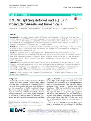
Menotta et al. Orphanet Journal of Rare Diseases (2017) 12:126In a recent investigation using lymphoblastoid cell lines(LCLs) derived from AT patients as a model, we showedthat the action of glucocorticoids may stem from theprompting of a non-canonical splicing event in the ATMmRNA precursor [14]. Specifically, we were able to identifya transcript of 1582 bp called ATMdexa1.The main aim of the present study was to investigate thepossible presence of ATMdexa1 in the blood cells of patients enrolled in the above-mentioned IEDAT clinical trial.In fact, we were not only able to identify the transcript,whose expression depends on drug administration, but alsoto show that it can act as a blood molecular marker togauge therapy responsiveness. Interestingly, in treated patients it was also possible to discover several additionalATMdexa1 variants.MethodsSample collection, RNA extraction and complementarycDNA synthesisThe blood samples were collected from 10 AT patients(6 males and 4 females, average age 9.7 years) enrolledin the IEDAT clinical trial (IEDAT-ERY01–2010) [10],from 6 untreated AT patients (4 females and 2 males,average age 17.5 years) and 6 untreated healthy volunteers (childhood blood samples, unknown gender). Forall the treated patients, blood was drawn 21 days afterdrug administration, and for 2 of those patients, it wasalso drawn 8 days after treatment administration.Blood samples were also collected from 5 AT participants (4 females and one male, average age 12.1 years)that were treated with EryDex (compassionate use).Briefly, 3 ml of blood were collected in appropriatevacutainers (Tempus Blood RNA tubes, Life Technologies)and handled and stored according to the manufacturer’sinstructions The blood samples were subsequently processed for total RNA extraction using the Tempus spin RNAisolation kit (Life Technologies) as described in the manufacturer’s instructions. A clean-up was performed using theQIAGEN RNA extraction kit to completely eliminate contaminating DNA. For each sample, 500 ng of RNA wasemployed as a template for cDNA synthesis using theSMARTScribe Reverse Transcriptase (Takara) in the presence of 50 ng of Random hexamers to prime the reaction.Quantitative PCR assaysTwo qPCR assays were set up for ATMdexa1 estimation.All PCR reactions were carried out using the ABI PRISM7500 sequence detection system platform (AppliedBiosystems). The first assay was based on SYBR greenchemistry by SYBR Premix Ex Taq (Tli RNaseH PlusTakara), with a final amount of MgCl2 3.5 mM, and300 nM of each primer (Forward 5′-ATCTAGATCGGCATTCAGATTCCA-3′ and Reverse 5′-GCAGACCAGCCAATTACTAAAC-3′). The thermal profile optimized toPage 2 of 7obtain a specific and linear amplification of ATMdexa1 wasset for 30 s at 95 C, followed by 40 cycles of denaturation(for 10 s at 94 C), annealing (for 20 s at 65 C) and extension at 72 C for 40 s. A melting curve analysis of theamplicons was performed at the end of each reaction.The second analysis consisted of a 5′ exonuclease-basedreal-time PCR assay performed with the same primers described above with the addition of 100 nM of the ATMdexa1 probe (5′6-FAM ATGCCACTCAGTGGAAGACTCAGAGAA-BHQ1 3′); the PCR reaction was performedusing the Hot-Rescue Real-Time PCR Kit–FP (Diatheva).The thermal profile was performed for 30 s at 95 C,followed by 40 10-s. cycles at 94 C and annealing/ extension at 72 C for 60 s. The FKBP5 and DUSP1 gene expression levels were measured using the TaqMan GeneExpression Assays kit (Life Technologies) according to themanufacturer’s instructions.Concomitantly with the targets, the cDNA of HPRT1was amplified using 5′-TATGCTGAGGATTTGGAAAGGGT-3′ and 5′-CCATCACATTGTAGCCCTCT-3′ asupper and lower primers, respectively. In the 5′ exonuclease qPCR, the 5′-JOE-TATGGACAGGACTGAACGTCTTGC-BHQ1–3′ probe was used and thereactions were also achieved in multiplex mode. Therelative quantification of the targets was calculated bythe ΔCT method [15] (HPRT1 as reference gene) andplotted as 1/2ΔCT.ResultsFollowing approval by the ethical committees and thepatients or their parents, blood samples of 10 out of 22patients enrolled in the IEDAT clinical study were obtained. Blood collection was carried out at the end of thetreatment, precisely 21 days after the sixth and final infusion of intra-erythrocitary Dexamethasone provided inthe clinical study.The qPCR assays were set up for qualitative and quantitative analysis, to identify the mRNA molecule,previously described in lymphoblastoid cell lines. Theassays were validated in terms of efficiency and Target toReference linearity (not shown).ATMdexa1 PCR amplification was carried out in all the10 IEDAT available samples and also in 6 untreated ATpatients and in 6 untreated healthy volunteers. Specificamplification of the target was obtained only in IEDATsamples, while in untreated AT patients and healthy controls, the expression levels were undetectable (Fig. 1a).The amplification carried out by SYBR green PCR onblood samples was similar to those obtained during theassay setup, but a small melting curve deviation was observed among the samples (Additional file 1: Fig. S1). Theresults obtained using the SYBR green PCR were confirmed by a 5′ nuclease assay as illustrated in Fig. 1b.These results suggest that the induction of the ATMdexa1
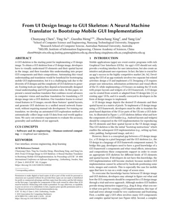
Menotta et al. Orphanet Journal of Rare Diseases (2017) 12:126Page 3 of 7also able to cluster the 10 AT participants into responder (5AT Dexa R) or non-responder groups (5 AT Dexa NR) usingthe ΔICARS score. Likewise, the matching ATMdexa1 expression data were assigned to the two clusters. Interestingly,the responders showed a statistically greater ATMdexa1 expression (t-test p 0.0425) than did non-responders (Fig. 2a).Furthermore, for one responder and one non-responder, it waspossible to evaluate the ATMdexa1 expression in blood samples collected at 8 and 21 days from EryDex administration. In this case as well the responder showed higherexpression levels than those of the non-responder at bothtimes (Fig. 2b). Moreover, by comparing intra-patient levels,it was observed that the ATMdexa1 transcript was expressedat greater levels at 8 day than it was at 21 day. This isconsistent with the gradual waning of the DEXA effect overtime after treatment, and underlines the need to resort toFig. 1 ATMdexa1 expression levels in patients treated with EryDex.Two qPCR assays were set up for ATMdexa1 estimation in wholeblood. AT, untreated patients with AT n 6; AT Dexa, patients withAT and treated with EryDex n 10; WT, healthy volunteers n 6.The first qPCR assay is based on SYBR green chemistry PCR (panel a)while a second one is built on 5′ exonuclease qPCR (panel b). Inboth settings, ATMdexa1 expression levels were detectable only inAT patients treated with EryDex. The transcript was undetectable inuntreated AT patients and in healthy volunteersRNA messenger is limited to AT patients and strictlydependent on treatment with the drug. The efficacy ofEryDex in treating neurological symptoms of AT patientshas been evaluated using the ICARS scale [16], and improvements were found to be related to the drug encapsulation efficiency, and therefore to the quantity of the drugadministrated [10]. Hence, a direct relationship betweendrug encapsulation efficiency and ATMdexa1 expressionfor the 10 available values was tested but the correlationwas found to be poor.In the same previous study by Chessa et al. [10] patientswere clustered as either responders or non-responders(AT Dexa R and NR). Responders (ΔICARS 10) weredesignated as loaders (Dexa 5 mg/bag), while the nonresponders (ΔICARS 9) were designated as poor loaders(Dexa 4.9 mg/bag). In the present investigation we wereFig. 2 ATMdexa1 expression in responders and non-responders. Inpanel a, AT patients treated with EryDex were divided into twoclusters (responders R n 5 and non-responders NR n 5, byaverage ICARS value decrement of at least 10 points, after sixmonths of therapy). Responders showed higher levels of ATMdexa1expression (p 0.05) than did non-responders. In panel b, for onepatient from each cluster, it was possible to evaluate the expressionof ATMdexa1 also at 8 days from EryDex administration. In bothcases, the extent of the target expression was time dependent
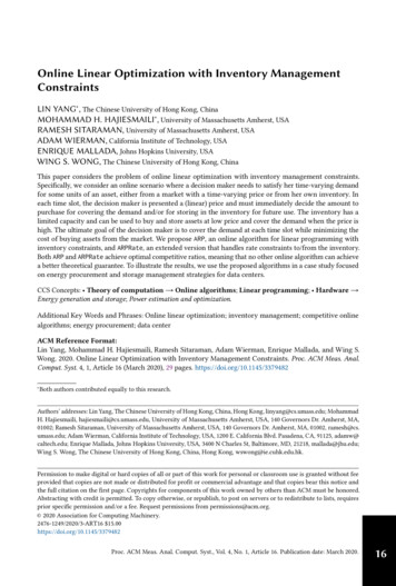
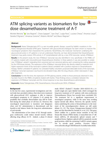
Menotta et al. Orphanet Journal of Rare Diseases (2017) 12:126repeated administrations in order to keep the drug effectiveness active. In order to assess if the improvement in responsiveness depended on glucocorticoid sensitivity the geneexpression of two targets (FKBP5 and DUSP1), known to beglucocorticoids modulated [17, 18], were tested (Fig. 3). Asexpected, the average expression levels of both targets werefound to be more elevated than those found in untreatedsubjects, but not statistically different in the two groups ofpatients (R and NR).The samples from patients treated with EryDex (compassionate usage), allowed a deeper analysis of ampliconsobtained by qPCR assays. In these samples the ATMdexa1was also detected and in order to verify the smalltemperature deviance observed during the qPCR meltingstep, as mentioned above, the amplicons were checked bygel electrophoresis. In Fig. 4 lanes 1–16 contain the qPCRproducts from 5 patient samples collected at different timeFig. 3 FKBP5 and DUSP1 mRNA expression. Gene expression levelsof the two evaluated prototypic targets. AT, untreated patients withAT n 6; AT Dexa, patients with AT and treated with EryDex n 10;WT, healthy volunteers n 6. As predicted, in treated AT patients animprovement in gene expression was observedPage 4 of 7points. The miniATM PCR product is indicated by whitearrows, while the other bands represent unattended amplicons. After the cloning and the DNA sequencing of someof the PCR products, it was possible to define five (currently) new ATM transcripts designated as “ATMdexa1variants” (Fig. 4 coloured arrows). Every variant containedthe qPCR assay probe domain and possessed the coding sequence for the kinase domain of the ATM native protein(Fig. 5). Three of the variants derived from the canonicalsplicing of the ATM mRNA (exons 3–52, 4–53 and 2–52),whereas two variants originated from a SDR (short directrepeat: 3–52 and 4–51) mediated splicing process. Theamplicon size differences from ATMdexa1 ranged from approximately 50 to 160 bp. Some of the described ampliconsmight explain the observed melting analysis discrepanciesduring the above-mentioned SYBR green qPCR tests.Samples from dissimilar patients or from the same patients,but at different blood collection times, showed indistinctlyvariegated ATMdexa1 variants and sometimes even morethan one simultaneously (Fig. 4). All the ATMdexa1variantspossess the MetMet starting site described by Menottaet al. [14] and can be translated in the miniATM protein byconceptual translation.DiscussionIn recent years, observational studies and a few clinicaltrials have shown that short-term treatment of AT patientswith oral glucocorticoids is able to improve theirneurological symptoms [1–5]. Unfortunately, this improvement is merely transitory and fades shortly after discontinuation of oral treatment with GC. Suspension of the GCtreatment is necessary since the hazards long-term therapywith steroids outweigh any potential benefits such a therapymay confer [11]. To overcome this limitation theadministration of very low GC doses by erythrocytesreduces steroid toxicity without compromising the drug’seffectiveness [6–9].In 2010 a phase II clinical study, envisaging the longterm treatment of AT patients by Dexa encapsulatedwithin autologous erythrocytes, was set up [10, 19]. Theproposed therapy brought about a significant improvement in neurological symptoms, while avoiding theonset of known side effects typical of GC. Although themechanism of action of glucocorticoids in AT patientsremains unclear, over the past few years several studieshave been conducted to shed light on mechanismsunderlying the effects of GC in AT patients. D’Assanteet al. reported the influence of Betamethasone on molecules involved in autophagosome degradation [20], whilethe authors of the present study described the influenceof Dexamethasone on gene expression, NRF2-mediatedantioxidant response by redox balance improvement andcellular nano-mechanics by cytoskeleton and nuclear dynamics [21–23]. Furthermore, despite the observation
Menotta et al. Orphanet Journal of Rare Diseases (2017) 12:126Page 5 of 7Fig. 4 ATMdexa1 transcript family. Amplicons obtained by 5′ nuclease qPCR of the compassionate EryDex samples analysed by electrophoreticgel. Lane M, DNA ladder. Lanes 1–16 contain the qPCR products from five patient samples collected at different time points. The white arrowsshow the ATMdexa1 PCR product, while the amplicon in all lanes at 115 bp is the HPRT1 PCR product. The other PCR products, with theexception of non-specific amplicons, represent ATMdexa1 splicing variants: green arrow splicing SDR 4–51; yellow arrows splicing 4–53; red arrows splicing 3–52; cyan arrow splicing SDR 3–52 and magenta arrow splicing 2–52reported by Savitsky et al. [24] regarding the possibilityof alternative splicing of ATM mRNA, we were able todiscover and report an alternative splicing event withinthe coding sequence of the ATM gene product. In fact,we have shown, in lymphoblastoid cell lines establishedfrom AT patients, that the action of the synthetic GCDexamethasone may also be exerted through theassembly of a new mRNA molecule generated by anon-canonical splicing of the ATM gene. The resultingtranscript, designated as ATMdexa1, has also beenidentified “in vivo” in the blood of AT patients receivingintra-erythrocyte Dexamethasone. The identified splicingprocedure is an SDR (short direct repeat) mediatedjoining, an occurrence described in plants as an environmental stress defense mechanism [25–27]. In mammals,this occurrence has only been described as aberrant andnonfunctional splicing [28, 29] while to our knowledge,the splicing involved in ATMdexa1 assembly representsthe only reported case of functional non-canonical SDRsplicing.ATMdexa1 transcript expression levels depend on thetreatment with Dexamethasone since, neither untreatedAT patients nor untreated healthy volunteers showeddetectable levels of the transcript, and its levels are partially correlated with the amount of the administereddrug. This correlation was observed in two of the available samples in a time dependent manner. Of considerable interest are the differences in ATMdexa1 expressionlevels between the two clusters of patients, respondersand non-responders. Indeed, on average, respondersshowed higher levels than non-responders. In light ofthe fact that the expression of genes such as FKBP5 andDUSP1 are known to be induced by glucocorticoid treatment, ATMdexa1 (miniATM RNA) may be a potentialFig. 5 Schematic of the ATMdaxa1 transcripts family. Five ATMdexa1 variants were characterised. Three components of the family derived fromcanonical splicing while two were from an SDR mediated splicing
Menotta et al. Orphanet Journal of Rare Diseases (2017) 12:126molecular blood marker to gauge treatment efficacy andpredict outcome in AT patients. Additional studies withlarger sample pools are needed to confirm thishypothesis.The analysis of the transcripts in
SMARTScribe Reverse Transcriptase (Takara) in the pres-ence of 50 ng of Random hexamers to prime the reaction. Quantitative PCR assays Two qPCR assays were set up for ATMdexa1 estimation. All PCR reactions were carried out using the ABI PRISM 7500 sequence detection sy stem platform (App