Transcription
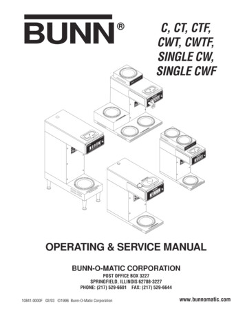
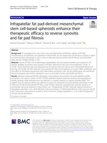
Preparation of Single-Cell RNA-SeqLibraries for Next Generation SequencingUNIT 4.22John J. Trombetta,1 David Gennert,1 Diana Lu,1 Rahul Satija,1Alex K. Shalek,2 and Aviv Regev1,31Broad Institute of MIT and Harvard, Cambridge, MassachusettsDepartment of Chemistry and Chemical Biology and Department of Physics, HarvardUniversity, Cambridge, Massachusetts3Howard Hughes Medical Institute, Department of Biology, Massachusetts Institute ofTechnology, Cambridge, Massachusetts2ABSTRACTFor the past several decades, due to technical limitations, the field of transcriptomicshas focused on population-level measurements that can mask significant differencesbetween individual cells. With the advent of single-cell RNA-Seq, it is now possible toprofile the responses of individual cells at unprecedented depth and thereby uncover,transcriptome-wide, the heterogeneity that exists within these populations. This unitdescribes a method that merges several important technologies to produce, in highthroughput, single-cell RNA-Seq libraries. Complementary DNA (cDNA) is made fromfull-length mRNA transcripts using a reverse transcriptase that has terminal transferaseactivity. This, when combined with a second “template-switch” primer, allows for cDNAsto be constructed that have two universal priming sequences. Following preamplificationfrom these common sequences, Nextera XT is used to prepare a pool of 96 uniquelyindexed samples ready for Illumina sequencing. Curr. Protoc. Mol. Biol. 107:4.22.1C 2014 by John Wiley & Sons, Inc.4.22.17. ⃝Keywords: SMART-Seq ! single-cell RNA-Seq ! transcriptomics ! cell type ! singlecell ! low-input RNA-Seq ! SMART-Seq2 ! Next-generation sequencing !template-switchingINTRODUCTIONThe development of single-cell RNA-Seq provides new opportunities to study complexcellular systems at unprecedented resolution. While previous RNA-Seq methods couldonly capture the average behavior of a population, single-cell methods now enable studiesof cellular heterogeneity, as well as the discovery of novel cell types and states. Thesemethods employ a variety of techniques to address the technical challenges posed bysingle cells—most notably, the ultra-low input quantity of RNA.SMART-Seq SINGLE-CELL RNA-Seq (METHOD)Here, the terminal transferase activity of the SMARTScribe reverse transcriptase isexploited in conjunction with a “template-switch” primer to make cDNAs, that havePCR priming sites on both ends, directly from full-length mRNA (Fig. 4.22.1). First,single cells are lysed in a guanidine thiocyanate solution. Second, a bead cleanup isused to isolate RNA. Third, cDNA is synthesized from the nearly full-length mRNAtranscripts. Fourth, these cDNA products are amplified by PCR and normalized. Fifth,the amplified cDNA is incubated with a Tn5 transposase to fragment nearly full-lengthdouble-stranded cDNAs and append adapters on each molecule. Sixth, each single-celllibrary is individually barcoded by PCR with index primers, and the sample set is pooled.Finally, the resulting pool is purified by a bead cleanup and is then sent for Illuminasequencing.Current Protocols in Molecular Biology 4.22.1-4.22.17, July 2014Published online July 2014 in Wiley Online Library (wileyonlinelibrary.com).DOI: 10.1002/0471142727.mb0422s107C 2014 John Wiley & Sons, Inc.Copyright ⃝BASICPROTOCOLPreparation andAnalysis of RNA4.22.1Supplement 107
polyadenylated RNAfirst strand synthesisand trailingSMARTer II Aoligonucleotide3 poly ASMART CD5primer35template switchingand extension5 blocked primer35amplify cDNA by PCRdouble-stranded cDNAtagmentation byTn5 transposaseNextera PCR with index primersFigure 4.22.1 Single-cell RNA-Seq library construction. Illustrated are the steps required toconvert mRNA to cDNA with sequencing adapters.In this protocol, Microseal F is used when sealing for long-term storage and MicrosealB when thermal cycling.MaterialsSingle-CellRNA-SeqLibraries for NGS2-Mercaptoethanol, !99.0%TCL buffer (Qiagen, cat. no. 1031576)Cell suspension in complete mediumDry iceRNeasy Plus Micro Kit (Qiagen)RNA-SPRI beads (Agencourt RNAClean XP RNA-SPRI beads, Beckman Coulter,cat. no. A63987)RNAse decontamination solution (RNaseZap, Life Technologies, cat. no. AM9780)RNase-free water80% ethanolSMARTer Ultra-Low Input RNA Kit for Illumina Sequencing-HV, 96 reactions(Clontech, cat. no. 634828) containing:Dilution buffer3′ SMART CDS Primer II A (12 µM): 5′AAGCAGTGGTATCAACGCAGAGTACT(30)VNRNase inhibitor (40 U/µl)5 first-strand buffer (RNase-free)Dithiothreitol (DTT; 100 mM)SMARTer dNTP Mix (dATP, dCTP, dGTP, and dTTP, each at 20 mM)SMARTer II A oligonucleotide (12 µM): 5′AAGCAGTGGTATCAACGCAGAGTACATrGrGrGSMARTScribe reverse transcriptase (100 U/µl)4.22.2Supplement 107Current Protocols in Molecular Biology
Nuclease-free waterIS PCR primer (12 µM): 5′ AAGCAGTGGTATCAACGCAGAGTPurification buffer (10 mM Tris·Cl, pH 8.5)Control total RNA (1 µg/µl)Advantage 2 PCR Kit, 100 reactions (Clontech, cat. no. 639206) containing:10 Advantage 2 PCR buffer50 dNTP Mix (dATP, dCTP, dGTP, and dTTP, each at 10 mM)50 Advantage 2 polymerase mixPCR-grade water10 Advantage 2 SA PCR bufferControl DNA template (100 ng/µl)Control primer mix, 5′ primer and 3′ primer (10 µM each)Agencourt AMPure XP SPRI beads (Beckman Coulter, cat. no. A63881)1 TE buffer (see recipe)Agilent High Sensitivity DNA Kit (Agilent Technologies, cat. no. 5067-4626)Qubit dsDNA HS assay kit, 100 reactions (Life Technologies, cat. no. Q32851)Taqman Fast Advanced Master Mix, 100 reactions (Life Technologies, cat. no.4444557)Taqman Probe set (choose housekeeping genes that are appropriate for specificsystem)Nextera XT DNA Sample Preparation Kit, 96 samples (Illumina, cat. no.FC-131-1096) containing:Amplicon Tagment Mix (ATM)Tagment DNA Buffer (TD)Nextera PCR Master Mix (NPM)Neutralize Tagment Buffer (NT)Resuspension BufferLibrary Normalization Additives 1Library Normalization Wash 1Hybridization BufferLibrary Normalization Beads 1Library Normalization Storage Buffer 1Nextera XT Index Kit, 96 indices, 384 samples (Illumina, cat. no. FC-131-1002)containing:Index primers, S501 to S508 (8 tubes)Index primers, N701 to N712 (12 tubes)96-well PCR plates, skirted (Eppendorf, cat. no. 951020401)1.5-ml RNAse- and DNAse-free microcentrifuge tubesMicroseal F foil (Bio-Rad, cat. no. MSF-1001)FACS machinePlate centrifugeVortexDynaMag-96 side-skirted magnet (Life Technologies, cat. no. 12027)8-channel pipettorThermal cyclerMicroseal B adhesive seals (Bio-Rad, cat. no. MSB-1001)2100 Electrophoresis Bioanalyzer Instrument (Agilent Technologies, cat. no.G2939AA)Qubit assay tubes (Life Technologies, cat. no. Q32856)Qubit 2.0 fluorometer (Life Technologies, cat. no. Q32866)TruSeq Index Plate Fixture Kit (Illumina, cat. no. FC-130-1005)DynaMag-2 magnet (Life Technologies, cat. no. 12321D)Illumina sequencerCurrent Protocols in Molecular BiologyPreparation andAnalysis of RNA4.22.3Supplement 107
Prepare single-cell lysates1. Prepare a 1% solution of 2-mercaptoethanol (by volume) in TCL buffer and distribute10 µl of this solution into each well of a skirted-side 96-well PCR plate, and 350 µlinto a 1.5-ml RNase-free microcentrifuge tube. Cover the plate with Microseal Fand keep at room temperature until ready for single-cell isolation.2. Prepare a cell suspension in complete medium and use a FACS machine to sort asingle cell into each well of the abovementioned 96-well plate containing TCL buffer,and an additional sample of !10,000 cells into the 1.5-ml microcentrifuge tube touse as a population control. Once sorting is complete, seal plate with Microseal Ffoil and centrifuge 1 min at 800 g, room temperature. Immediately freeze plateand population control on dry ice and keep up to 1 year at 80 C until ready forlysate cleanup.To improve yield, sorting single cells on both the presence of a positive viability indicator(e.g., Calcein AM, Life Technologies) and the absence of a cell death marker (e.g., themembrane-impermeant DNA stain EthD-1, Life Technologies) is recommended.Perform lysate cleanup and reverse transcription of mRNA speciesThe following steps can be performed inside a biosafety cabinet or a RNA workstation(if available); otherwise, they can be carefully performed on a standard benchtop.3. Purify RNA from the population control sample using the RNeasy Plus Micro Kitaccording to the manufacturer’s recommendations. Keep purified RNA at 4 C untilstep 18 ( 80 C for up to 1 year).4. Bring RNA-SPRI beads to room temperature (allow 30 min) and use RNAseZap toclean workbench and all equipment used to process RNA.5. Thaw lysate plate on ice, and then centrifuge 1 min at 800 g, room temperature.6. Thoroughly vortex RNA-SPRI beads to ensure a uniform suspension. Add 2.2 volRNA-SPRI beads to each well of lysate (2.2 10 µl cell lysate 22 µl of beadsare added, and 2.2 15 µl 33 µl) and mix well by pipetting up and down tentimes. If each solution is not in the bottom of its respective well, centrifuge plate 1min at 300 g, room temperature.The following steps are done at room temperature.Do not centrifuge plates containing SPRI beads at 300 g. Beads will adhere to thewell wall and impair RNA recovery.7. Incubate lysate and bead suspension for 10 min on bench.If this procedure is performed on a standard benchtop (i.e., not in an RNA fume hood),keep the plate covered (e.g., using the lid from a fresh box of pipet tips, placed slightlyajar) to prevent dust and debris from falling into samples during this and subsequentincubation steps.8. Place plate on 96-well plate magnet (e.g., DynaMag-96 side-skirted magnet) andincubate for 5 min. Remove supernatant from each column of the plate, being carefulnot to aspirate the beads collected on the side of each well.The magnet described draws the beads to alternating sides of each column of the plate.It is therefore recommended to use an eight-channel pipettor to remove the supernatant.Single-CellRNA-SeqLibraries for NGS9. Wash beads by adding 75 µl of 80% ethanol (prepared same day with nuclease-freewater) to each well. Wait "30 sec. Aspirate ethanol and repeat wash two additionaltimes.4.22.4Supplement 107Current Protocols in Molecular Biology
10. Aspirate final ethanol wash. Leave the plate on the magnet and allow beads to dryfor "10 min at room temperature. Keep plate loosely covered with the lid from afresh box of pipet tips, placed slightly ajar.Allowing beads to dry for too long will impair recovery of RNA. Immediately proceedwith protocol when visible cracks appear in bead pellet.11. Once bead pellet has dried, remove plate from magnet and elute RNA from beadsby resuspending dried beads in 4.5 µl of the following mix, using reagents from theSMARTer Ultra-Low Input RNA Kit (volumes per sample):2.375 µl dilution buffer1 µl RNase-free H2 O1 µl 24 µM 3′ SMART CDS Primer II A0.125 µl RNase inhibitor.Do not remove eluent from original plate.For population control, substitute water with 1 µl purified RNA.12. Seal and incubate 1 min at room temperature.13. Centrifuge 1 min at 800 g, room temperature.14. Incubate 3 min at 72 C to anneal 3′ SMART CDS Primer II A.15. Place plate on ice immediately following incubation.16. Remove seal and add the following as a mastermix (volumes per sample):2 µl first strand buffer0.25 µl 100 mM DTT1 µl 10 mM dNTP mix1 µl 12 µM SMARTer II A oligonucleotide0.25 µl RNase inhibitor1 µl SMARTScribe reverse transcriptase.Mix well by pipetting up and down, reseal with a new Microseal B adhesive seal,and centrifuge 1 min at 800 g, room temperature.17. Carry out the following reverse transcription in a thermal cycler using the followingconditions:1 cycle:90 min10 minHold42 C (initial step)72 C (inactivate)4 C (cool).Following this step, the product can be stored overnight at 4 C.Residual SPRI beads remain in each well with the libraries until step 28.The protocol provided with the SMARTer kit describes an SPRI cleanup at this stageof the protocol, which is omitted here. It is important to avoid losing material beforeamplification, and it has been found that the cleanup is not essential to generate ahigh-quality sequencing library (Picelli et al., 2013; Shalek et al., 2013).Perform whole transcriptome amplification and post-PCR cleanup18. Centrifuge plate 1 min at 800 g, room temperature and add the following fromthe Advantage 2 PCR kit as a mastermix (volumes per sample):5 µl 10 Advantage 2 PCR buffer2 µl 10 mM dNTP mix2 µl 12 µM IS PCR primer (from Clontech Ultra-Low Input RNA Kit)2 µl 50 Advantage 2 polymerase mix29 µl H2 O.Preparation andAnalysis of RNA4.22.5Current Protocols in Molecular BiologySupplement 107
Mix well by pipetting up and down, seal with Microseal B adhesive seal, andcentrifuge 1 min at 800 g, room temperature.19. Carry out the whole transcriptome amplification in a thermal cycler using the following conditions:1 cycle:5 cycles:9 cycles:7 cycles:1 cycle:1 min20 sec4 min6 min20 sec30 sec6 min20 sec30 sec7 min10 minHold95 C (initial denaturation)95 C (denaturation)58 C (annealing)68 C (extension)95 C (denaturation)64 C (annealing)68 C (extension)95 C (denaturation)64 C (annealing)68 C (extension)72 C (final extension)4 C (cool).Following this step, the product can be stored overnight at 4 C.For this amplification, a few modifications to the thermal cycling steps described in theoriginal SMARTer protocol have been made. These modifications improve annealing,denaturation, and extension for the diverse transcript sets with which the authors work.20. Bring DNA SPRI beads (Agencourt AMPure XP) to room temperature.21. Centrifuge whole transcriptome amplification plate 1 min at 800 g, room temperature.22. Unseal and add 0.8 vol of DNA SPRI beads to each well and mix well by pipettingup and down.23. Cover with a clean lid from a pipet tip box and incubate bead suspension for 5 minon bench.24. Place plate on 96-well plate magnet and incubate for an additional 5 min. Carefullyremove supernatant.25. Wash beads by adding 100 µl of 80% ethanol to each well and shift the plateleft and right on the magnet to move the beads from side to side in each well.Continue shifting plate on magnet for "30 sec. Aspirate ethanol and repeat washtwo additional times.26. Aspirate final ethanol wash. Leave the plate on the magnet and allow beads to dry"10 min at room temperature. Keep plate covered with the lid of a pipet tip box(left slightly ajar) to prevent dust and debris from falling into samples.27. Once bead pellet has dried, remove plate from magnet and elute DNA from beadsby resuspending dried beads with 20 µl of 1 TE buffer. Transfer eluent to a new96-well plate.Following this step, the new product plate can be sealed and stored overnight at 4 C orfor months at 20 C.Single-CellRNA-SeqLibraries for NGS28. Use 1 µl of the purified PCR product to measure the fragment size distributionusing the Agilent HS DNA BioAnalyzer or similar instrument according to themanufacturer’s instructions and 1 µl to estimate the library concentration using theQubit dsDNA HS Assay kit with Qubit assay tubes and the Qubit fluorometer orsimilar instrument according to the manufacturer’s instructions.4.22.6Supplement 107Current Protocols in Molecular Biology
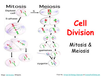
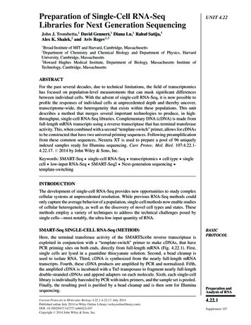
29. Use 1 µl of the purified PCR product to measure single-cell viability with theTaqman qPCR assay.In most single-cell sample sets, a small subset of libraries will have low complexity andwill lack expression of highly expressed housekeeping genes. Since there are, in mostsystems, a set of housekeeping genes that should be expressed in every cell (Shalek et al.,2013), use qPCR of two housekeeping genes (usually ACTB and B2M) to identify viablesamples. Samples with low or no expression of these genes can be removed from Nexteralibrary construction to reduce cost.30. For each sample, dilute the purified PCR product to a concentration between 0.1and 0.2 ng/µl with TE buffer in preparation for Nextera XT library construction.Construct Nextera XT sequencing library31. Before beginning library construction, thaw index primers and mix by brief vortexingfollowed by centrifugation. Arrange Nextera index tubes in the TruSeq Index PlateFixture such that each slot in the fixture holds one index tube. The 12 index 1 (i7)primers, which have orange caps, should be arranged horizontally in order, suchthat each tube corresponds to a column (Fig. 4.22.2). The column 1 slot should holdN701 and the column 12 slot should hold N712. Similarly, the index 2 (i5) primersshould be arranged vertically in order, such that S501 is in the slot for row A andS508 is in the slot for row H. Using a multichannel pipettor, distribute 10 µl of eachindex into the corresponding row/column (i7 primers will be distributed down rows,while i5 primers will be distributed across columns). Seal, vortex mildly to mix, andthen centrifuge 1 min at 800 g, room temperature.These mixed index primers can be used immediately, or stored long-term at 20 C.There is enough combined index primer solution in each well to generate 8 96 singlecell libraries.32. Using a new 96-well plate, add to each well:2.5 µl TD Buffer1.25 µl ATM1.25 µl diluted PCR product from SMART RT-PCR.Figure 4.22.2 Layout to prepare the combined dual-index primer plate that is used in NexteraXT library construction.Current Protocols in Molecular BiologyPreparation andAnalysis of RNA4.22.7Supplement 107
Mix well by pipetting up and down, seal, and centrifuge 1 min at 800 g, roomtemperature.33. Carry out tagmentation using the following conditions:Initial step:Cool:10 minhold55 C10 C.34. Unseal and immediately add 1.25 µl NT Buffer to each well and mix well bypipetting up and down. Wait 5 min at room temperature.35. Add 3.75 µl NPM and 2.5 µl combined Index primer solution to each well. Mix wellby pipetting up and down, seal, and centrifuge 1 min at 800 g, room temperature.36. Carry out the amplification in a thermal cycler using the following conditions:1 cycle:1 cycle:12 cycles:1 cycle:3 min30 sec10 sec30 sec60 sec5 minhold72 C (initial annealing)95 C (denaturation)95 C50 C72 C72 C4 C.Following this step, the product can be stored overnight at 4 C.Pool DNA and perform DNA SPRI bead cleanup37. Bring DNA SPRI beads (Agencourt AMPure XP) to room temperature.38. Centrifuge plate 1 min at 800 g, room temperature, unseal, and pool 2.5 µlfrom each well into a single 1.5-ml microcentrifuge tube. Measure total volume ofresulting pool, add 0.9 volumes SPRI beads to tube, and mix well by pipetting upand down.39. Incubate bead suspension for 5 min on bench.40. Place tube on DynaMag-2 magnet, and incubate for 5 min. Carefully remove supernatant.41. Wash beads by adding 500 µl of 80% ethanol to the tube and rotate it on the magnetto move the beads from front to back across the tube. Aspirate ethanol and repeatwash one additional time.42. Aspirate final ethanol wash. Leave the tube on the magnet and allow beads to dry"10 min at room temperature. Keep the tube covered to prevent dust and debrisfrom falling into the sample.43. Once bead pellet has dried, remove the tube from the magnet and elute DNAfrom beads using 30 µl TE buffer (consider smaller volumes if working with 96samples). Transfer eluent to a new 1.5-ml tube and repeat 0.9 SPRI bead cleanupone additional time.44. Use 1 µl of purified PCR product to measure the fragment size distribution using theAgilent HS DNA BioAnalyzer or similar instrument according to the manufacturer’sinstructions and 1 µl to estimate the library concentration using the Qubit dsDNAHS Assay kit with Qubit assay tubes and Qubit fluorometer or similar instrumentaccording to the manufacturer’s recommendations.Single-CellRNA-SeqLibraries for NGSPerform sequencing45. Sequence libraries on an Illumina sequencer using paired-end reads.4.22.8Supplement 107Current Protocols in Molecular Biology
The authors typically run two times 25 bp for expression quantification.Expression estimates will saturate "1 106 aligned reads, so one lane of HiSeq shoul
Here, the terminal transferase activity of the SMARTScribe reverse transcriptase is exploited in conjunction with a “template-switch” primer to make cDNAs, that have PCR priming sites on both ends, directly from full-length mRNA (Fig. 4.22.1). First, single cells are lysed in a