Transcription
Stern et al. BMC Gastroenterology 2014, 6RESEARCH ARTICLEOpen AccessMagnetic resonance enterography in pregnantwomen with Crohn’s disease: case series andliterature reviewMyriam D Stern1, Uri Kopylov2*, Shomron Ben-Horin2, Sarah Apter1 and Marianne Michal Amitai1AbstractBackground: Evaluation of pregnant women with known or suspected Crohn’s disease (CD) remains a challenge.Magnetic Resonance Enterography (MRE) is a promising diagnostic tool in these patients; however, the clinical dataon MRE utilization in pregnancy is scarce. The aim of the study was to describe the experience with MRE inpregnant CD patients in a tertiary referral center.Methods: We retrospectively reviewed MRE studies performed in pregnant women with known or suspected CDthat were performed between January 2007 and November 2012. Imaging findings, clinical management andoutcome were extracted from patient’s file and electronic records. Image quality was evaluated.Results: Ten studies of 9 patients were included. MRE protocol was modified to maximize maternal and fetal safety,and intravenous gadolinium was not used. In 7 patients, CD diagnosis was previously established; six were admittedwith clinical symptoms consistent with CD exacerbation, and an additional patient with a recurrent groin abscesswithout apparent luminal symptoms. In all seven patients, imaging features consistent with active CD weredetected; new penetrating complications were detected in 4 patients. Two patients underwent MRE for suspectedCD which was not comforted by study results. The clinical management was significantly impacted by MRE resultsin all positive cases. The image quality of the fast MRE sequences obtained without gadolinium was satisfactory andallowed meaningful interpretation.Conclusion: MRE with an adapted protocol for pregnancy is a reliable imaging modality to manage in pregnantwomen with known or suspected CD.Keywords: Crohn’s disease, Pregnancy, Magnetic resonance enterography, MRIBackgroundPregnant women with Crohn’s disease (CD) pose a rangeof diagnostic and clinical challenges [1]. When a pregnantwoman presents with a clinical picture consistent with suspected new-onset CD or exacerbation of previously established CD, diagnostic options are somewhat limited due tomaternal and fetal safety concerns. Endoscopy can be performed if needed for evaluation of colonic or distal ileal inflammation with necessary procedural precautions andcareful selection of a sedation regimen, however, evaluationof small-bowel disease and penetrating complications usually requires cross-sectional imaging [2]. Prompt evaluation* Correspondence: ukopylov@gmail.com2Department of Gastroenterology, Sheba Medical Center, Ramat Gan, IsraelFull list of author information is available at the end of the articleand management are required, as active CD or penetratingcomplications carry a significant risk of morbidity such aspreterm delivery and low birth weight [3-6]. Accurate anatomical information and evaluation of CD complications ismandatory in considering and planning surgical intervention, when necessary, in severely ill patients. Moreover, imaging may be valuable in planning the mode of delivery, ascesarean section is usually recommended in patients withactive perianal disease [5,7].Both computer tomography enterography (CTE) andmagnetic resonance enterography (MRE) are commonlyused to evaluate disease activity and complications inCD patients. However, CTE is associated with a significant exposure to ionizing radiation potentially harmfulfor the fetus [8,9]. 2014 Stern et al.; licensee BioMed Central Ltd. This is an Open Access article distributed under the terms of the CreativeCommons Attribution License (http://creativecommons.org/licenses/by/4.0), which permits unrestricted use, distribution, andreproduction in any medium, provided the original work is properly credited. The Creative Commons Public DomainDedication waiver ) applies to the data made available in this article,unless otherwise stated.
Stern et al. BMC Gastroenterology 2014, 6Page 2 of 9MRE is not associated with radiation exposure andtherefore is the preferred cross-sectional diagnostic modality in pregnancy. However there is a paucity of dataoutlining the utilization of MRE in pregnant CD patients. In particular, safety data pertaining to the intravenous contrast material (gadolinium) routinely utilizedin MRE is very limited, and it’s use in pregnancy recommended only when absolutely necessary [10].Several typical MRE findings reflect disease activity inCD [11]. Bowel wall thickness, relative contrast enhancement, presence of edema, and mucosal ulcerations havebeen shown to be independently correlated with endoscopic disease activity. An index incorporating these parameters (Magnetic resonance index of activity, MaRIA)was significantly correlated with the Crohn’s DiseaseEndoscopic Index of Severity (CDEIS) [12].An additional MRE scoring system based on muralthickness and T2 signal intensity was demonstrated tobe significantly correlated with a histological score of theterminal ileum inflammation [13].The purpose of the present study is to report our experience in with MRE utilizing a modified protocoladapted to maximize patient and fetus safety in the context of suspected CD or CD exacerbation in a tertiary referral center setting.MethodsStudy populationThe study cohort included all pregnant patients whounderwent MRE studies for suspected or established CDbetween 01/2007-12/2012. Relevant demographic andclinical data was retrieved from the patient’s files andelectronic records. This retrospective study was approvedby ethics review board of Chaim Sheba medical center.MRE protocolAll MRE studies were performed on a 1.5 T MRSystem (General Electric Healthcare, Little Chalfont,Buckinghamshire, United Kingdom) with an 8 channels cardiac coil. Our standard MRE protocol wasadapted for pregnant patients. Oral contrast with Manitol 5% (1000 ml) was administered 60 minutes priorto the examination. However, neither Gadolinium chelate nor glucagon (used to minimize bowel motility)were administered.The obtained MRE sequences included:FIESTA (Fast Imaging Employing Steady StateAcquisition) in axial, coronal and sagittal planes (repetitiontime (TR)/echo time (TE) 4.2-5/1.2-2.3 milliseconds(ms), slice thickness 6 mm, without interslice gap, field ofview (FOV) of 36–44 cm2, acquisition matrix of 384 384,number of excitations (NEX) o 1; flip angle 60 ):FSE (Fast spin Echo) T2 with fat suppression, in axialand coronal planes (TR/TE 1555-3500/88-93 ms, slicethickness 6 mm without interslice gap, FOV of 44 cm2,acquisition matrix of 384 224, NEX 1):T1 weighted 2D FSPGR (Fast spoiled Gradient RecalledAcquisition in the Steady State) with fat suppression andbreath hold in axial and coronal planes (TR/TE 75-155/1.3-4.2 ms, slice thickness 6 mm, without interslice gap,FOV of 36 cm2, acquisition matrix of 512 256, NEX 1,flip angle 75 ).T1 weighted 3D LAVA (Liver Acquisition with VolumeAcceleration) (without contrast) with fat suppressionTable 1 Summary of MRE findings in pregnant patients with known (1–7) or suspected CD (8, 9), NA: not availablePatient number1a1b23456789Mural signsSmall bowel mural thickening -- - --Large bowel mural thickening -- - Mural high T2 signal NANA NA -NAStenosis & prestenotic dilatation -- - Ulcers - - Comb sign --Creeping fat ---Lymphadenopathy-- - - -Phlegmon -- - --Abscess- --------Mesenteric signsComplicationsFistula -- - ---Free fluid------ ---
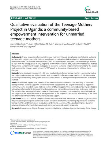
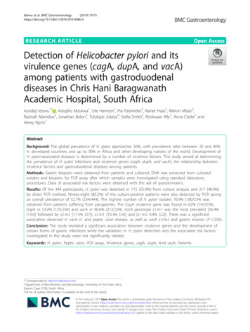
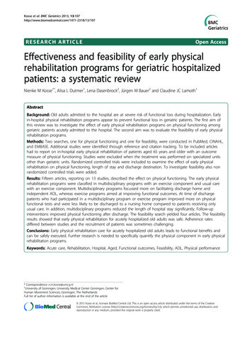
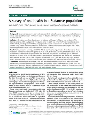
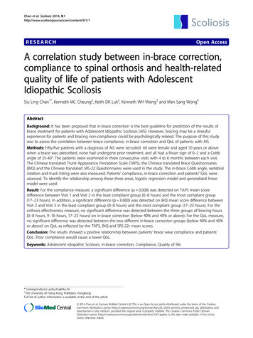
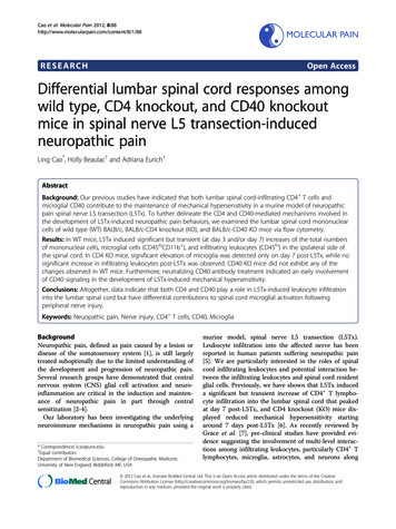
Stern et al. BMC Gastroenterology 2014, 6Page 3 of 9and breath hold in coronal and axial planes (TR/TE 3.6-4.3/1.7-2.1 ms, coronal: slice thickness 4 mm, nointerslice, axial: slice thickness 6 mm and overlap of3 mm, FOV of 44 cm2, acquisition matrix of416-320*192, NEX 0.75, flip angle 15 ).Analysis of MRE findingsMRE studies were reviewed by two experienced abdominalradiologists (MMA, SA) for signs of active and chronicCD and extraluminal complications. The main recordedfindings included:Mural findings: mural thickening of 3 mm or more,ulcerations, wall edema (high T2 signal on FSEsequence), luminal stenosis, prestenotic dilatation.Mesenteric findings: congestion (comb sign), hypertrophy(creeping fat), and lymphadenopathy. (lymph nodeshortest diameter 10 mm).Extra luminal findings: phlegmon, abscess, fistula, free fluid.Bowel obstruction, bowel perforation or acute hemorrhagewere considered as findings requiring urgent surgicalattention.Image quality assessmentThe image quality of each sequence for every MRE studywas evaluated using a score varying from poor to good(1–3): 1 blurred image, 2 fairly satisfactory image, 3 Figure 2 A 20 weeks pregnant patient with CD, Coronal FIESTA:signs of active disease: small bowel mural thickening and ulcer(arrow), note free fluid (dashed arrow).clear image. A mean score was obtained for each sequenceby both reviewers.The clinical course and outcomes of the included patients was described.ResultsThe study population included nine pregnant CD patients who underwent a total of ten MRE studies. Theindications for MRE studies were as follows:Clinical exacerbation of known CD (n 7), includingexacerbation of luminal disease in 6 patients (in one accompanied by a new-onset cholestasis and in anotherone- by preeclampsia) and a recurrent groin abscess inone patient; suspected CD (n 2).The demonstrated MRE findings for each patient aredescribed in detail in Table 1. In all patients with knownTable 2 Sequences quality score of MRE protocol adaptedto pregnancyFigure 1 A 19 weeks pregnant patient with CD, Coronal FIESTA:signs of active disease: mesenteric congestion (arrow) andlarge bowel mural thickening and edema (dotted arrow).MRE sequenceQuality scoreFIESTA coronal2.8FIESTA sagittal2.8FIESTA axial3Fast SE T2 coronal2.8Fast SE T2 axial2.2FSPGR 2D T1 coronal fat sat1.6FSPGR 2D T1axial fat sat1.5LAVA 3D T1 coronal fat sat1.6LAVA 3D T1 axial fat sat2FIESTA- Fast Imaging Employing Steady State Acquisition.SE-spin echo.FSPGR- Fast spoiled Gradient Recalled Acquisition in the Steady State.LAVA- Liver Acquisition with Volume Acceleration.
Stern et al. BMC Gastroenterology 2014, 6Page 4 of 9Table 3 Indications, MRE findings and clinical outcome of pregnant CD patientsPatient CD status priornumber to pregnancyPregnancynumber/weekIndication for Principal MREMRfindings11/23Clinicalexacerbationof known CD1/2620 yearsdurationClinicalmanagementClinical outcomePhlegmon, sinustract and fistulaPrednisone therapyand IV antibioticsNo improvement onmedical treatmentClinicalexacerbationSmall abscess3 weeks laterAbscess not accessibleto drainage, conservativetreatment with steroidsand IV antibioticsClinical deterioration,Spontaneous VDsurgical intervention one at 34 weeks,month post-delivery,healthy newbornincluding ileostomy andcecectomy.2/19Clinicalexacerbationof known CDActive disease, no Addition of IV steroidscomplications, noobstructionClinical response anddischargeSpontaneous VDat 38 weeks,healthy newborn1/31Clinicalexacerbationof known CDnew onset ofcholestasisUDCA and prednisoneScant signs ofactive disease, no added to maintenancecomplications, no treatment with 6 MPobstructionImprovement of CDsymptoms, persistentcholestasisInduced pretermvaginal deliveryfor cholestatsis at35 weeks healthynewborn2/22Clinicalexacerbationof known CDActive diseasephlegmon andfistulaeEnteral nutritionmodulation aPartial responsephlegmon and fistulaein CT post- delivery,antibiotics: Adalimumabwas after deliverySpontaneousvaginal delivery at38 weeks, healthynewborn1/37Clinicalexacerbationof known CDpreeclampsiaSome signs ofactive disease,no complicationsIV steroids andantibioticsPreeclampsia UrgentdeliverySpontaneous onsetof labor, vaginaldelivery convertedto C/S, at 37 weekshealthy newborn2/20Recurrentabscess inright groin,fistula?Phlegmon in RLQ IV and PO antibioticsand abscess drainagefistula to rightprior to MRgroinClinical improvementSpontaneousdelivery, healthynewborn atweek distributionNo currenttreatment29 ibutionTx: Azathioprine316 yIleocolonicdistributionTx: 6-MP415 IleocolonicdistributionTx: Azathioprine54 yearsdurationInflammatoryphenotypeInactiveperianal diseaseCrohn’s colitisTx: infliximab610 ibutionTx: Azathioprine
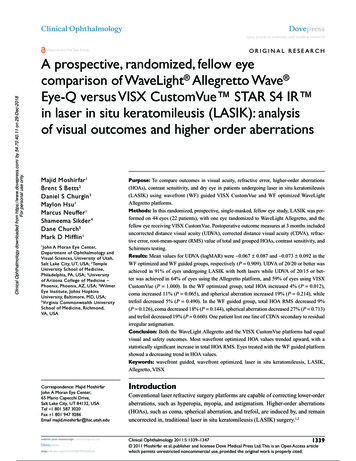
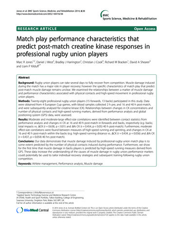
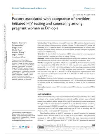
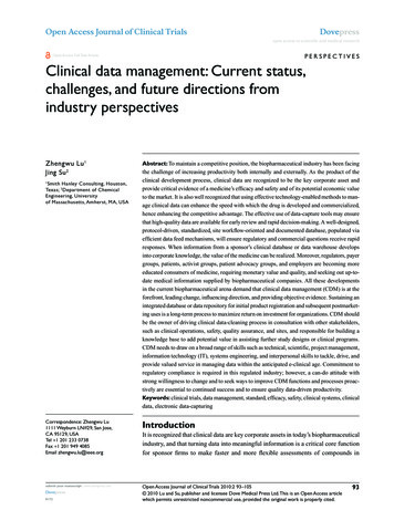
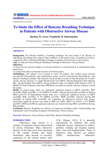
Stern et al. BMC Gastroenterology 2014, 6Page 5 of 9Table 3 Indications, MRE findings and clinical outcome of pregnant CD patients (Continued)72 yearsduration2/25Clinicalexacerbationof known CDInflammatoryphenotypeSigns of activeConservative treatmentdisease, newwith steroids and IVphlegmon in RLQ antibiotics emergencycerclageTemporary clinicalimprovementhypoalumiemia &anasarcaSpontaneousvaginal delivery at28 weeks healthyvery low birthweight newbornIleocolonicdistributionTx: 6-MP andadalimumab8No knowndisease1/26Suspected CD Bowel normalNo treatmentAbdominal symptomsresolvedHealthy twinsnewborns C/Sat 32 w9No knowndisease?/11MRE signs of UCUncertaindiagnosis ofUC, suspectedCDNANASpontaneousdelivery with ahealthy newbornat week 41SUC-ulcerative colitis, VD- vaginal delivery, RQ-right lower quadrant, 6-mp- 6-mercaptopurine, UDCA-ursodeoxycholic acid.CD, pathognomonic signs of active disease were demonstrated on MRE. Wall thickening and ulcers were demonstrated in 6 patients (86%), high T2 signal in bowelwall in 4/4 patients who had T2 sequences performed,phlegmon in 4/7 (57%) and abscess in 1/7 (14%). Positivecomb sign (a hallmark of mesenteric inflammation) wasdemonstrated in all patients. Fistulae were seen in 3(43%), while stenosis and prestenotic dilatation wereseen in 5 (71%).In two patients, MRE was performed for clinical suspicion of CD. In one patient, no radiological findings compatible with this diagnosis were demonstrated. Thesecond patient was diagnosed with ulcerative colitis inthe past. Her study demonstrated colonic findings compatible with UC and not CD (large bowel mural thickening, submucosal edema).The typical radiographic signs of active CD identified onour modified protocol for pregnancy included mural thickening of 3 mm or more, ulcers, wall edema, comb sign,phlegmon, abscess and fistula are presented in Figures 1and 2.Image quality assessmentFIESTA sequences were of very good quality and enabled accurate assessment of the bowel anatomy, as wellas the presence of a phlegmon, an abscess or free fluid.The images did not have significant motion artifacts.Fast spin echo T2 sequences, although not availablefor all patients, were useful for evaluation of edematousbowel loops and in ruling out the presence of free fluid.Gradient echo (GRE) T1 weighted 3D LAVA sequenceswere blurred by motion artifacts. GRE T1 weighted 2DFSPGR had motion artifacts and their added diagnosticvalue was relatively low. Coronal planes were easier tointerpret than axial planes (Table 2).Clinical impact of the MRE findingsIn all the 9 patients, MRE findings have contributed usefulclinical information and impacted the clinical management(Table 3). The findings of the MRE were valuable in rulingout the diagnosis in the two patients suspected of CD: onepatient (n 8) had no radiological findings suggesting CDand did not require anti-inflammatory treatment. In theother patient, the diagnosis of UC was confirmed and CDwas ruled out, as no signs of small bowel or mesenteric disease and no penetrating complications were demonstrated(Figure 3).In all patients with known CD, no complications requiring emergent surgical interventions such as obstruction, perforation or hemorrhage were demonstrated. Thepatients were treated conservatively and did not requiresurgical intervention prior to the delivery.Extraluminal complications such as phlegmon, abscess,fistula, free fluid were ruled out in 3 patients (n 2,3,5). Inone patient with preeclampsia (n 5), MRE was performedone day before delivery, and assisted the decision to perform a cesarean section approach since no intra-abdominalcomplications were detected. In two others (n 2 & 3), medical treatment was adjusted with good response of CDsymptoms.In four patients (n 1,4,6,7), extraluminal complicationswere newly diagnosed by MRE .In one of them (n 1) thevisualization of a phlegmon transforming into an abscess,inaccessible for drainage, was detected on a second MREexamination, and impacted a physician decision to undertake a surgical intervention after a spontaneous vaginaldelivery at 34 weeks. MRE findings were confirmed duringsurgery (Figure 4).In patient n 4, phlegmon and fistula were demonstrated. A partial clinical response was obtained withintravenous antibiotics until spontaneous vaginal delivery at 38 weeks. Due to persistence of the clinical
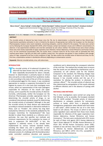
Stern et al. BMC Gastroenterology 2014, 6Page 6 of 9Figure 3 A 11 weeks pregnant patient diagnosed with UC, a: Coronal FIESTA, large bowel mural thickening with a thumb printingpattern (arrow), b: Coronal FIESTA, ileo-cecal stenosis (arrow), c: Axial SSFSE T2 submucosal edema (arrow).exacerbation after the delivery, an additional postdelivery CT was performed, which confirmed the MRfindings.Patient n 6 presented with a recurrent groin abscess thatrequired multiple drainage procedures, without clinical exacerbation of the luminal disease. An MRE was performedat week 12 of pregnancy. The study demonstrated activedisease in terminal ileum and cecum accompanied by aphlegmon and a new inflammatory tract communicatingto another phlegmonous subcutaneous zone corresponding to the location of the drained abscess.In the fourth patient (n 7) who presented with CD exacerbation, cervical widening and early contractions at17 week of pregnancy, a phlegmon was demonstrated.The patient did not respond to medical treatment, andspontaneus delivery occurred at 28 week. On presentation this patient was treated with a combination of6-mercaptopurine and adalimumab. There is no clearevidence to suggest that a combination of adalimumabwith a thiopurine is superior to adalimumab alone in CD(as opposed to Infliximab where such benefit was clearlydemonstrated [14]) and pregnancy-related data are evenmore limited.DiscussionMRE has long been established as an accurate and safemodality for diagnosis of CD, monitoring of disease activity and detection of complications [2]. In our series, itappears that, despite the technical limitations (mainlycaused by avoidance of intravenous contrast materialand by motion artifacts), MRE was able to demonstratesignificant and clinically relevant findings impacting theclinical management in pregnant patients. Importantclassical stigmata of CD were demonstrated and confirmed on subsequent studies in some of the patients;non-emergent surgical procedures were deferred untilafter delivery and additional clinical decisions such asmode and timing of planned delivery were impacted byMRE. Thus, the information provided by MRE plays akey role in the clinical and surgical management of thepatients.To the best of our knowledge, our study is the largestcase series describing the utility of MRE in pregnant CDpatients published to date. In addition, in our series wehave utilized oral contrast material, which has not beenreported in pregnant CD patients in current literature.In an early article, Shoenut et al. [15] reported two casesFigure 4 A pregnant patient with CD at 23 weeks (a) and 26 weeks (b) of pregnancy: a: Coronal FIESTA fistula (dotted arrow) and sinustracts (arrow), b: Axial heavily weighted T2, absc
pregnant CD patients in a tertiary referral center. Methods: We retrospectively reviewed MRE studies performed in pregnant women with known or suspected CD that were performed between January 2007 and November 2012. Imaging findings, clinical management and outcome were extracted from pat