Transcription
Clinical OphthalmologyDovepressopen access to scientific and medical researchO r i g i n al R e s e a r c hClinical Ophthalmology downloaded from https://www.dovepress.com/ by 54.70.40.11 on 29-Dec-2018For personal use only.Open Access Full Text ArticleA prospective, randomized, fellow eyecomparison of WaveLight Allegretto Wave Eye-Q versus VISX CustomVue STAR S4 IR in laser in situ keratomileusis (LASIK): analysisof visual outcomes and higher order aberrationsThis article was published in the following Dove Press journal:Clinical Ophthalmology19 September 2011Number of times this article has been viewedMajid Moshirfar 1Brent S Betts 2Daniel S Churgin 3Maylon Hsu 1Marcus Neuffer 1Shameema Sikder 4Dane Church 5Mark D Mifflin 11John A Moran Eye Center,Department of Ophthalmology andVisual Sciences, University of Utah,Salt Lake City, UT, USA; 2TempleUniversity School of Medicine,Philadelphia, PA, USA; 3Universityof Arizona College of Medicine –Phoenix, Phoenix, AZ, USA; 4WilmerEye Institute, Johns HopkinsUniversity, Baltimore, MD, USA;5Virginia Commonwealth UniversitySchool of Medicine, Richmond,VA, USACorrespondence: Majid MoshirfarJohn A Moran Eye Center,65 Mario Capecchi Drive,Salt Lake City, UT 84132, USATel 1 801 587 3020Fax 1 801 947 9286Email majid.moshirfar@hsc.utah.edusubmit your manuscript 7/OPTH.S24316Powered by TCPDF (www.tcpdf.org)Purpose: To compare outcomes in visual acuity, refractive error, higher-order aberrations(HOAs), contrast sensitivity, and dry eye in patients undergoing laser in situ keratomileusis(LASIK) using wavefront (WF) guided VISX CustomVue and WF optimized WaveLightAllegretto platforms.Methods: In this randomized, prospective, single-masked, fellow eye study, LASIK was performed on 44 eyes (22 patients), with one eye randomized to WaveLight Allegretto, and thefellow eye receiving VISX CustomVue. Postoperative outcome measures at 3 months includeduncorrected distance visual acuity (UDVA), corrected distance visual acuity (CDVA), refractive error, root-mean-square (RMS) value of total and grouped HOAs, contrast sensitivity, andSchirmers testing.Results: Mean values for UDVA (logMAR) were 0.067 0.087 and 0.073 0.092 in theWF optimized and WF guided groups, respectively (P 0.909). UDVA of 20/20 or better wasachieved in 91% of eyes undergoing LASIK with both lasers while UDVA of 20/15 or better was achieved in 64% of eyes using the Allegretto platform, and 59% of eyes using VISX CustomVue (P 1.000). In the WF optimized group, total HOA increased 4% (P 0.012),coma increased 11% (P 0.065), and spherical aberration increased 19% (P 0.214), whiletrefoil decreased 5% (P 0.490). In the WF guided group, total HOA RMS decreased 9%(P 0.126), coma decreased 18% (P 0.144), spherical aberration decreased 27% (P 0.713)and trefoil decreased 19% (P 0.660). One patient lost one line of CDVA secondary to residualirregular astigmatism.Conclusion: Both the WaveLight Allegretto and the VISX CustomVue platforms had equalvisual and safety outcomes. Most wavefront optimized HOA values trended upward, with astatistically significant increase in total HOA RMS. Eyes treated with the WF guided platformshowed a decreasing trend in HOA values.Keywords: wavefront guided, wavefront optimized, laser in situ keratomileusis, LASIK,Allegretto, VISXIntroductionConventional laser refractive surgery platforms are capable of correcting lower-orderaberrations, such as hyperopia, myopia, and astigmatism. Higher-order aberrations(HOAs), such as coma, spherical aberration, and trefoil, are induced by, and remainuncorrected in, traditional laser in situ keratomileusis (LASIK) surgery.1,2Clinical Ophthalmology 2011:5 1339–13471339 2011 Moshirfar et al, publisher and licensee Dove Medical Press Ltd. This is an Open Access articlewhich permits unrestricted noncommercial use, provided the original work is properly cited.
Clinical Ophthalmology downloaded from https://www.dovepress.com/ by 54.70.40.11 on 29-Dec-2018For personal use only.Moshirfar et alAlthough conventional LASIK surgery is mostly accurate,abundant research has shown reoperation rates for primarymyopic keratorefractive surgery are between 5.5% and19.2%.3–9 When HOAs cannot be corrected, image qualitymay suffer.1,2,10–13 The HOAs call for more advanced opticalmeasurements and more sophisticated laser algorithms. Theselaser algorithms are found in wavefront (WF) based treatments, which have been shown to diminish induced HOAscompared to traditional LASIK, and increase predictabilityof visual outcomes.14–18 As WF based methods have evolvedrapidly over the years, an important question is whether thereare significant differences in the induced HOAs and visualoutcomes between specific WF guided and WF optimizedplatforms used in LASIK.To date, multiple studies have compared WF guidedand WF optimized platforms in LASIK surgery.19–24 Thedata shows varied results, with some studies suggesting anadvantage to WF guided platforms,19,22–24 and others showingno significant difference between the two platforms.20,21 In thisprospective, randomized, fellow eye study, we compared WFguided VISX CustomVue platform (Abbott Medical Optics[AMO], Santa Ana, CA) with WF optimized WaveLightAllegretto platform (Alcon, Inc, Hüenberg, Switzerland) in thesame patient undergoing LASIK, with respect to visual acuity,refractive error, HOAs, contrast sensitivity, and dry eyes.Patients and methodsThis prospective, single masked, randomized, fellow eyestudy evaluated and compared the outcomes of LASIK performed on 44 eyes (22 patients) using the VISX CustomVuelaser system and the WaveLight Allegretto laser system.Patients were recruited and enrolled at the John A Moran EyeCenter, Department of Ophthalmology and Visual Science,University of Utah, between December 2010 and July 2011.All patients were over the age of 21.The University of Utah Hospital Institutional Review Boardapproved the research protocol in accordance with the tenetsof the Declaration of Helsinki. All patients provided informedconsent after they received an explanation of the procedure,including all risks and benefits. All patients had a preoperativediscussion of relevant medical history, including history ofherpetic eye disease and family history of keratoconus.Patients were excluded if they had a history of clinicallysignificant lens opacity, previous corneal or intraocular surgery, thin corneas, keratoconus, unstable refraction, amblyopia, or autoimmune disease; were pregnant or breastfeeding;or were on immunosuppressive therapy. Patients desiringmonovision were not included in the study.1340Powered by TCPDF (www.tcpdf.org)submit your manuscript www.dovepress.comDovepressDovepressEligible patients were scheduled for bilateral LASIK andcorrection for distance in both eyes. The patients were randomly assigned (Research Randomizer software – Urbaniak,www.randomizer.org) to treatment in one eye with theWaveLight Allegretto system (WF optimized group), whichutilizes the WaveLight Allegretto 400 Hz Wave Eye-QLaser. The fellow eye was assigned treatment with the VISXCustomVue STAR S4 IR Excimer Laser with ActiveTrack iris registration (WF guided group). The patientswere blinded as to which laser was used for treatment ineach eye. No patient had LASIK enhancement at any timeduring the study.Soft contact lenses were discontinued 2 weeks beforescreening and rigid gas permeable contact lenses werediscontinued 6 weeks before screening. All patients hada preoperative examination including manifest refraction(MRX) and cycloplegic refraction, uncorrected distancevisual acuity (UDVA), corrected distance visual acuity(CDVA), tonometry, slitlamp examination, and dilatedfundus evaluation. Corneal topography and thicknesswere measured using the Pentacam (OCULUS OptikgeräteGmbH, Wetzlar, Germany) and Humphrey Atlas (CarlZeiss Meditec, Inc, Jena, Germany) systems. Pupil sizewas measured in the dark using the Colvard pupillometer(Oasis Medical, Inc, Glendora, CA). Contrast sensitivitywas performed using the VectorVision CSV-1000 (Dayton, OH) chart in controlled mesopic (70 lux) conditionsat three, six, twelve, and 18 cycles per degree. Schirmerstesting evaluation for dry eye was measured in millimeterswith topical anesthetic after 5 minutes. MRX and WF measurements were repeated on two separate visits to ensurerefractive stability.All eyes received five preoperative wavefront analysesusing the VISX CustomVue WaveScan aberrometer v 3.62(Fourier) (AMO), without pharmacologic intervention,under mesopic conditions, with a minimum pupil diameterof 6.0 mm. The emmetropic correction target was based onMRX, topography, and wavefront analysis for the WF guidedgroup. For the WF optimized group, emmetropic correctiontarget was based on topography and MRX. Iris registration was obtained for eyes receiving WF guided treatment. Previously established Moran Laser Center WF guidedLASIK nomograms were followed for both the Allegretto andthe CustomVue treatments. The nomograms were generatedwith Datagraph-med refractive outcomes software (v 3.20a;Ingenieurbüro Pieger GmbH, Wendelstein, Germany) usinga minimum of 50 eyes for each wavefront platform with aminimum follow-up of 3 months.Clinical Ophthalmology 2011:5
Clinical Ophthalmology downloaded from https://www.dovepress.com/ by 54.70.40.11 on 29-Dec-2018For personal use only.DovepressWavefront optimized vs wavefront guided LASIKAll LASIK flaps were created with an iFS AdvancedFemtosecond Laser (AMO) at 150 kHz in a raster pattern withbed energy of 1.15 µJ, side cut energy of 2.00 µJ, and pocketenabled. Flaps were created with an intended thickness of110 µm, diameter of 8.4 to 9.0 mm, superior hinge angle of55 , and a side cut angle of 70 . If the 8.0 mm maximumintended ablation diameter exceeded the flap diameter, thehinge and flap were shielded during ablation. All surgerieswere performed by Majid Moshirfar and Mark D Mifflin.Postoperatively, each eye received one drop of gatifloxacin 0.3% (Allergan Inc, Irvine, CA) and prednisolone acetate1% (Allergan, Inc). The prednisolone acetate was continuedhourly on postoperative day 1, and then four times daily for6 additional days. The gatifloxacin was continued four timesdaily for 1 week.Patients were seen and data was collected at 1 day, 1 week,and 1 and 3 months after surgery. UDVA and CDVA weretested using a standard Snellen eye chart. Visual acuitywas recorded in both Snellen notation and logarithm of theminimum angle of resolution (logMAR) format. Evaluation ofcontrast sensitivity, dry eye, and HOAs were performed. HOAsincluding total HOA root mean square (RMS), coma Z(3,1),trefoil Z(3,3), and spherical aberration Z(4,0), were measuredusing the WaveScan aberrometer. Undilated scans of botheyes were taken at 3 months postoperatively regardless of thewavefront platform used for treatment. Subjective outcomeswere recorded using the Quality of Life Impact of RefractiveCorrection (QIRC) instrument. The ratings from the QIRCwere interpreted using the guidelines set by Pesudovs et al.25StatisticsAfter the study was completed, the results were compiled andthe data unmasked for statistical analysis. MRX, refractive error,visual acuity, contrast sensitivity, HOA values for coma, trefoil,and sphere, and HOA RMS wavefront aberration values weretreated as continuous variables and analyzed for significanceby using a paired Student’s t-test. A P value of 0.05 or less wasconsidered statistically significant. Data analysis was performedusing Microsoft Excel (Microsoft Corp, Redmond, WA).Results22 patients (44 eyes) with 3 months of postoperative carewere evaluated in this study. The sample population consisted of 15 women and seven men with a mean age of 31.2years 6.3 standard deviation (SD) (range 23 to 53 years).Baseline data showed no significant differences in preoperative CDVA or refractive error (Table 1).Visual acuityUDVA and CDVA were not statistically different betweenthe groups at 1 or 3 months (Table 2). Postoperatively, 20/15UDVA was achieved in 64% and 59% of the eyes in the WFoptimized and WF guided groups, respectively. Three monthsafter surgery, 91% of the eyes in both groups achieved 20/20UDVA (Table 3). Postoperatively, spherical equivalent andsphere measurements decreased significantly in the WFoptimized group compared to the WF guided group at 1 and3 months (P 0.020, P 0.017). There was no significantdifference in cylinder measurements in either group beforeor after surgery.Safety, efficacy, and predictabilityAt 1 month, 21 eyes (95%) in the WF optimized group and18 (81%) in the WF guided group were within 0.50 diopters(D) of emmetropia. In addition, ten eyes (45%) in the WFoptimized group and six eyes (27%) in the WF guided groupwere within 0.25 D of emmetropia (P 0.035). Althoughthere was a significant difference in emmetropia at 1 month,this did not remain significant at 3 months (Table 3).At 3 months, one eye (5%) in the WF optimized group lostone line of CDVA. The vision loss was attributed to residualirregular astigmatism confirmed by topography. No other eyesTable 1 Preoperative group comparisonsParameterAgeMale/femaleMean LogMAR CDVACDVA (20/X)Spherical equivalent (D)Sphere (D)CylinderPlatformP-value*Allegretto (n 22)VISX (n 22)31.7 6.307/15-0.03 0.06 (-0.125 to 0)18.6 2.28 (15 to 20)-3.61 1.98 (-7.25 to 1.50)-3.97 2.10 (-7.75 to 1)0.70 0.97 (0 to 4.25)-0.03 0.06 (-0.125 to 0)18.6 2.28 (15 to 20)-3.86 1.81 (-7.25 to -0.375)-4.17 1.99 (-7.5 to -0.375)0.63 0.78 (0 to 2.25)1.0001.0000.1370.1910.461Notes: Values represented as mean standard deviation (range); *paired Student’s t-test.Abbreviations: UDVA, uncorrected distance visual acuity; CDVA, corrected distance visual acuity; logMAR, logarithm of the minimum angle of resolution.Clinical Ophthalmology 2011:5submit your manuscript www.dovepress.comDovepressPowered by TCPDF (www.tcpdf.org)1341
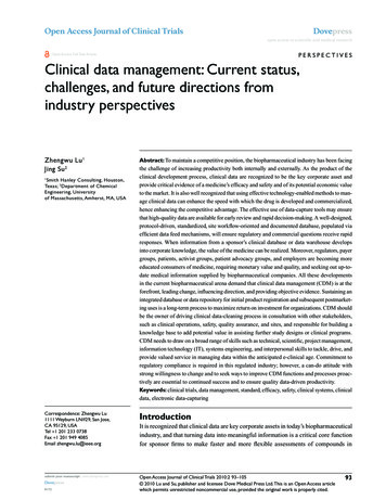
DovepressMoshirfar et alTable 2 One month and three month visual acuity comparisonsClinical Ophthalmology downloaded from https://www.dovepress.com/ by 54.70.40.11 on 29-Dec-2018For personal use only.ParameterPostoperative month 1UDVA (LogMAR)UDVA (20/x)CDVA (LogMAR)CDVA (20/x)Spherical equivalent (D)Sphere (D)Cylinder (D)Postoperative month 3UDVA (LogMAR)UDVA (20/x)CDVA (LogMAR)CDVA (20/x)Spherical equivalent (D)Sphere (D)Cylinder (D)Allegretto (n 22)VISX (n 22)P-value*-0.04 0.10 (-0.125 to 0.176)19.3 5.19 (15 to 30)-0.05 0.08 (-0.125 to 0.097)18.1 3.35 (15 to 25)0.02 0.24 (-0.05 to 0.625)-0.08 0.29 (-0.75 to 0.5)0.19 0.27 (0 to 0.75)-0.05 0.11 (-0.125 to 0.301)19.3 7.29 (15 to 40)-0.06 0.07 (-0.301 to 0.097)17.6 3.01 (10 to 25)0.32 0.51 (-0.375 to 1.125)0.25 0.48 (-0.5 to 1.75)0.14 0.17 (0 to 0.5)0.5151.0000.7450.3290.0200.0120.234-0.07 0.09(-0.125 to 0.301)17.5 4.01 (15 to 30)-0.11 0.08 (-0.301 to 0.097)15.9 2.94 (10 to 25)0.04 0.27 (-0.375 to 0.75)-0.1 0.31 (-0.5 to 0.75)0.26 0.28 (0 to 0.75)-0.07 0.09 (-0.301 to 0.097)17.3 3.69 (10 to 25)-0.10 0.09 (-0.301 to 0)16.1 3.06 (10 to 20)0.30 0.43 (-0.25 to 1.75)0.18 0.39 (-0.25 to 1.25)0.23 0.28 (0 to 1)0.7140.7470.9090.7470.0170.0490.623Notes: Values represented as mean standard deviation (range); *paired Student’s t-test.Abbreviations: UDVA, uncorrected distance visual acuity; CDVA, corrected distance visual acuity; logMAR, logarithm of the minimum angle of resolution.in the study population lost any lines of CDVA. Nine eyes(41%) in the WF optimized group maintained equal CDVAcompared to twelve eyes (55%) in the WF guided group.Eleven eyes (50%) in the WF optimized group and nine eyes(41%) in the WF guided group gained one line of CDVA.One eye in both groups gained two lines (P 1.000).20.3 mm 8.52 and 21.1 mm 10.2 for the WF optimizedgroup and the WF guided group, respectively. The 3 monthspostoperative values were 21.1 mm 7.83 for the WF optimized group and 20 mm 8.12 for the WF guided group.There was no significant difference in Schirmers testingbetween both groups during the study period (P 0.081).Schirmers testingContrast sensitivityThe preoperative average Schirmers test value for the WFoptimized group was 19.9 mm 9.95 and 22.1 mm 9.98for the WF guided group. At 1 month the values wereContrast sensitivity did not change significantly in eithergroup when preoperative measurements were compared topostoperative measurements (P 0.07) (Figure 1).Table 3 Efficacy, predictability, and safety comparison of allegretto and VISX laser platformsPostoperative month 1ParameterEfficacy (UDVA)20/15 or better20/20 or better20/30 or better20/40 or better20/50 or betterPredictability 0.25 D of emmetropia 0.50 D of emmetropia 1.00 D of emmetropiaSafety (CDVA)Loss of 2 lines or moreLoss of 1 lineNo loss of linesGain of 1 lineGain of 2 lines or morePostoperative month 3Allegretto (n 22)VISX (n 22)P-value*Allegretto (n 22)VISX (n 22)0.77810 (45%)18 (82%)22 (100%)22 (100%)22 (100%)12 (55%)19 (86%)20 (91%)22 (100%)22 (100%)10 (45%)21 (95%)22 (100%)6 (27%)18 (81%)20 (91%)0 (0%)3 (14%)13 (62%)5 (24%)0 (0%)0 (0%)2 (10%)13 (62%)6 (29%)0 (0%)P-value*1.00014 (64%)20 (91%)21 (95%)22 (100%)22 (100%)13 (59%)20 (91%)22 (100%)22 (100%)22 (100%)17 (77%)21 (95%)22 (100%)15 (68%)18 (82%)22 (100%)0 (0%)1 (5%)9 (41%)11 (50%)1 (5%)0 (0%)0 (0%)12 (55%)9 (41%)1 (5%)0.0350.1991.0001.000Notes: Values represented as number of eyes (percentege); *paired Student’s t-test.Abbreviations: UDVA, uncorrected distance visual acuity; CDVA, corrected distance visual acuity; logMAR, logarirhm of the minimum angle of resolution.1342Powered by TCPDF (www.tcpdf.org)submit your manuscript www.dovepress.comDovepressClinical Ophthalmology 2011:5
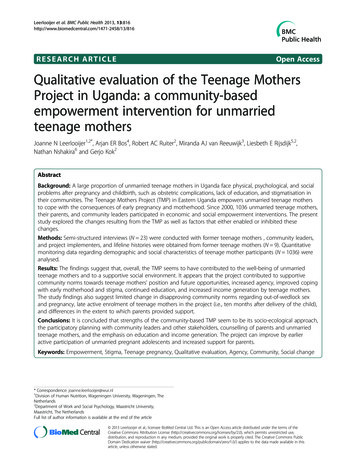
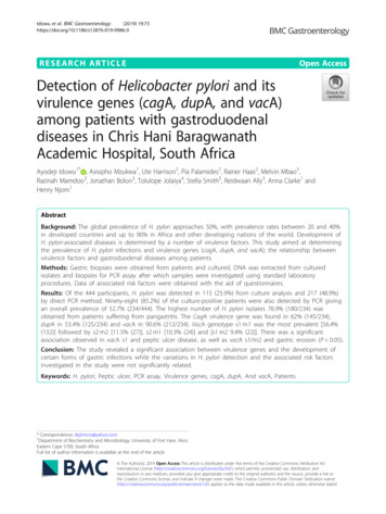
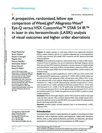
DovepressWavefront optimized vs wavefront guided LASIK2.50log contrast sensitivityClinical Ophthalmology downloaded from https://www.dovepress.com/ by 54.70.40.11 on 29-Dec-2018For personal use only.2.001.50Preoperative3 3CPD6CPD12CPD18CPDIog spatial frequency (cycles/degree or CPD)Figure 1 Comparison of contrast sensitivity between allegretto and VISX laser platforms at 3, 6, 12, and 18 cycles per degree (CPD) preoperatively (n 22) and at 3 monthspostoperatively (n 22) using the Vectrovision CSV-1000E chart (P 0.066 for all measurements at any time).Higher-order aberrationsAt 3 months postoperatively, 15 eyes (68%) in eachgroup completed CustomVue WaveScan analysis. Theonly statistically significant change discovered was a 4%increase in total HOA RMS (P 0.012) in the WF optimized group. In this group, other HOAs had nonsignificantchanges, which showed the following: coma increased 11%(P 0.065), trefoil decreased 5% (P 0.239) and sphericalaberration increased 19% (P 0.214) (Figure 2). The WFguided group showed a decreasing trend in HOA values,with total HOA RMS, coma, trefoil, and spherical aberration decreasing 9% (P 0.126), 18% (P 0.144), 19%(P 0.660), and 28% (P 0.713) respectively (Figure 3).There were no significant differences in preoperative orpostoperative HOA values between the WF guided or WFoptimized groups.0.400.35*Preoperative0.30RMS HOA (µm)3 months0.250.200.150.100.050.00Total HOAComaTrefoilSpherical aberration 0.05Figure 2 Comparison of higher-order aberrations before surgery (n 16) and 3 months postoperatively (n 16) in the wavefront optimized platform. Total, coma, Z(3, 1),trefoil, Z(3, 3), and spherical aberration, Z(4, 0) were measured using the WaveScan at a mean diameter of 6 mm.Note: *Statistically significant (P 0.012).Abbreviation: RMS HOA, root-mean-square higher order aberration.Clinical Ophthalmology 2011:5submit your manuscript www.dovepress.comDovepressPowered by TCPDF (www.tcpdf.org)1343
DovepressMoshirfar et al0.600.50PreoperativeRMS HOA (µm)0.400.300.200.100.00Total HOAComaSpherical aberrationFigure 3 Comparison of higher-order aberrations
(LASIK) using wavefront (WF) guided VISX CustomVue and WF optimized WaveLight Allegretto platforms. Methods: In this randomized, prospective, single-masked, fellow eye study, LASIK was per-formed on 44 eyes (22 patients), with one eye randomized to WaveLight Allegretto, and