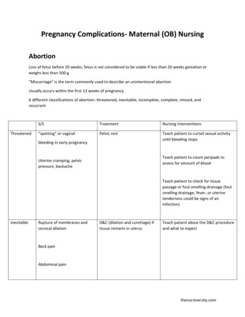
Transcription
Proceedings - 8th World Rabbit Congress –September 7-10, 2004 – Puebla, MexicoULTRASONOGRAPHY STUDY OF RABBITS PREGNANCYGUTIERREZ H. E., ZAMORA F. M. M.Centro de Enseñanza Agropecuaria, Facultad de Estudios Cuautitlán UNAMjema81@prodigy.net.mx, mag1956@servidor.unam.mx, n this assay an ultrasonography study of pregnant uterus was performed in 100 femalerabbits of New Zealand, California and Chinchilla breeds, belonging to the RabbitProduction Module at FESC - Facultad de Estudios Superiores Cuautitlán, UNAM,Universidad Nacional Autónoma de México. Fetal development was measured andrecorded after the 5th day post copulation and on 7th , 12 th, 15 th, 20th 27th and 29th dayssuccessively, watching closely the changes appeared in every one of these steps andcomparing with the in vivo measurements of the slaughtered obtained products of 4pregnant useless female rabbits in the same pregnancy periods. Observation andmeasurement of the ultrasonography images led to establish the characteristics of thefetal development of rabbits. The above data let us to determine the gestational age ofinternal products and demonstrated the ability to make diagnosis as early as 7 days ofpregnancy, which is important in rabbit production, because it is possible to know thenumber of carried embryos and finally to establish standard data in rabbit production toevaluate the gestational age.Key words: rabbits, pregnancy, ultrasound.INTRODUCTIONUltrasonography as a medical tool in Veterinary Medicine is really used for diagnosis inmany animal species, whereas in rabbit production is focused in early pregnancy studies(YPSILANTIS, 1999). The ultrasonography pregnancy diagnosis in female rabbits is madeafter the 7th day of pregnancy, when ultrasound given images are clear and sharp evenin the blastocyte step of the embryos (MEXICAN RADIOLOGY AND IMAGE FEDERATION,1992).This ultrasonography diagnosis method is precise and accurate at the 7th day ofintrauterine development embryos, establishing the parameters or indicators units of thefetus size, this tool may be useful in rabbit production at near future when its applicationbecome widely used in a practical way, but even now the most usual way for detectingpregnancy is manual examination.276
Proceedings - 8th World Rabbit Congress –September 7-10, 2004 – Puebla, MexicoIn this assay some results are show about ultrasonography measurements of embryosin-uterus in pregnant rabbits comparing the in vivo respective sizes of products in killedrabbits.MATERIAL AND METHODSMaterial:1. 100 female rabbits of different ages and races (New Zealand White, Californiaand Chinchilla) all of them coming from a productive rabbit module at FES-CUNAM.2. An ultrasound trasductor (5 megahertz) for rectal human use was employed(ALOKA SSD500)3. Alcohol solution 96 º GL.MethodologyAll the animals included in this experiment were lodged under the same food andreproductive management conditions. Feeding process consisted in a balancedcommercial product furnished ad libitum. Natural mating was performed in a natural wayat 11th days post partum.Measures of structural embryo images were initiated at the 5th day post coitum andafterwards at 7th, 12 th, 15th, 20th, 27th and 29th days, overlapping of ultrasonographyimages after day the 20th did not permit to measures fetal development.Rabbits were positioned in a leg up position, abdomen area was moisturized with 96 ºGLalcohol and the trasductor was applied to the area moving gently but firmly from skull totail areas or side to side up to get clear images on screen.Once the images were clear and sharp on screen, they were kept and saved inelectronic media, to be measured later.During this period (7, 12, 15, and 20 days) of pregnancy, 4 useless rabbits were killed inorder to measure the fetal vesicles through a surgical dissection.Measures size records were valid after taking an ellipse figure control (standardultrasonography data apparatus) and were: Major axis, Measure Area, Circumference,Minor axis.RESULTS AND DISCUSSIONMeasures of the embryonic vesicles are given as follows: 104 observations of 7 days,120 observations of 12 days, 193 observations of 15 days and 137 observations of 20days of pregnancy, and as previous mentioned, it was not possible to get measures of27th and 29 th days of pregnancy, due to the fact that vesicles are overlapping and noaccurate neither objective data are available277
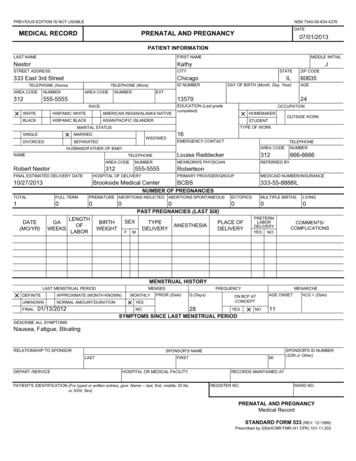
Proceedings - 8th World Rabbit Congress –September 7-10, 2004 – Puebla, MexicoThe following data (Table 1) correspond to obtained averages gotten from the measuresduring the study (subsequent observations were registered just to follow gestationperiod).In this study it has been proved that images of 7th day are only available for pregnancydiagnosis and not for measuring.The differences between measures obtained by ultrasonography of vesicles and realfetus sizes in killed male rabbits, probably was ought to kind of ultrasonography system,it used to a sectorial tube, that affect the measuring angle.Table 1. Differences between obtained by ultrasonography and vesicles and realfetus sizes in killed rabbit does.UltrasonographyDissection7 th day1.080.912 th day1.481.515 th day1.892.020 th day2.852.5Image 1:7 th pregnancy days.Image 2:12th pregnancy days.Image 3:15th pregnancy days.Image 4:20th pregnancy days.278
Proceedings - 8th World Rabbit Congress –September 7-10, 2004 – Puebla, MexicoImage 5: 27 th pregnancy days .Image 6: 29th pregnancy days .At 7 days of pregnancy (image: 1) embryonic vesicles (EV) are clearly seen. In thisgestation step the identified images are built by round areas that may be hyperopicor isoecoic, and these images indeed permit diagnose pregnancy in this period.Later, on 12th day of gestation (image 2), it is possible to identify hypoecoicstructures in the inner part of vesicles tha t correspond to the placenta formation (P)and the embryo (E).At the 15 th day (image 3), the embryo is clearly seen and the placenta formationtoo occupying a third part of the vesicle, in this pregnancy step; heart beating canbe noticed. At the 20th day of pregnancy (image 4), the placenta-fetus proportion is1 to 1, and fetus movements are appreciated; heart beating may be seen alsowhich is very notorious this time, besides, fetus image is whiter (hyperecoic).On 27th and 29th day fetus structures (image 5 and 6), are clearly seen and fetalmovements and heart beating may be recorded but in this period it is hard to saythe difference that are for single fetus and the one that is for a neighbor productdue to the overlapping of the osseous structures as spinal column (SC) and jaw(J).As YPSILANTIS (1999) showed the above data let us determine the gestational ageof internal products and demonstrated the ability to make diagnosis as early as at 7days of pregnancy, which is important in rabbit production, because it is possible toknow the number of carried embryos and finally to establish standard data in rabbitproduction to evaluate the gestation age (RADIOLOGY AND IMAGE MEXICANF EDERATION, 1992).CONCLUSIONSThe use of ultrasound test in rabbits is an alternative method of pregnancydiagnosis in rabbits.279
Proceedings - 8th World Rabbit Congress –September 7-10, 2004 – Puebla, MexicoA 7 th day pregnancy diagnosis can be obtained by ultrasound tests.The use of ultrasound tests as a diagnostic method doesn’t allow us to know thenumber of embryos that the rabbit carries.The number of births, breed and age of the animals don’t affect in any way the sizeof the vesicles.REFERENCESA LVARIÑO M. 1993. Control de la reproducción en el conejo. Edit. Mundi-prensaEspaña.B IRCHARD 1996. Manual clínico de pequeñas especies. Traducción Socorro Lara.Edit McGraw-Hill Interamericana. México Vol. 2.C OMARCK D. 1987. Histología de Ham. Traducción J.R. Blengio 9a Edición Edit.Harla, México .C UNNINGHAM J. 1999. Fisiología Veterinaria Traducción Víctor O. Fuentes 2aEdición Edit. McGraw-Hill Interamericana. MéxicoD UCKES H. 1981. Fisiología de los animales domésticos Edit. Aguilar España Vol.2.F EDERACIÓN MEXICANA DE RADIOLOGÍA E IMAGEN 1992 Ultrasonografía en obstetricia.Edit. Interamericana. México.HAN 1997 Diagnóstico por imagen: guía práctica de radiografía y ecografía.Traducción Diorki. Edit. Hercourt Brace, España.IMAGENOLOGÍA II 2003 (ultrasonografía) Curso de actualización módulopropedéutico II Reg CON EVET (PG016/03) AMMVEPE.K OLB E. 1987. Fisiología veterinaria. Edit. Acribia España Vol. 2.MACD ONALD L. E. 1989. Veterinary endocrinology and reproduction. 4a Edition, Edit.Lea & Febiger Philadelphia, London.MANUAL PARA EDUCACIÓN AGROPECUARIA . 1985. Conejos área de producciónanimal. Edit. Trillas,P ROCURADURÍA FEDERAL DEL CONSUMIDOR Revista del consumidor. Número 304México, junio 2002RUCKEBUSH Y 1994. Fisiología de pequeñas y grandes especies. Traductor AnaFelícitas Edit. Manual Moderno, MéxicoROCA MARTÍNEZ T. J. 1989. Ecografía Clínica del Abdomen. 2a Edición Edit. Jims ,BarcelonaYPSILANTIS P., S ARATSIS PH. 1999 Early pregnancy diagnosis in the rabbit by realtime ultrasonography. World Rabbit Science. Vol. 7 pp 95-99280
Centro de Enseñanza Agropecuaria, Facultad de Estudios Cuautitlán UNAM jema81@prodigy.net.mx, mag1956@servidor.unam.mx, ghelisa2000@yahoo.com. ghelisa@hotmail.com ABSTRACT In this assay an ultrasonography study of pregnant uterus was performed in 100 female rabbits of New Zealand,