Transcription
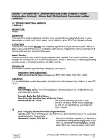
Measure #147: Nuclear Medicine: Correlation with Existing Imaging Studies for All PatientsUndergoing Bone Scintigraphy – National Quality Strategy Domain: Communication and CareCoordination2017 OPTIONS FOR INDIVIDUAL MEASURES:CLAIMS ONLYMEASURE TYPE:ProcessDESCRIPTION:Percentage of final reports for all patients, regardless of age, undergoing bone scintigraphy that include physiciandocumentation of correlation with existing relevant imaging studies (e.g., x-ray, MRI, CT, etc.) that were performedINSTRUCTIONS:This measure is to be reported each time bone scintigraphy is performed during the performance period. There is nodiagnosis associated with this measure. It is anticipated eligible clinicians who perform the professional component ofthe bone scintigraphy study will report on this measure.Measure Reporting:The listed denominator criteria is used to identify the intended patient population. The numerator quality-data codesincluded in this specification are used to submit the quality actions allowed by the measure. All measure-specific codingshould be reported on the claim(s) representing the eligible encounter.DENOMINATOR:All final reports for patients, regardless of age, undergoing bone scintigraphyDenominator Criteria (Eligible Cases):Patient encounter during the performance period (CPT): 78300, 78305, 78306, 78315, 78320NUMERATOR:Final reports that include physician documentation of correlation with existing relevant imaging studies (e.g., x-ray, MRI,CT, etc.)Definition:Relevant Imaging Studies – Relevant imaging studies are defined as studies that correspond to the sameanatomical region in question.ORNumerator Quality-Data Coding Options:Bone Scintigraphy Report Correlated with Existing StudiesPerformance Met: CPT II 3570F:Final report for bone scintigraphy study includescorrelation with existing relevant imaging studies (eg, xray, MRI, CT) corresponding to the same anatomicalregion in questionBone Scintigraphy Report not Correlated for System ReasonsAppend a modifier (3P) to CPT Category II code 3570F to report documented circumstances that appropriatelyexclude patients from the denominator.Denominator Exception: 3570F with 3P:Documentation of system reason(s) for not documentingcorrelation with existing relevant imaging studies in finalreport (eg, no existing relevant imaging study available,patient did not have a previous relevant imaging study)Version 1.112/15/2016CPT only copyright 2016 American Medical Association. All rights reserved.1 of 6
Note: Correlative studies are considered to be unavailable if relevant studies (reports and/or actualexamination material) from other imaging modalities exist but could not be obtained after reasonable efforts toretrieve the studies are made by the interpreting physician prior to the finalization of the bone scintigraphyreport.ORBone Scintigraphy Report not Correlated, Reason not Otherwise SpecifiedAppend a reporting modifier (8P) to CPT Category II code 3570F to report circumstances when the actiondescribed in the numerator is not performed and the reason is not otherwise specified.Performance Not Met: 3570F with 8P:Bone scintigraphy report not correlated in the final reportwith existing relevant imaging studies, reason nototherwise specifiedRATIONALE:Radionuclide bone imaging plays an integral part in tumor staging and management; the majority of bone scans areperformed in patients with a diagnosis of malignancy, especially carcinoma of the breast, prostate gland, and lung. Thismodality is extremely sensitive for detecting skeletal abnormalities, and numerous studies have confirmed that it isconsiderably more sensitive than conventional radiography for this purpose. However, the specificity of bone scanabnormalities can be low since many other conditions may mimic tumor; therefore, it is important that radionuclide bonescans are correlated with available, relevant imaging studies. Existing imaging studies that are available can help informthe diagnosis and treatment for the patient. Furthermore, correlation with existing radiographs is considered essential toinsure that benign conditions are not interpreted as tumor. While there are no formal studies on variations in care in howoften correlation with existing studies is not performed, there is significant anecdotal information from physicianspracticing in the field that there is a gap in care and that correlation is not occurring frequently when images areavailable.Literature suggests that as many as 30% of Radiology reports contain errors, regardless of the imaging modality,radiologists’ experience, or time spent in interpretation. Evidence has also suggested that Radiology reports are largelynon-standardized and commonly incomplete, vague, untimely, and error-prone and may not serve the needs of referringphysicians. Therefore, it is imperative that existing imaging reports be correlated with the Nuclear Medicine bonescintigraphy procedure to ensure proper diagnosis and appropriate patient treatment.CLINICAL RECOMMENDATION STATEMENTS:Bone scintigraphic abnormalities should be correlated with appropriate physical examination and imagingstudies to ascertain that osseous or soft-tissue abnormalities, which might cause cord or other nervecompression or pathologic fracture in an extremity, are not present. (SNM, 2003)Interpretation criteriaBone scans are very sensitive for disease, but specificity of findings is low and must be interpreted in light ofother information1.2.3.4.HistoryPhysical ExamOther test resultsComparison with previous studies(SNM, 2003)Reporting1.2.3.4.Description of techniqueDescription of abnormal tracer uptakeCorrelation with other studiesComparison with previous studiesVersion 1.112/15/2016CPT only copyright 2016 American Medical Association. All rights reserved.2 of 6
5. Interpretation(SNM, 2003)Comparisons with previous examinations and reports, when possible, should be a part of theimaging consultation and report. Integrated PET/CT studies are more valuable when correlated withprevious diagnostic CT, previous PET, previous PET/CT, previous MRI, and all appropriate imagingstudies and clinical data that are relevant. (SNM, 2010)As bone tracer concentration reflects osteoblastic activity which is a common response to a widerange of pathologies, a focus of abnormal tracer concentration should not be confidently assigned toa particular pathology without a typical pattern of tracer distribution such as multiple randomly placedfoci in metastatic bone disease or multiple aligned foci of rib uptake in trauma. In the absence of this,correlation of foci or uptake with alternative modality images such as plain radiographs, MR or CTimages should be reviewed when available as this can significantly increase the accuracy of bonescintigraphy interpretation. (BNMS, 2014)COPYRIGHT:The Measures are not clinical guidelines, do not establish a standard of medical care, and have not been tested for allpotential applications.The Measures, while copyrighted, can be reproduced and distributed, without modification, for noncommercialpurposes, e.g., use by health care providers in connection with their practices. Commercial use is defined as the sale,license, or distribution of the Measures for commercial gain, or incorporation of the Measures into a product or servicethat is sold, licensed or distributed for commercial gain.Commercial uses of the Measures require a license agreement between the user and the PCPI Foundation (PCPI )or the Society of Nuclear Medicine and Molecular Imaging (SNMMI). Neither SNMMI, nor the American MedicalAssociation (AMA), nor the AMA-convened Physician Consortium for Performance Improvement (AMA-PCPI), nowknown as PCPI, nor their members shall be responsible for any use of the Measures.The AMA’s and AMA-PCPI’s significant past efforts and contributions to the development and updating of theMeasures is acknowledged. SNMMI is solely responsible for the review and enhancement (“Maintenance”) ofthe Measures as of August 8, 2014.SNMMI encourages use of the Measures by other health care professionals, where appropriate.THE MEASURES AND SPECIFICATIONS ARE PROVIDED “AS IS” WITHOUT WARRANTY OF ANY KIND. 2016 PCPI Foundation and Society of Nuclear Medicine and Molecular Imaging. All Rights Reserved.Limited proprietary coding is contained in the Measure specifications for convenience. Users of the proprietary codesets should obtain all necessary licenses from the owners of these code sets. SNMMI, the AMA, the PCPI and itsmembers and former members of the AMA-PCPI disclaim all liability for use or accuracy of any Current ProceduralTerminology (CPT ) or other coding contained in the specifications.CPT contained in the Measures specifications is copyright 2004-2016 American Medical Association. LOINC copyright 2004-2016 Regenstrief Institute, Inc. SNOMED CLINICAL TERMS (SNOMED CT ) copyright 2004-2016 TheInternational Health Terminology Standards Development Organisation (IHTSDO). ICD-10 is copyright 2016 WorldHealth Organization. All Rights Reserved.Version 1.112/15/2016CPT only copyright 2016 American Medical Association. All rights reserved.3 of 6
Version 1.112/15/2016CPT only copyright 2016 American Medical Association. All rights reserved.4 of 6
2017 Claims Individual Measure Flow#147: Nuclear Medicine: Correlation with Existing Imaging Studies for All Patients Undergoing BoneScintigraphyPlease refer to the specific section of the Measure Specification to identify the denominator and numerator informationfor use in reporting this Individual Measure.1. Start with Denominator2. Check Encounter Performed:a. If Encounter as Listed in the Denominator equals No, do not include in Eligible Patient Population. StopProcessing.b. If Encounter as Listed in the Denominator equals Yes, include in the Eligible population.3. Denominator Population:a. Denominator population is all Eligible Patients in the denominator. Denominator is represented as Denominatorin the Sample Calculation listed at the end of this document. Letter d equals 8 procedures in the samplecalculation.4. Start Numerator5. Check Final Report for Bone Scintigraphy Study Includes Correlation with Existing Relevant Imaging StudiesCorresponding to the Same Anatomical Region in Question:a. If Final Report for Bone Scintigraphy Study Includes Correlation with Existing Relevant Imaging StudiesCorresponding to the Same Anatomical Region in Question equals Yes, include in Data Completeness Metand Performance Met.b. Data Completeness Met and Performance Met letter is represented in the Data Completeness andPerformance Rate in the Sample Calculation listed at the end of this document. Letter a equals 3 procedures inSample Calculation.c. If Final Report for Bone Scintigraphy Study Includes Correlation with Existing Relevant Imaging StudiesCorresponding to the Same Anatomical Region in Question equals No, proceed to Documentation of SystemReason(s) for Not Documenting Correlation with Existing Relevant Imaging Studies in Final Report.6. Check Documentation of System Reason(s) for Not Documenting Correlation with Existing Relevant ImagingStudies in Final Report:a. If Documentation of System Reason(s) for Not Documenting Correlation with Existing Relevant ImagingStudies in Final Report equals Yes, include in Data Completeness Met and Denominator Exception.b. Data Completeness Met and Denominator Exception letter is represented in the Data Completeness in theSample Calculation listed at the end of this document. Letter b equals 2 procedures in the Sample Calculation.c. If Documentation of System Reason(s) for Not Documenting Correlation with Existing Relevant ImagingStudies in Final Report equals No, proceed to Bone Scintigraphy Report Not Correlated in the Final Reportwith Existing Relevant Imaging Studies, Reason not Specified.Version 1.112/15/2016CPT only copyright 2016 American Medical Association. All rights reserved.5 of 6
7. Check Bone Scintigraphy Report Not Correlated in the Final Report with Existing Relevant Imaging Studies,Reason not Specified:a. If Bone Scintigraphy Report Not Correlated in the Final Report with Existing Relevant Imaging Studies, Reasonnot Specified equals Yes, include in the Data Completeness Met and Performance Not Met.b. Data Completeness Met and Performance Not Met letter is represented in the Data Completeness in theSample Calculation listed at the end of this document. Letter c equals 2 procedures in the Sample Calculation.c. If Bone Scintigraphy Report Not Correlated in the Final Report with Existing Relevant Imaging Studies, Reasonnot Specified equals No, proceed to Data Completeness Not Met.8. Check Data Completeness Not Met :a. If Data Completeness Not Met equals No, Quality Data Code or equivalent not reported. 1 procedure has beensubtracted from the data completeness numerator in the sample calculation.Version 1.112/15/2016CPT only copyright 2016 American Medical Association. All rights reserved.6 of 6
2017 Claims Individual Measure Flow #147: Nuclear Medicine: Correlation with Existing Imaging Studies for All Patients Undergoing Bone Scintigraphy . Please refer to the specific section of the Measure Specification to identify the denominator and numerator information for use in reporting this Individu
![First Revision No. 147-NFPA 20-2013 [ Global Input ]](/img/4/20-a15-fim-aaa-fd-frstatements.jpg)


