Transcription
Veterinary Dentistry BasicsIntroductionThis program will guide you, step by step, through the most important features of veterinary dentistry incurrent best practice.This chapter covers the basics of veterinary dentistry and should enable you to:ü Describe the anatomical components of a tooth and relate it to location and functionü Know the main landmarks important in assessment of dental diseaseü Understand tooth numbering and formulae in different species. 2002 eMedia Unit RVC1 of 10
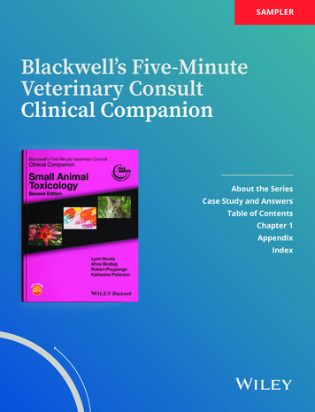
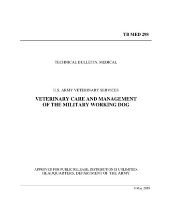
Dental AnatomyCrownThe crown is normally covered by enamel and meets the root at an importantlandmark called the cemento-enamel junction (CEJ). The CEJ is anatomically theneck of the tooth and is not normally visible.RootTeeth may have one or more roots. In those teeth with two or more roots the pointwhere they diverge is called the furcation angle. This can be a bifurcation or atrifurcation.At the end of the root is the apex, which can have a single foramen (humans), amultiple canal delta arrangement (cats and dogs) or remain open as in herbivores. Insome herbivores the apex closes eventually (horse) whereas whereas in others itremains open throughout life. The apical area is where nerves, blood vessels andlymphatics travel into the pulp.Alveolar BoneThe roots are encased in the alveolar processes of the jaws. The process comprisesalveolar bone, trabecular bone and compact bone. The densest bone lines thealveolus and is called the cribriform plate. It may be seen radiographically as a whiteline called the lamina dura.Lamina DuraSight of an uninterrupted lamina dura is interpreted radiographically as a sign of goodperiodontal health.EnamelEnamel is 96% inorganic, mainly hydroxyapatite crystals, with 4% water and fibrousorganic material. It is the hardest substance in the body and covers the exteriorsurface of the crowns only. The enamel consists of hexagonal prisms or rods ofhydroxyapatite crystals held together by a cementing organic matrix. Enamel isformed by ameloblasts within the tooth bud before eruption. It is capable of only verylimited repair when damaged, once the tooth has erupted. 2002 eMedia Unit RVC2 of 10
DentineDentine is the main supporting structure of the tooth and is the second hardesttissue in the body after enamel. It is 70% mineral and acellular, as hydroxyapatitecrystals, and 30% organic as water, collagen and mucopolysaccharide. The mainstructure is the dentinal tubule, which extends from the external surface to the pulp.2There are approximately 30,000 - 40,000 tubul es per mm , which can transmit painto the pulp if the dentine is exposed.Dentine Types Primary dentine forms before tooth eruption. Secondary dentine forms after eruption, as the tooth develops with age. Itdevelops from the odontoblasts living within the pulp and is laid down in layerswithin the pulp cavity. Reparative or tertiary dentine forms as a result of trauma to the odontoblasts;this can be thermal, chemical, bacterial or mechanical. Tertiary dentine has fewtubules and is darker in colour and very dense in structure.CementumCementum covers the enamel free roots and provides a point of attachment for theperiodontal ligament. Similar in composition to woven bone it is 45-50% inorganic,primarily as hydroxyapatite crystals, and 50-55% organic material.Cementum is capable of formation, destruction and repair and remodels continuallythroughout life. It is nourished from vessels within the periodontal ligament.PeriodontalLigamentThe periodontal ligament is comprised of taut collagen fibre bundles (calledSharpey’s fibres where they are inserted in cementum and alveolar bone), whichare anchored to the cementum of the tooth and the alveolar bone.There are three distinct categories of periodontal fibres- gingival, trans-septal andalveolodental.There are blood vessels within the periodontal ligament (PL), which are evenlydistributed. There are also nerves that are capable of transmitting heat, cold, painand pressure in addition to proprioception in some species. 2002 eMedia Unit RVC3 of 10
PulpThis living tissue within the tooth is located in the pulp chambers and root canals. Itis well innervated and vascularised and comprises connective tissue, nerves, lymphand blood vessels, collagen and undifferentiated reserve mesenchymal cells (e.g.odontoblasts).Odontoblasts line the pulp cavity and branch into the dentine tubules. Thesebranches, together with the fine nerve endings, cause the dentine to be sensitive totemperature and pain. The odontoblasts lay down dentine and reduce the pulpcavity in size as the animal ages. The pulp is nourished via vessels entering andleaving the root canal at the apical delta and, occasionally, via accessory canals.Potential Dangers Physical trauma: may cause bruising, haemorrhage or pulpitis. Accidental over-heating from polishing or scaling: may cause pulp necrosis. Pulp exposure after tooth fracture: may cause pulpitis and possibly pulpnecrosis. Loss of blood supply following trauma: will cause ischaemic necrosis. 2002 eMedia Unit RVC4 of 10
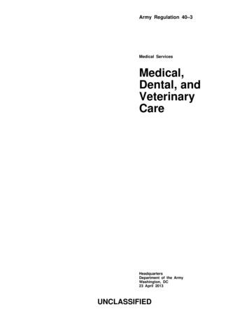
Gingival LandmarksAttached GingivaThe attached gingiva is tightly adherent to the subgingival connective tissue andbone via deep rete pegs. It is keratinised to withstand the stress of ripping andtearing food.MucogingivalJunctionThe mucogingival junction (MGJ) is the junction between the soft, fleshy mucusmembrane of the oral cavity and the tough, collagen rich gingiva. The MGJ remainsstationary throughout life although the gingiva around it may change in height, dueto hyperplasia, recession or attachment loss.Free GingivaFree gingiva forms the gingival margin, which is visible during examination. Itsurrounds the crown of the tooth.Gingival SulcusThe gingival sulcus is located between the tooth and the free gingival margin and isthe crevice that surrounds the tooth.The sulcus lining epithelium renews itself rapidly every 4-6 days - compared to 6 12 days for oral epithelium. The sulcus is bathed in crevicular fluid, which containsmany of the elements of immunity – antibodies, neutrophils, lymphocytes etc.The normal depth of the sulcus is 0.5mm to 1mm in cats and 1-3mm in dogs. As arule 1mm for cats and 1 -2mm in dogs can be considered normal. Sulcus depth ofmore than 4mm in the dog means that the tooth is in danger!During active disease and attachment loss it is common for the sulcus to deepen. Asthe junctional epithelium becomes inflamed and oedematous it separates from theroot surface. The tissues become infiltrated with the cells normally found in thisresponse – both primary and secondary. The junctional epithelium allows for themigration of polymorphonuclear granulocytes and fluids. 2002 eMedia Unit RVC5 of 10
JunctionalEpitheliumAt the bottom of the sulcus is the junctional epithelium (or epithelial attachment),which is so important in the control of periodontal disease. This attaches the gingivaltissues to the tooth using hemidesmosomes. The apical (towards the root) extent ofthe JE is usually the cemento-enamel junction.Cemento-enamelJunctionThe cemento-enamel junction (CEJ) is the junction between the anatomical crownand root.In health the junction is not visible. If it is visible, the division between the shinyenamel and the slightly dull root cementum is very clear. The CEJ is normally theapical (towards the root) extent of the junctional epithelium. Therefore sight of theCEJ indicates recession of the attachments of the tooth and i s highly significant.Interdental PapillaThe gingival peak between closely adjacent teeth is called the interdental papilla.This structure prevents impaction of food and debris between closely adjacent teeth(e.g. incisors in dogs) and is a structure that should be preserved during surgerywhenever possible.When viewed from the coronal aspect there is an indentation called the col. Theepithelium of the col is not keratinised.Function of TeethIncisorsIncisors are used for cutting, scooping, picking up objects and grooming.Incisors are small, single-rooted teeth that commonly become mobile when affected by periodontal disease.CaninesCanines are used for holding prey, display, slashing and tearing when fighting and act as a cradle for thetongue.Canines form important structures at the front of the mouth. Loss of lower canines is serious as it weakensthe rostal mandibles significantly. It also allows the tongue to fall out of the mouth. This can lead to excessivedrying and/or trauma to the tongu e.Loss of upper canines, especially in cats, causes the upper lip to fall inward. The lower canine can thenocclude lateral to the displaced lip and cause excoriation or punctures. 2002 eMedia Unit RVC6 of 10
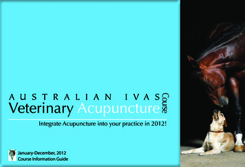
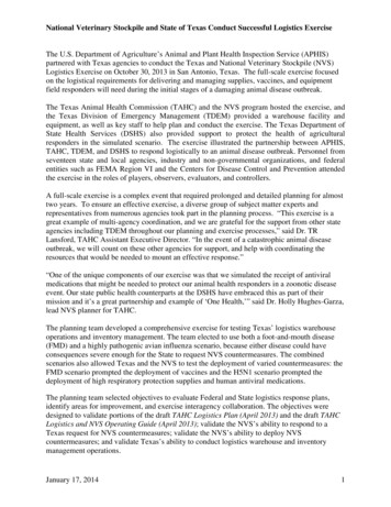
PremolarsPremolars are used for holding, carrying and breaking food into small pieces.Looked at from the side, premolars should show a “pinking shear” appearance with the tip of an upper toothpointing into an interdental space on the lower jaw and vice versa. If they do not do so, it may indicate a bitedefect such as mandibular prognathism or brachygnathism.MolarsMolars are used for grinding food into small pieces with flat occlusal tables.The masticatory forces in the molar zone of the dog have been estimated to be 300 to 800 psi as passive biteforce with a sudden localised bite force when snapping the jaws shut of as much as 30,000 to 80,000 psi.Molars can be affected by dental caries. This condition has a reported 7% incidence in mature dogs.The Modified Triadan SystemTooth Numbering in the DogThe modified Triadan system provides a consistent method of numbering teeth across different animalspecies. The system is based on the permanent dentition of the pig, which has 11 teeth in each quadrant three incisors, one canine, four premolars and three molars. The grand total is 44 teeth.QuadrantThe first digit of the modified Triadan system denotes the quadrant. 2002 eMedia Unit RVC7 of 10
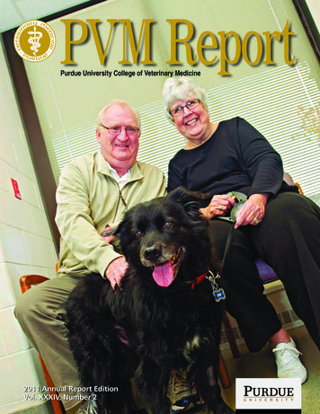
Tooth PositionThe second and third digits denote the tooth position within the quadrant, with the sequence always startingat the midline.The following diagram shows tooth numbering in the dog.Tooth Numbering in Other SpeciesLandmarksUsing definite landmarks we can number animals with less teeth, such as domestic cats, horses and rabbits.The central incisor is always 01 with the following incisors 02 and 03.The canines are always 04 .The premolars are 05 to 08 and the last premolar present is always 08.The first molar is always 09 with the following molars 010 and 011.Domestic CatIn cats, where tooth numbers are reduced, the use of the carnassials as landmarks will help considerably.The upper carnassials in carnivores are always the last premolars, which are 108 and 208. The lowercarnassials in carnivores are always the first molars, 309 and 409. 2002 eMedia Unit RVC8 of 10
Horse 2002 eMedia Unit RVC9 of 10
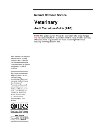
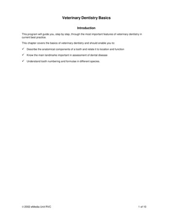
RabbitKey PointsüThe tooth structure and its surroundings contain key landmarks which are important in assessing the levelof disease.üDiseased oral structures will generally follow the same pathological pathways as other damaged tissues inthe body.üTooth numbering systems form the basis of accurate charting and clinical recording.Further Reading American Veterinary Dental Society, Journal of Veterinary Dentistry, 1988 to present. Crossley DA and Penman S (eds), BSAVA Manual of Small Animal Dentistry, 1995. Holmstrom SE, Frost P and Eisner ER, Veterinary Dental Techniques , 2nd edition, Saunders 1998. Lobprise HB and Wiggs RB, The Veterinarian's Companion for Common Dental Procedures , AAHA Press2000. Verstraete FJM, Self-assessment Colour Review of Veterinary Dentistry, Manson 1999. Wiggs RB & Lobprise HB, Veterinary Dentistry - principles and practice, Lippincott 1997. 2002 eMedia Unit RVC10 of 10
Dental Anatomy Crown The crown is normally covered by enamel and meets the root at an important landmark called the cemento -enamel junction (CEJ). The CEJ is anatomic ally the neck of the tooth and is not normally visible. Root Teeth may have one or more roots. In those teeth with two or more roots the point where they diverge is called the furcation angle. This can be a bifurcation or a .