Transcription
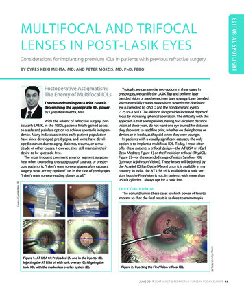
EDITOR IA L S POTL IG H TMULTIFOCAL AND TRIFOCALLENSES IN POST-LASIK EYESConsiderations for implanting premium IOLs in patients with previous refractive surgery.BY CYRES KEIKI MEHTA, MD; AND PETER MOJZIS, MD, PhD, FEBOPostoperative Astigmatism:The Enemy of Multifocal IOLsThe conundrum in post-LASIK cases isdetermining the appropriate IOL power.By Cyres Keiki Mehta, MDCourtesy of Cyres Keiki Mehta, MDWith the advent of refractive surgery, particularly LASIK, in the 1990s, patients finally gained accessto a safe and painless option to achieve spectacle independence. Many individuals in this early patient populationhave since developed presbyopia, and some have developed cataract due to aging, diabetes, trauma, or a multitude of other causes. However, they still maintain theirdesire to be spectacle-free.The most frequent comment anterior segment surgeonshear when counseling this subgroup of cataract or presbyopic patients is, “I don’t want to wear glasses after cataractsurgery; what are my options?” or, in the case of presbyopes,“I don’t want to wear reading glasses at all.”ABCDTHE CONUNDRUMThe conundrum in these cases is which power of lens toimplant so that the final result is as close to emmetropiaFigure 2. Injecting the FineVision trifocal IOL.Courtesy of Cyres Keiki Mehta, MDFigure 1. AT LISA tri: Preloaded (A) and in the injector (B).Injecting the AT LISA tri with toric overlay (C). Aligning thetoric IOL with the markerless overlay system (D).Typically, we can exercise two options in these cases. Inpresbyopes, we can lift the LASIK flap and perform laserblended vision or another excimer laser strategy. Laser blendedvision essentially creates monovision, wherein the dominanteye is corrected to -0.50 D and the nondominant eye to-1.25 to -1.50 D. The ablation also provides increased depth offocus by increasing spherical aberration. The difficulty with thisapproach is that some patients, having had excellent distancevision all these years, do not want one eye blurred for distance;they also want to read fine print, whether on their phones ordevices or in books, as they did when they were younger.In patients with a visually significant cataract, the onlyoption is to implant a multifocal IOL. Today, I most oftenoffer these patients a trifocal design—the AT LISA tri (CarlZeiss Meditec; Figure 1) or the FineVision trifocal (PhysIOL;Figure 2)—or the extended range of vision Symfony IOL(Johnson & Johnson Vision). These lenses will be joined bythe AcrySof IQ PanOptix (Alcon) once it is available in mycountry. In India, the AT LISA tri is available in a toric version, but the FineVision is not. In patients with more than0.50 D cylinder, I always opt for a toric lens.JUNE 2017 CATARACT & REFRACTIVE SURGERY TODAY EUROPE 15
PREOPERATIVE ASSESSMENTSBefore cataract surgery or refractive lens exchange (RLE)for presbyopia, post-LASIK eyes should undergo the following workup.Corneal topography. Accurate corneal topographymeasurements allow the surgeon to place the phaco incision on the steep axis and, thus, minimize postoperativeastigmatism—the enemy of multifocal IOLs. Cornealtopography can also reasonably estimate central cornealpower, and this value can be used in IOL power calculation formulas. Simulated K is of less relevance. The AtlasCorneal Topography System (Carl Zeiss Meditec) has beennoted in the literature to be a useful tool for measuringcentral corneal power.6Ray-tracing software such as that of the iTrace (TraceyTechnologies) can provide additional information about corneal aberrations and can distinguish between those on thecorneal surface and those caused by cataract formation.16 CATARACT & REFRACTIVE SURGERY TODAY EUROPE JUNE 2017Figure 3. Presbyopic RLE.Accurate axial length measurement. Immersion A-scanbiometry (Axis; Quantel Medical) or optical biometry withthe IOLMaster 700 (Carl Zeiss Meditec) or the Lenstar(Haag-Streit) should be used to calculate axial length. Thismeasurement is important because errors in axial length leaddirectly to errors in IOL power.Dilated fundus examination. If there are problems atthe macula, such as an impending macular hole or dry agerelated macular degeneration, multifocal IOL implantationshould be avoided. In addition to dilated fundus exam, OCTshould be used to evaluate the macula in these patients.Anterior segment OCT. Anterior segment OCT can revealthe exact corneal thickness, LASIK flap thickness, and qualityof the stromal bed. This technology is also helpful if the lenspower calculation goes awry and we have to lift the flap anduse the excimer laser for a postoperative adjustment.IOL power calculation. Typically, I prefer to use theHaigis-L formula (myopic or hyperopic), which is included inthe software of the IOLMaster 700. The IOLMaster 700 hasmany more keratometric points than the IOLMaster 500, andit uses swept-source OCT for axial length measurement ascompared with laser interferometry in the IOLMaster 500.7The Barrett True-K formula has been shown to be eitherequal to or better than alternative methods available on theASCRS online calculator for predicting IOL power in eyeswith previous myopic LASIK or PRK.The Barrett True-K formula is based on the BarrettUniversal II formula. It calculates a modified K value forpatients who have had myopic or hyperopic LASIK or PRK orRK, and it requires the measured K reading and the pre- andpostlaser refraction values. The Barrett True K formula canpredict the required power of the IOL when no refractivehistory is available (no-history formula). It can also be usedwith or without considering the surgically induced change inrefraction (change in manifest refraction) because it uses aninternal regression formula to calculate an estimated changein manifest refraction when those data are not entered.Courtesy of Cyres Keiki Mehta, MDEDITOR IA L S POTL IG H Tas possible. All of the lenses mentioned here are aspheric.Studies show that spherical aberration increases significantlyafter myopic LASIK, degrading overall image quality. Anaspheric IOL helps to compensate for the increased sphericalaberration after myopic LASIK.1In planning for cataract surgery in post-LASIK eyes, traditional IOL power calculation formulas no longer apply.Refractive outcomes after cataract surgery in eyes with previous myopic excimer laser surgery are unpredictable. Thelaser ablation for a myopic correction makes the anteriorcorneal surface flatter than that in an untreated eye. The keratometry (K) readings for this flatter cornea are thus lowerthan in a normal eye. The ratio between anterior and posterior corneal curvatures is also altered. Simultaneously, theasphericity of the anterior cornea is changed from prolateto oblate. In an oblate cornea, the K reading in the paracentral area is higher than at the center; it resembles a tabletopshape, so IOL power is underestimated by standard powercalculation formulas. With third-generation theoretical formulas such as the SRK-T and Hoffer-Q, the underestimationsin IOL power in these eyes can be significant.2These formulas calculate the effective lens position (ELP)under the assumption that the cornea has a spherical shape.The ELP is estimated as a function of the presumed anteriorchamber depth, which is geometrically determined by theK reading. In eyes with previous myopic LASIK, the ELP isunderestimated because of the flattened cornea, whereasthe actual anterior chamber depth does not change. As aresult, the IOL power is underestimated, and the postoperative refractive outcome is a hyperopic error.3 This hyperopicsurprise, in which the patient presents after cataract surgerywith an error between 1.50 and 3.00 D, or even more, wascommon in the past after IOL calculation with the standardSRK-T method, but it is now rare.4,5
EDITOR IA L S POTL IG H TFor all postrefractive surgery cases, I use the ASCRSonline calculator. This online calculator has recently beenrevised to include new, innovative formulas such as theBarrett True-K and the Potvin-Hill formula, which usesinput from a Scheimpflug device (Pentacam; Oculus)and OCT.PRESBYOPIC RLEFor presbyopic RLE (Figure 3), patients must be 48 yearsor older; below this, the degree of presbyopia does notjustify the procedure. Patients aged 43 or 44 years do nothave a sufficient degree of presbyopia and, therefore, afterthe procedure often feel that their preoperative vision withtheir residual accommodation was better. Additionally,the rate of retinal detachment after Nd:YAG capsulotomyis unacceptably high in this young group of presbyopes(personal communication from Sheraz M. Daya, MD,FACP, FACS, FRCS(Ed), FRCOphth). Patients undergoingpresbyopic RLE must consent that some amount of glareand halos is tolerable. It is advisable to avoid performingthe procedure in low myopes; for example, a patient with-1.00 D will be happier without surgery.Surgeons should exercise caution when performing presbyopic RLE. Remember that you are operating on patientswho have 20/20 CDVA and N5 CNVA. Any shortcoming insurgery will be immediately picked up by the patient. Thefemtosecond laser is an efficient tool in this setting, as it canbe used to perform limbal relaxing incisions if a toric IOL isnot available; further, the capsulorrhexis position and sizewill be perfect in every case.1. Tzelikis PF, Akaishi L, Trindade FC, Boteon JE. Spherical aberration and contrast sensitivity in eyes implanted withaspheric and spherical intraocular lenses: a comparative study. Am J Ophthalmol. 2008;145:827-833.2. Wang L, Booth MA, Koch DD. Comparison of intraocular lens power calculation methods in eyes that have undergoneLASIK. Ophthalmology. 2004;111:1825-1831.3. Alio JL, Pinero D, Muftuoglu O. Corneal wavefront-guided retreatments for significant night vision symptoms aftermyopic laser refractive surgery. Am J Ophthalmol. 2008; 145:65-74.4. Hoffer KJ. Intraocular lens power calculation after previous laser refractive surgery. J Cararact Refract Surg. 2009;35:759-765.18 CATARACT & REFRACTIVE SURGERY TODAY EUROPE JUNE 2017Cyres Keiki Mehta, MD Surgical Chief, Dr. Cyres Mehta’s International Eye Centre,Mumbai, Indian Director, CKM Eye Specialities Pvt Ltdn cyresmehta@yahoo.comn Financial disclosure: None acknowledgednThe Ideal Surgical SolutionThese lenses can provide excellentspectacle independence and improveintermediate visual acuity withoutsacrificing distance and near vision inthe post-LASIK eye.By Peter Mojzis, MD, PhD, FEBOOver the past decade, the number of patients who presentfor cataract surgery with a history of LASIK has increased dramatically. These patients are familiar with refractive procedures,are of presbyopic age, and seek options for improving theirvisual acuity across all distances. In my experience, trifocal IOLsare an ideal surgical solution in these challenging cases.EVALUATING PREVIOUS LASIKMany patients with a history of refractive surgery present without any preoperative data. Often, they do noteven know whether they underwent hyperopic or myopicLASIK. Thus, the first step in planning cataract/IOL surgeryis identifying the type of LASIK correction they received.This is done by looking for evidence of central steepeningABFigure 4. MTF (A) and PSF (B) corneal values in a patientwith previous myopic LASIK scheduled for trifocal IOLimplantation, measured in a 4-mm pupil. The Strehl ratio forHOAs was high at 0.179.Courtesy of Peter Mojzis, MD, PhD, FEBOCONCLUSIONThe happiest patients after lens surgery for presbyopia are: Hyperopes, as spectacle independence for far andnear can be achieved; Patients with cataracts and poor vision preoperatively,as the improvement in vision is gratifying; and Patients who refuse to wear spectacles for cosmeticreasons and are presbyopic before surgery.Refractive surprises after monovision LASIK can occurbecause the surgical corneal power is underestimated orbecause the IOL position was not accurately predicted.Before cataract surgery, post-LASIK eyes should undergocorneal topography, accurate axial length measurement,dilated fundus examination, and anterior segment OCT. IfIOL power calculation goes grossly wrong, the option toexchange the lens always exists.5. Gimbel H, Sun R, Kaye GB. Refractive error in cataract surgery after previous refractive surgery. J Cataract Refract Surg.2000;26:142-144.6. Wang L, Hill WE, Koch DD. Evaluation of intraocular lens power prediction methods using the American Societyof Cataract and Refractive Surgeons Post-Keratorefractive Intraocular Lens Power Calculator. J Cataract Refract Surg.2010;36:1466-1473.7. Haigis W. Intraocular lens calculation after refractive surgery for myopia: Haigis-L formula. J Cataract Refract Surg.2008;34:1658-1663.
Courtesy of Peter Mojzis, MD, PhD, FEBOEDITOR IA L S POTL IG H Terror in an eye with ahigh degree of cornealastigmatism, I implant atrifocal IOL with a toriccomponent (Figure 6).ANGLE KAPPAAngle kappa is theangle between the visualand pupillary axes. Ahigh angle kappa canFigure 5. Low induced HOAs, coma, and spherical aberration are demonstrated in this eye afterplay a significant rolemyopic LASIK, measured in a 4-mm pupil. Corneal topography shows a well-centered optical zone.in postoperative lensdecentration, resulting inincreased glare and halos.Hyperopes especiallytend to have large anglekappa values. I conducted a study in which anglekappa was measuredin 60 eyes (30 patients).In 42 hyperopic eyes,mean angle kappa was0.43 0.18; in six emmetropic eyes, mean anglekappa was 0.34 0.12;and in 10 myopic eyes,Figure 6. A trifocal toric IOL centered in the capsular bag in a patient with previous hyperopic LASIK.mean angle kappa was0.2 0.1.(indicating hyperopic LASIK) or central flattening (indicatingIn patients with a small angle kappa after myopic LASIK, Imyopic LASIK).recommend centering a trifocal lens on the corneal vertex;I use the OPD-Scan III (Nidek) to evaluate corneal topograin patients with large angle kappa after hyperopic LASIK,phy, LASIK flap centration, higher-order aberrations (HOAs)—the lens should be centered halfway between the pupillarymainly coma and spherical aberration—and angle kappa.and corneal reflexes. In some cases, at the end of surgery,Additionally, I find it useful to assess the preoperative opticalintracameral carbachol intraocular solution may be usedquality of the whole optical system, the lens, and especially theto produce miosis. Induced miosis is a simple and effectivecornea by looking at point spread function (PSF) and modulamethod for centering a multifocal IOL in patients with smalltion transfer function (MTF). PSF measures the intensity of lightangle kappa, wherein the visual axis is close to the pupillaryin the image of a point source, and MTF evaluates the loss ofaxis. The best point for perfect multifocal IOL centrationcontrast when light passes through an optical system (Figure 4).in patients with a high degree of angle kappa is the firstDecentered LASIK flaps, postoperative ectasia, and highPurjkinje image, which is close to visual axis. The AT LISAdegrees of HOAs can decrease optical performance after lenstri has a large central optical zone of 1.04 mm, which issurgery. Corneal refractive surgery modifies the shape of thean excellent solution for patients with large angle kappacornea and can change or induce HOAs. Use of a multifo(Figure 7).cal IOL in an aberrated cornea could worsen optical qualityand cause symptoms of double vision, blurred vision, halos,IOL CALCULATIONor glare. The magnitude of HOAs is determined by, amongIOL calculation in patients with previous refractive surgery isother factors, ablation profile, flap quality, and pupil size. Theless predictable than in virgin eyes. Alterations of the anteriorsize and severity of corneal aberrations should be evaluatedcorneal curvature induced by flap creation and excimer laserusing wavefront aberrometry before multifocal IOL implanablation may lead to miscalculations. The main error in IOLtation is planned (Figure 5).calculations in post-LASIK eyes arises from the use of a formulaAn abnormal corneal shape can also induce regular or irregwith a normal anterior:posterior corneal curvature ratio. Afterular corneal astigmatism. To minimize postoperative refractivemyopic LASIK, this calculation overestimates corneal curvature20 CATARACT & REFRACTIVE SURGERY TODAY EUROPE JUNE 2017
EDITOR IA L S POTL IG H TCourtesy of Peter Mojzis, MD, PhD, FEBOand leads to implantation of an IOL with toolow dioptric power anda resultant hyperopicshift. Residual hyperopiaworsens visual acuity at alldistances. In such an event,previously myopic patientsare highly dissatisfied.Figure 7. An eye with a largeMany methods haveangle kappa. The white pointbeen presented to helpcalculate IOL power more indicates the corneal vertex andthe blue point the pupil center.precisely. I recommendusing the Wang-KochMaloney method. This method is simple: The formula is (CPx 1.114) - 6.1 estimated corneal power, where CP is the valueof central corneal power obtained upon placing the cursorin the center of the axial topography map.Hyperopic LASIK modifies the peripheral cornea, increasingthe anterior:posterior corneal radius; thus, calculations afterhyperopic LASIK are easier than those after myopic LASIK,in which the central corneal curvature is modified. In mypractice, in patients without access to their previous medicalreports, I exclusively use the hyperopic Haigis-L formula.Regarding surgical approach, the placement of incisionsshould avoid the LASIK flap. Corneal incisons should beplaced outside the flap to minimize damage by means ofphaco tip and/or cannulas or accidental instillation of balanced saline solution or OVD. In the event postoperativefine-tuning is needed, there is no problem lifting the damaged LASIK flap.CONCLUSIONCataract surgery in patients with previous myopic or hyperopic LASIK is challenging for a number of reasons. PreciseIOL calculation is essential, and assessments of HOAs, cornealastigmatism, angle kappa, and preoperative optical quality areall key factors for obtaining excellent postoperative results.In my experience, the AT LISA tri and AT LISA tri toric andthe Alsafit trifocal and Alsafit trifocal toric (both from Alsanza)can provide excellent spectacle independence and improveintermediate visual acuity without sacrificing distance andnear vision in post-LASIK patients. These lenses are stable,they center well in the capsular bag, and they provide normalcontrast sensitivity—all notable advantages in these complexsurgical scenarios. nPeter Mojzis, MD, PhD, FEBO edical Director, Premium Clinic Teplice, Czech RepublicM mojzispeter@hotmail.comn Financial disclosure: None acknowledgednnJUNE 2017 CATARACT & REFRACTIVE SURGERY TODAY EUROPE 25
ASCRS online calculator for predicting IOL power in eyes with previous myopic LASIK or PRK. The Barrett True-K formula is based on the Barrett Universal II formula. It calculates a modified K value for patients who have had myopic or hyperopic LASIK or PRK or RK, and it requires the measure