Transcription
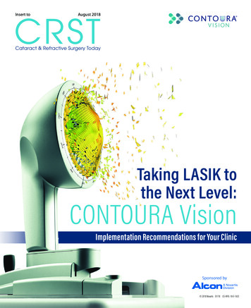
CRSTInsert toAugust 2018Cataract & Refractive Surgery TodayTaking LASIK tothe Next Level:CONTOURA VisionImplementation Recommendations for Your ClinicSponsored by 2018 Novartis 07/18 US-WVL-18-E-1635
OPTIMIZING OUTCOMES WITH CONTOURA VISIONOPTIMIZING OUTCOMESWITH CONTOURA VISIONImplemented correctly, this major advancement in refractive surgery enables better outcomes for patients.BY MARK LOBANOFF, MDThe CONTOURA Vision (Alcon) systemis changing LASIK. The technology enablessurgeons to treat each cornea’s uniquetopographic irregularities in a broad rangeof LASIK patients, which may produce a better quality of vision. By removing each cornea’s topographicirregularities CONTOURA Vision treatments also benefitpatients by reducing night vision problems such as glare andhalos.1 That is unique to CONTOURA Vision; a LASIK technology that creates vision higher in quality than anything thathas come before. Vision for many patients that is superior towhat glasses, contacts, or traditional LASIK can offer. Today,CONTOURA Vision is a practice differentiator, garnering highpatient satisfaction and referrals for surgeons who use it.Outcomes with CONTOURA Vision can be spectacular. In FDA clinical trials and today in open use,CONTOURA Vision outcomes are measurably superiorand show noticeably better patient satisfaction compared to standard LASIK. After the procedure in 249eyes, 92.6% of patients have 20/20 or better uncorrectedvisual acuity—better than preoperative best-correctedvisual acuity in more than 30% of eyes.2 An astounding1 in 3 patients achieved 20/12.5 or better uncorrectedvision. CONTOURA Vision also has lower rates of symptoms associated with LASIK, such as light sensitivity,night driving problems, reading difficulty, glare, halos,and starbursts.1 Given the higher quality of vision thatCONTOURA Vision delivers to patients, in a survey of 124patients, 98% said they would have the procedure again.2Figure 1. The WaveLight Topolyzer VARIO Diagnostic Device.NEXT-LEVEL TECHNOLOGY WITH BROAD APPLICATIONCONTOURA Vision takes customized refraction a stepfurther by addressing very specific problems related tocorneal topography that had never been fully addressedbefore. New cutting-edge topography must be meticulouslyCONTOURA VISION ALSO HAS LOWER RATES OF SYMPTOMS ASSOCIATED WITHLASIK, SUCH AS LIGHT SENSITIVITY, NIGHT DRIVING PROBLEMS, READINGDIFFICULTY, GLARE, HALOS, AND STARBURSTS.1 GIVEN THE HIGHER QUALITYOF VISION THAT CONTOURA VISION DELIVERS TO PATIENTS, IN A SURVEY OF 124PATIENTS, 98% OF PATIENTS SAID THEY WOULD HAVE THE PROCEDURE AGAIN.22 INSERT TO CATARACT & REFRACTIVE SURGERY TODAY AUGUST 2018Please see page 7 for Important Product Information.Trademarks are property of their respective owners.
OPTIMIZING OUTCOMES WITH CONTOURA VISIONCONTOURA VISION IS NOT A PREMIUMSURGERY FOR JUST A FEW PATIENTS.PATIENT SELECTION IS SIMILAR TOLASIK OR PRK, AND ANYONE WHOFALLS WITHIN THE ESTABLISHEDGUIDELINES CAN POTENTIALLY BE ACONTOURA VISION PATIENT.accurate, without shadows, tear film disruption, or otherfactors that could impair its quality. What used to be anassessment tool is now a surgical guide because the topographic corneal data are sent to the Alcon WaveLight laserand actually used to improve the accuracy of the excimerlaser treatment. That’s a true paradigm shift—topographicdata not just examined by the surgeon but actually sentdirectly to the laser as part of the treatment. And as always,the quality of the outcomes starts with the quality of inputinto the laser.Once practices master capturing WaveLight TopolyzerVARIO Diagnostic Device (Alcon) images, the rest of theCONTOURA Vision process is seamless (Figure 1). Through ahighly visual process, the software helps technicians and surgeons evaluate the quality of the images, so it is easy to pickthe four images required for surgery. We can now look atexquisitely detailed corneal topography, isolate the impact ofany topographic aberrations on the cornea, and decide howto integrate those aberrations into the refraction. Finally,the laser will have the actual 22,000 topographic data pointsfor each eye’s treatment, a huge improvement over simplyimputing a steep K and a flat K. The 22,000 keratometricdata points are much better than 2 simple K readings.CONTOURA Vision is not a premium surgery for justa few patients. Patient selection is similar to LASIK orPRK, and anyone who falls within the established guidelines (up to -8.00 D sphere, up to -3.00 D cylinder, and aspherical equivalent no greater than -9.00 D) can potentially be a CONTOURA Vision patient. Even patients withlow refractions benefit from CONTOURA Vision, becauseeveryone has topographic aberrations in the cornea thataffect visual performance. However, we sometimes needto rule out patients if we think that corneal scarring oranother challenge will make it impossible to acquire reliable images. I use CONTOURA Vision for all of my eligiblepatients and on most days 80% to 90% of my patientsare treated with CONTOURA Vision. Yet 100% of myPlease see page 7 for Important Product Information.Trademarks are property of their respective owners.patients benefit from it. How so? Because even if a patientcan’t have CONTOURA Vision, we are still able to capture iris recognition data, which is sent to the WaveLightlaser’s tracker. This allows perfect axis placement ofastigmatic treatments in WAVEFRONT OPTIMIZED treatments. Hyperopic patients benefit because the TopolyzerVARIO data can identify the true corneal vertex withnearly perfect precision. Hyperopic treatments can thusbe centered on the pupil center or the corneal apexdepending on surgeon preference for each eye. Tangiblebenefits from having your diagnostic technology “talk”directly to your laser.CAPTURE THE HIGH-QUALITY MEASUREMENTS YOU NEEDTo optimize the outcomes of refractive surgery withCONTOURA Vision, we need to capture high-quality images with minimal shadowing and healthy tear film. My staffand I follow these five steps:1. Ensure the ocular surface is clear and consistent.Because the Topolyzer VARIO is a Placido disc measurement that relies on rings reflected onto the corneal surface,it is important to have a healthy, uncompromised ocularsurface. In preparation for these measurements, we havepatients stop wearing soft contact lenses for 1 week, orgas permeable lenses for 3 weeks. We also perform theTopolyzer VARIO measurements first, before any othertesting is done that requires staring or other actions thatcan disrupt or distort the tear film or corneal surface.2. Position and coach the patient. Patients have a rolein capturing the best CONTOURA Vision images, so ourtechnicians give patients a 30-second overview of what toexpect before they begin. Technicians explain that nothinguncomfortable or surprising (no puff of air) will take placeduring the measurements.Figure 2. A technician using the Topolyzer VARIO system with a patient.AUGUST 2018 INSERT TO CATARACT & REFRACTIVE SURGERY TODAY 3
OPTIMIZING OUTCOMES WITH CONTOURA VISIONPATIENTS CAN HELP OPTIMIZE THEIR OCULAR SURFACE BY KEEPING THEIREYES CLOSED BETWEEN MEASUREMENTS. WHEN THE TECHNICIAN IS READYTO CAPTURE A MEASUREMENT, THEY INSTRUCT PATIENTS TO DO THREE QUICKBLINKS, OPEN THEIR EYES WIDE WITH THEIR EYEBROWS UP, AND FOCUS INTHE CENTER OF A LITTLE YELLOW “DONUT” (FIXATION TARGET).Body positioning is very important, because if patients areuncomfortable, they will move a lot, which makes it difficultto capture a good image. Patients should be sitting uprightin a comfortable position, and we do not want them to sitback in between measurements (Figure 2). Patients can helpoptimize their ocular surface by keeping their eyes closedbetween measurements. When the technician is ready tocapture a measurement, they instruct patients to do threequick blinks, open their eyes wide with their eyebrows up,and focus in the center of a little yellow “donut” (fixationtarget). If it takes longer than 4 to 5 seconds to capture animage, technicians tell the patients, “blink, blink, blink, andbrows up,” and they try again.3. Position the eyes for optimal image capture. The rightpositioning helps ensure that we get the most data possiblefor CONTOURA Vision treatment. First, we adjust the tableheight so patients are comfortable because uncomfortablepatients naturally shift around and need to be repositioned.To prevent shadowing from the nose, we measure the righteye with the head turned (never tilted) slightly to the left,and measure the left eye with the head turned slightly right.Using the forehead spacer is also important because it helpspush the forehead back ever so slightly for better superiorFigure 3. The right positioning helps ensure that we get the most data possible forCONTOURA Vision treatment.4 INSERT TO CATARACT & REFRACTIVE SURGERY TODAY AUGUST 2018exposure. Technicians do a quick eye-to-eye test beforetaking the first measurement on the right eye or left eye, inorder to help ensure there is no cyclotorsion related to headtilt. Sometimes having a second technician lift the eyelidfrom the side helps as well (Figure 3).4. Capture six images for the CONTOURA Visionprocedure. Optimally, we need four images to performthe CONTOURA Vision procedure. For patients whosemeasurements fall within the criteria for CONTOURA Visionsurgery (up to -8.00 D sphere, up to -3.00 D cylinder, and aspherical equivalent no greater than -9.00 D), my techniciansaim to capture six images. This allows me to rule out outliers,such as images with variations from compromised tearfilm or missing data (lids, lashes, or nose shadows) duringthe comparison process. However, I will still proceed withCONTOURA Vision in many cases if we capture fewer than6 images but those that are captured are of a high quality.5. Gather measurements for other refractive surgeries.When patients fall outside the CONTOURA Visionindication, the Topolyzer VARIO measurements are stillvaluable. Iris registration can be used for other procedures.I also export data from the Topolyzer VARIO (patientFigure 4. Once we capture images for the CONTOURA Vision procedure, mytechnicians follow an interactive image analysis process, eliminating outliers andverifying that measurements are consistent and reliable.Please see page 7 for Important Product Information.Trademarks are property of their respective owners.
OPTIMIZING OUTCOMES WITH CONTOURA VISIONdemographics, iris registration, keratometry, and cornealvertex measurement) for any WAVEFRONT OPTIMIZEDprocedure I perform using the WaveLight EX500 ExcimerLaser (Alcon). This increases our efficiency on OR days as thedata will not need to be entered into the laser “on the fly.”ANALYZE AND SELECT THE BEST IMAGESOnce we capture images for the CONTOURA Vision procedure, my technicians follow an interactive image analysisprocess, eliminating outliers and verifying that measurementsare consistent and reliable (Figure 4). From the patient’s sixcaptures, they select four high-quality images for either theCONTOURA Vision procedure or for iris registration alone.First, we compare measurements for consistency andreliability of /- 0.75 D. Using comparison maps of thetreatment area in the Compare Images Display on theTopolyzer VARIO, we look at four images on the screen.We choose the scan with the best tear film/topographyimage as a baseline for comparing Topolyzer VARIO images.To compare other images to that baseline, we check fromthe pupil to the mid-periphery to make sure that the difference is less than 0.75 D. After discarding the outliers, we pullup additional scans and make the same comparison.Next, the Analyzer Area tool helps us ensure we have thedata we need. It calculates the data available for 5.50 mmand 6.50 mm optical zones. The Optical Zone Analyzer section on the Overview page shows the optical zone for theeye, split into inferior, superior, temporal, and nasal quadrants (Figure 5). The software marks each quadrant in redor green. Green check marks tell us that there is enoughdata in the optical zone for CONTOURA Vision and irisregistration. In addition, a green “P” means preferred (forthe CONTOURA Vision procedure) and a green “R” indicates good iris registration.If the examination is less than perfect, the screen willFigure 5. The Optical Zone Analyzer section on the Overview page shows the opticalzone for the eye, split into inferior, superior, temporal, and nasal quadrants.Please see page 7 for Important Product Information.Trademarks are property of their respective owners.display a red “X” and show which quadrant or quadrantshave insufficient data. For example, data may be missingfrom the superior region if the eyelashes cast a shadow, orthe nose may have a shadow in the nasal quadrant.Images should have 90% or higher AA OZ in the 6.50OZ and pupil border recognition within 0.1 mm or 100μm of the baseline image. Patients typically will have several scans with sufficient data, but if, for example, the 5.50mm optical zone has 100% of the data, while 6.50 mm ismissing data but is still over 90%, we can manually changethe image to “preferred” for the CONTOURA Vision procedure. Any scans below those thresholds are not usable.Alcon’s trainers are excellent and can help explain all of thisthoroughly. In practice, it is not difficult.CALCULATE CONTOURA VISION TREATMENTOnce the technicians have a good set of images forsurgery, I calculate the laser refractive treatment for theCONTOURA Vision procedure. It begins with checkingthe reproducibility of imported measurements. Once mytechnicians send the selected scans to the laser, I havethe opportunity to double-check them for accuracy. Atthe planning station, I look at the images in the Raw DataSagittal view, comparing the appearance of the cornealtopographies and eliminating any extreme outliers. Inspectthe K values, axes, Q values, and pupil sizes, if necessary.This usually takes me about 30 seconds or less.I can dig deeper into the data behind the images if needed, but generally, this simplified approach is very effective.Once I approve the scans and removed any scans that thetechnician may have missed, I am ready for the next step.Advancing to the next screen, I input the patient’s specifictreatment data: manifest refraction, vertex distance, K values,pupil size, and pachymetry results (Figure 6). The third pagein the planning process is where I can calculate the patient’sfinal treatment. In the table at the top of the page, the top linedisplays the manifest refraction that was inputted on the previous screen. The second line shows the highly accurate measured refraction and cylinder axis of the astigmatism found byFigure 6. This screen shows the patient’s specific treatment data: manifestrefraction, vertex distance, K values, pupil size, and pachymetry results.AUGUST 2018 INSERT TO CATARACT & REFRACTIVE SURGERY TODAY 5
OPTIMIZING OUTCOMES WITH CONTOURA VISIONFigure 7. This ablation profile shows only the anterior corneal topographicaberrations.THE ALCON TRAINER SPENT A FULLWORKDAY WITH OUR TECHNICIANS,TRAINING THEM TO CAPTURE ANDANALYZE SCANS, OBSERVINGTHEIR WORK WITH PATIENTS, ANDANSWERING QUESTIONS.the Topolyzer VARIO. (Note: the Topolyzer VARIO is incapableof determining the sphere, so this figure should be disregarded.)On the third line, known as the “Modified” line, I enter the final,nomogram-adjusted treatment chosen for the eye.In making my final choice for treatment, I considerwhich nomogram to use and whether I should make anage adjustment. I can also dig deeper into the topographicpattern found on the cornea and think about how thiscan affect the refraction. To see how the CONTOURAVision procedure will remove these aberrations to providea smoother, more regular corneal surface, I enter 0 in themodified sphere and cylinder boxes. The resulting ablationprofile shows only the anterior corneal topographic aberrations (Figure 7). Understanding how the aberrations inthis profile can influence vision, I feel confident in treatingpatients with CONTOURA Vision.Because I have been using CONTOURA Vision for morethan 2 years, and we track our outcomes, I am able to individualize patients’ treatment calculations, when needed,based on various criteria. This is the “art” of workingwith CONTOURA Vision, and it comes with experience.However, for surgeons who are new to the procedure, I recommend following the straightforward calculations givenby Alcon, which are based on the company’s most currentdata. Start simple and straightforward, then advance withmore complex treatments as you gain experience and confidence with the technology.better outcomes with higher quality of vision leads to happierpatients and more referrals. We take pride that we are offeringexcellent refractive treatments for our patients. It is how weseparate ourselves in a competitive market.Like any paradigm shifting technology, CONTOURAVision required a period of transition. The Alcon teamtrained our surgeons and staff very thoroughly and conveniently. The Alcon trainer spent a full workday withour technicians, training them to capture and analyzescans, observing their work with patients, and answering questions. Next, Alcon practiced surgical planning forCONTOURA Vision procedures with our surgeons, andthey were there on surgery day to make sure everythingwent smoothly. The trainers are available to answer questions, visit additional surgery days, or do refresher trainingfor technicians and new staff. The instruction and supporthelped us implement the CONTOURA Vision measurement process and procedure very quickly and confidently.After just a few weeks of practice, CONTOURA Visiondid not have a major impact on patient flow at the clinicor surgery center. Because CONTOURA Vision is a practicedifferentiator, Alcon has also given us some pointers so wecould position ourselves to turn patient satisfaction intoreferrals for the procedure. CONTOURA Vision helps usdeliver a game-changing procedure that reduces side effectscommonly associated with LASIK, and we want patients inour community to know that we offer those advantages.CONTOURA VISION TRAINING AND IMPLEMENTATION1. Stulting RD, Fant BS, The T-CAT Study Group. Results of topography-guided laser in situ keratomileusis customablation treatment with a refractive excimer laser. J Cataract Refract Surg. 2016;42:11–18.2. At 12 months: Summary of safety and effectiveness data, WaveLight ALLEGRETTO WAVE Eye-Q Excimer LaserSystem and the ALLEGRO Topolyzer. September 27, 2013. Available online: http://www.accessdata.fda.gov/cdrh docs/pdf2/P020050S012b.pdfWith CONTOURA Vision, there are some additional stepsversus standard WAVEFRONT OPTIMIZED LASIK, where wesimply enter refractions and two K values into the laser toguide surgery. We are going from using 2 K values to 22,000 toachieve our better results. Our technicians have to take quality,reproducible images, and surgeons have to take a more indepth look at the corneal topographic irregularities. Once webegan using this approach in my practice, my staff and I quicklybecame confident, and now it adds only a little extra time. Butthat extra time and effort are worth it in my practice, because6 INSERT TO CATARACT & REFRACTIVE SURGERY TODAY AUGUST 2018MARK LOBANOFF, MDn D irector of Refractive Surgery at North Suburban Eye Specialists, M inneapolis, Minnesotan m lobanoff@gmail.comn F inancial disclosure: consultant to AlconPlease see page 7 for Important Product Information.Trademarks are property of their respective owners.
OPTIMIZING OUTCOMES WITH CONTOURA VISIONThis information pertains to all WaveLight Excimer Laser Systems, including the WaveLight ALLEGRETTO WAVE, theALLEGRETTO WAVE Eye-Q, and the WaveLight EX500.Caution: Federal (U.S.) law restricts the WaveLight Excimer Laser Systems to sale by or on the order of a physician.Only practitioners who are experienced in the medical mangement and surgical treatment of the cornea, who havebeen trained in laser refractive surgery (including l
BY MARK LOBANOFF, MD The CONTOURA Vision (Alcon) system is changing LASIK. The technology enables surgeons to treat each cornea’s unique topographic irregularities in a broad range of LASIK patients, which may produce a bet-ter quality of vision. By removing each cornea’s topographic irregularities CONTOURA Vision treatments also benefit


![[Insert Logos] Creating a New National Leader in Adult .](/img/10/ir-deck-for-ndrs-meetings-3-9-17-final.jpg)