Transcription
Vet. World, 2012, Vol.5(12): 742-747RESEARCHHypothyroidism - A cause for dilated cardiomyopathy in dogs;four year study (2008-2011)12Satish Kumar Karlapudi , Devarakonda Srikala , D S Tirumala Rao1. Department of Veterinary Clinical Medicine, College of Veterinary Science, Rajendranagar,Sri Venkateswara Veterinary University, Hyderabad-30, Andhra Pradesh, India. 2. Department of VeterinaryClinical Medicine, College of Veterinary Science, Tirupati-517502, Andhra Pradesh, India. 3. Director ofResearch, Sri Venkateswara Veterinary University, Hyderabad-30, Andhra Pradesh, India.Corresponding author: K. Satish Kumar, email: dr petlover@yahoo.co.inReceived: 02-06-2012, Accepted: 03-07-2012, Published online: 07-11-2012doi: 10.5455/vetworld.2012.742-747AbstractAim: The study was carried out to understand the thyroid dysfunction and its association with dilated cardiomyopathy in dogs.Materials and Methods: The study was done at Teaching Veterinary Hospital, Bhoiguda, College of Veterinary Science,Hyderabad for four years, i.e., from 2008 to 2011. A total of 256 dogs of various breed, age and sex were presented with typicalskin and coat abnormalities. Few were also exhibiting signs of low metabolic rate. Skin sample analysis was done to rule outthe causes of dermatitis. Thyroid profile was estimated to diagnose hypothyroidism. Selected cases were also subjected forechocardiography to study the association of cardiomyopathy. Based on thyroid profile, hypothyroid dogs were treated withlevothyroxine @20mcg/kg wt, once daily, orally on empty stomach and dilated cardiomyopathy (DCM) associated patientswere additionally supplemented with enalapril @0.5 mg/kg, twice daily, orally for 6 months. The hemato biochemical andechocardiographic aspects are discussed.Results: The classical signs that were recorded in almost all the thyroid dysfunction dogs (231) were bilateral alopecia, rat tailand pigmentation and whereas, dyspnoea at rest, exercise intolerance, obesity, pale mucosae and corneal lipidosis were thesignificant low metabolic rate signs noticed in 42 dogs. However, syncope and seizures were also recorded in 31 of thesehypothyroid dogs. Echocardiographic evaluation revealed significantly (P 0.01) increased LVEDd and LVEDs along withdecreased IVS and LVPW both at systole and diastole among 33 dogs. Normocytic, normochromic and non-regenerativeanemia and significantly (P 0.05) low T3, fT4, tT4 along with elevated serum cholesterol, triglycerides, TSH, CKMB, LDHand ALP were the hemato – biochemical findings among these dogs. After the initiation of therapy, improvement in clinicalsigns was noticed from day 7 and complete clinical recovery by the end of therapy. However, a non significant improvementwas recorded with respect to left ventricle dimensions.Conclusions: From the present study it may be concluded that thyroid dysfunction is presented by manifestations of twodifferent systems, i.e., skin and coat and metabolic abnormalities. Echocardiography is the more sensitive diagnostic aid toascertain the involvement of cardiovascular system and to assess the therapeutic response. Further, supplementation of ACEinhibitors along with the specific thyroid supplements helps to resolve the disorder.Key words: dilated cardiomyopathy, dogs, enalapril, hypothyroidism, levothyroxineAbbreviations: DCM-Dilated cardiomyopathy, T4-Thyroxine, T3-Tri-iodothyronine, tT4-Total thyroxine, fT4-Freethyroxine, TSH-Thyroid stimulating hormone, LVEDd-Left ventricle end diameter at diastole, LVEDs-Left ventricle enddiameter at systole , IVSd-Inter ventricular septum at diastole, IVSs-Inter ventricular septum at systole, LVPWd-Leftventricle free wall at diastole, LVPWs-Left ventricle free wall at systole, EF- Ejection fraction, FS-Fractional shortening,ACEi-Angiotensin converting enzyme inhibitorsTo cite this article:Karlapudi SK, Srikala D, Rao DST (2012) Hypothyroidism - A Cause for dilated cardiomyopathy in dogs; four yearstudy (2008-2011), Vet World, 5(12): 742-747, doi: idism results in decreased productionof thyroid hormones, thyroxine (T4) and 3, 5, 3 triiodothyronine (T3) from the thyroid gland. Naturallyoccurring hypothyroidism is a common disease indogs but rare in cats [1]. Thyroid hormones havepositive inotropic and chronotropic effects on the heartand have catabolic effects on muscle and adiposewww.veterinaryworld.orgtissue, stimulate erythropoiesis and regulate bothcholesterol synthesis and degradation. Thyroiddysfunction is characterised by slow, lazy behaviour,bilateral alopecia, dry skin, obesity, lethargy andrespiratory distress. Uncommonly, cardiovascularabnormalities are also associated with hypothyroidism. Further, cardiac changes that develop in dogswith hypothyroidism are important because of theVeterinary World, Vol.5 No.12 December 2012742
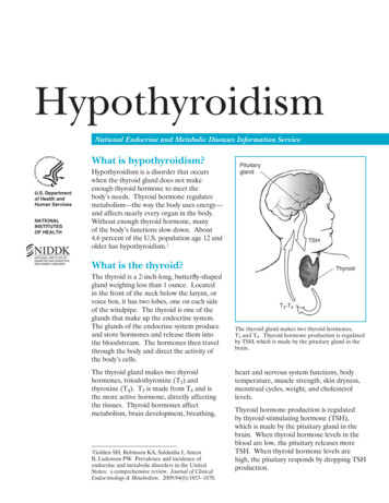
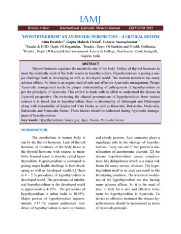
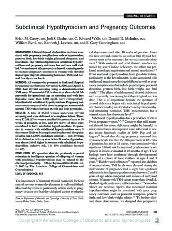
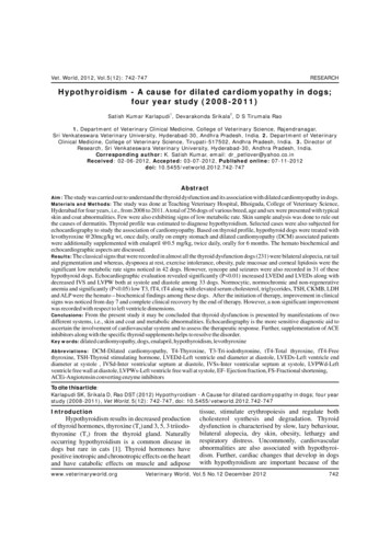
Hypothyroidism - A cause for dilated cardiomyopathy in dogs; four year study (2008-2011)high incidence of hypothyroidism and heart disease insome breeds [2]. Any alteration in cardiac functioninduced by hypothyroidism could result in worseningof pre-existing disease [3]. Interest in the role ofthyroid hormones on cardiac failure has increased inrecent years. The driving considerations can besummarized as follows: (a) the known effects ofthyroxine on contractile and relaxation properties ofthe heart; (b) experimental findings offering strongsupport for the hypothesis that thyroid hormonesignaling is critical in preserving cardiac structure andperformance under normal conditions and after cardiacinjury; and (c) evidence that thyroid dysfunction isstrongly associated with a worsening prognosis incardiac patients in general and in cardiac failurepatients in particular [4,5]. Diastolic function andsystolic function are clearly influenced by variousthyroid hormones [6]. Ventricular contractile functionis also influenced by changes in hemodynamic conditionssecondary to thyroxine effects on peripheral vasculartone [7]. The homeostasis of these hormones preservespositive ventricular-arterial coupling, leading to afavorable balance for cardiac work. Other studiessuggest reduced cardiac tissue triiodothyronine levelsafter myocardial infarction (MI) or with developmentof hypertension by upregulating type 3 deiodinase(D3), which leads to deactivation of triiodothyronineand thyroxine [8].Keeping this hypothesis in view the presentstudy was carried out to evaluate the association ofcardiomyopathy with thyroid dysfunction in dogs.Materials and MethodsA total of 256 dogs of various breed, age and sexthat were presented from 2008 to 2011 to theVeterinary Hospital, Bhoiguda, College of VeterinaryScience, Hyderabad, with the classical signs of skinand coat and metabolic abnormalities were consideredfor the study. Almost all of these dogs were treated fordermatitis by local Veterinarian, but the condition wasreported to be recurring when therapy discontinued.The owners further reported that, there was noimprovement with respect to the signs associated withlow metabolic rate in few of the cases. Thoroughclinical examination was followed by blood and serumanalysis. Further, the hypothyroid patients presentingsigns associated with reduced metabolic rate (cyanotictongue, dyspnoea at rest, lethargy and exerciseintolerance) were suspected for cardiac involvementand were subjected for electrocardiography, thoracicradiography and 2-d echocardiography. Echocardiogram was carried out using a microconvex array c5-2www.veterinaryworld.orgR13 cardiac probe placed on right precardium, withdogs positioned in right lateral recumbancy. Access tothe right side of the thorax was facilitated by use of atable with a special cut-out to allow the transducer tobe directed upward towards the site of maximalcardiac pulsation. Transducer is located parasternallybetween right third and sixth intercostal spaces betweensternum and costochondral junction. M-mode recordingswere taken at the high papillary level. However,echocardiogram measurements were also recorded inapparently healthy dogs to establish normal values.Based on these findings 33 hypothyroid patientsdiagnosed for associated dilated cardiomyopathy wereselected and were treated with levothyroxine @ 20mcg/kg, once daily on empty stomach and enalapril @0.5 mg/kg once daily for 6 months. However, levothyroxinewas also supplemented for the remaining hypothyroidcases for 6 months. The blood and sera samples werecollected as per the animal ethics and the therapy wasalso carried out as per the ethical treatment protocol.ResultsAlmost all the hypothyroid dogs revealed similarclinical manifestations viz., bilateral symmetricalalopecia, rat tail appearance, pigmentation, bald thigh,weakness, cold intolerance and lethargy. Whereas,dyspnoea at rest, exercise intolerance, corneal lipidosisand cyanotic tongue were recorded in 41 cases andseizures and syncope among 31. The mean weight andage was 45 kg and 8 yrs, respectively. Hematologically,non-regenerative anemia and biochemically, significantincrease (P 0.05) in serum cholesterol, triglycerides,alkaline phosphatase (ALP) with a significantly decreased(P 0.05) levels of tri-iodothyronine (T3), total thyroxine(tT4), free thyroxine (fT4) and increased thyroidstimulating hormone (TSH) were recorded in 231/256dogs, along with a significantly increased (P 0.05)creatine kinase MB (CKMB) and lactate dehydrogenase (LDH) in 33 hypothyroid dogs in comparisonwith apparently healthy dogs (Table-1). These dogswith abnormal cardiac markers were suspected forcardiomyopathy and subjected for further detailedinvestigations. Low voltage QRS complexes andventricular premature complexes were the significantelectro cardiographic (ECG) findings and whereas,cardiomegaly was the radiographic abnormality. Bmode echocardiography revealed round greatlyenlarged globose shaped heart (fig. 1). A significantly(P 0.01) increased left ventricle end diameter (LVED)along with significantly (P 0.05) decreased interventricular septum (IVS) and left ventricle free wall(LVWP) dimensions both at diastole (d) and systole (s)Veterinary World, Vol.5 No.12 December 2012743
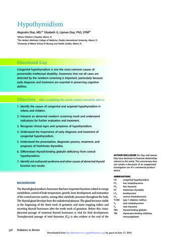
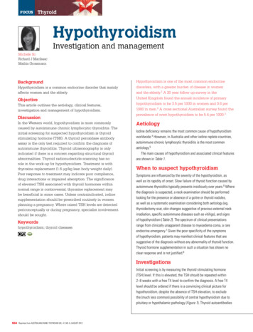
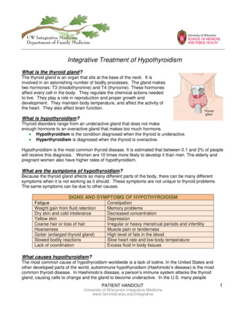
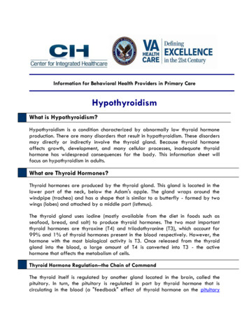
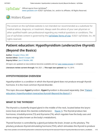
Hypothyroidism - A cause for dilated cardiomyopathy in dogs; four year study (2008-2011)Fig.1. B-mode echocardiogram showing round globose leftventricle (day 0)Fig.2. M-mode echocardiogram showing abnormal leftventricle dimensions and indices (day 0)Table.1 Serum profile hypothyroid associated Cardiomyopathy dogsSl. No.ParameterHealthy dogsDCM dogsday 0Day 90day 1801.2.3.4.5.6.7.8.9.10.Cholesterol (mmol/L)Triglycerides (mmol/L)TT4 (mcg/dl)fT4 (ng/dl)T3 (ng/dl)TSH (ng/ml)ALT (U/L)ALP (U/L)CKMB (U/L)LDH (U/L)4.36 0.201.40 0.843.20 0.122.72 0.5069.20 0.282.12 0.2233.88 0.5464.10 0.0424.50 0.84286.46 1.8014.12* 0.226.10* 0.240.88* 0.100.70* 0.0831.50* 0.7626.00* 0.8035.94 0.74154.10** 1.0060.18** 1.50696.50** 2.2211.80 0.445.20 0.481.02 0.301.14 0.0843.88 0.7019.00 0.7436.00 0.06131.36 0.0855.20 0.20622.18 1.206.00* 0.222.32* 0.042.60* 0.722.12* 0.2061.08** 1.244.88** 0.2235.80 0.4074.50** 1.5424.2** 1.16318.44** 1.98* significant at 5 per cent (P 0.05); ** significant at 1 per cent (P 0.01)and Ejection fraction (EF) and Fractional Shortening(FS) were the significant m-mode echocardiographicmeasurements. Thus, confirming associated dilatedcardiomyopathy among 33 hypothyroid dogs of whichGerman shepherd (14) followed by Labrador retriever(09) were commonly affected breeds (fig. 2; table. 2).Improvement in skin and coat associated signswere noticed from day 7 followed by signs associatedwith low metabolic rate and complete clinical recoverywith alleviation of all the signs by the end of therapy. Anon- significant improvement in haematological,radiological and electrogardiographic findings werenoticed by day 180. Whereas, significant improvementin biochemical and thyroid profile was noticed fromday 90 and reached to near normal by the end of thetherapy (Table-1). With respect to left ventricledimensions on m-mode echocardiography, a nonsignificant alteration was noticed on day 90, but asignificant (P 0.05) improvement was recorded withrespect to LVEdD, LVEsD and EF and FS by the end ofwww.veterinaryworld.orgtherapy (day 180). However, a non- significant increasein IVSd, IVSs, LVPWd and LVPWs were alsorecorded (fig. 3&4; table. 2).DiscussionHypothyroidism is the common endocrinological disease suspected mostly in canine sufferingfrom non-pruritic alopecia, bald thigh syndrome, rattail appearance with brittle, dry and change in coatcolour and scaling of skin [2]. In the present studycommon manifestations include abnormalitiesassociated with skin and coat followed by those relatedto decreased metabolic rate. Dermatological changesoccur in 60 to 80 per cent of hypothyroid dogs [3]. Thedegree of obesity is usually moderate, althoughobesity is the presenting complaint in dogs ofdecreased thyroid function [9]. These abnormalitiescould be attributed to general metabolic derangementsthat occur in impaired thyroid function or may be anindication of a neuropathy or myopathy [10]. TheVeterinary World, Vol.5 No.12 December 2012744
Hypothyroidism - A cause for dilated cardiomyopathy in dogs; four year study (2008-2011)Fig.3. M-mode echocardiogram showing partially improvedleft ventricle dimensions and indices (day 180)Fig.4. B-mode echocardiogram showing partially contractingleft ventricle (day 180)Table.2. Left Ventricle dimensions and Indices of DCM dogsSl. No.1.2.34.5.6.7.8.ParameterLVEdD (mm)LVEsD (mm)LVPWd (mm)LVPWs (mm)IVSd (mm)IVSs (mm)EF (%)FS (%)Healthy dogs38.72 0.2229.68 0.448.58 0.269.48 0.809.00 0.4610.10 0.0663.42 0.2432.74 0.22DCM dogsday 0Day 90day 18064.52* 1.8255.20* 1.505.10* 0.426.00* 0.525.10* 0.046.00* 0.3232.20* 1.1215.00* 0.9061.10 0.9650.10 0.245.50 0.686.80 0.206.00 0.267.12 0.0838.50* 0.9819.20* 0.5055.30** 1.2043.70* 1.846.28* 0.487.98* 0.287.90* 0.228.88* 0.4248.00** 1.5027.16** 1.00* significant at 5 per cent (P 0.05); ** significant at 1 per cent (P 0.01)signs associated with low metabolic rate viz., generalweakness, exercise intolerance, lethargy and listlessness observed in hypothyroidism might be attributedto poor conduction of electrical impulses in hypothyroid patients [11]. Ocular changes are not commonin hypothyroidism but the high levels of bloodcholesterol and circulating fat can sometimes leads tocorneal dystrophy. This finding is usually representedas a small white spot on the eye surface [12,13]. Nonregenerative anaemia, hypercholesterolemia andhypertriglyceridemia, with mild increase in alkalinephosphatase, alanine amino transferase and creatinekinase activities were the common hemato-biochemicalalterations noticed in the present study were inagreement with the previous authors [14,15].The significant ECG findings among hypothyroid patients include bradycardia, low voltage QRScomplexes and ventricular premature complexes [16].Experimental studies of hypothyroid dogs haveconfirmed that atrioventricular conduction time,functional refractory period of the atrioventricularnode and duration of ventricular action potential arewww.veterinaryworld.orgprolonged. Many of these abnormalities reverse withthyroid hormone supplementation and there appears tobe a direct correlation between the electrocardiographical changes and the severity of thyroid deficiency[17].Significantly decreased left ventricle dimensionsand indices with poor left ventricle function wasnoticed among the DCM affected hypothyroid dogs. Asignificant increase in LVEDd and LVEDs anddecrease in IVSd, IVSs, LVPWd, LVPWs andfractional shortening and ejection fraction wererecorded in clinically affected hypothyroid humanbeings and dogs [3,18]. Whereas, decreased leftventricle end diameter during diastole, left ventricleposterior wall during diastole and systole andfractional shortening as compared with baselinevalues were noticed in experimentally inducedhypothyroid dogs [19].The difference in these dimensions betweenexperimentally induced and clinically affected hypothyroid dogs could be due to a difference in the severityor duration of hypothyroidism in the experimental andVeterinary World, Vol.5 No.12 December 2012745
Hypothyroidism - A cause for dilated cardiomyopathy in dogs; four year study (2008-2011)clinically affected dogs. In the present investigationthe DCM dogs were reported to be showing clinicalsigns suggestive of hypothyroidism for a long periodbefore diagnosis.Infiltration of the myocardium with mucopolysacharides or impaired coronary arterial vasodilatoryability, possibly secondary to increased alphaadrenergic activity or atherosclerosis, might accountfor prolonged impairment of cardiac function [11]. Inaddition alterations in peripheral circulation mayreturn to normal slowly. Some markers of hypothyroidismat tissue level change slowly following levothyroxinesupplementation and normalisation of many clinicalabnormalities often requires months of thyroidhormone supplementation [20]. Because a completeturnover of protein occurs in the heart approximatelyevery three weeks [21], the 6 months period oflevothyroxine administration in the present studyseems adequate to allow resolution of most of thecardiac changes.Following therapy with angiotensin convertingenzyme inhibitors (ACEi) and levothyroxine,improvement in clinical signs were recorded.Although non-significant, but improvement in P and Rwave amplitudes with normal heart rate was alsonoticed. With respect to echocardiographicabnormalities noticed in DCM associated hypothyroiddogs, significant decrease in LVEDd and LVEDs withan increase in EF and FS was noticed. In themyocardium, thyroid hormone controls myosinenzyme production, sarcolemma calcium ATPaseactivity, sodium-potassium ATPase activity, calciumchannel activity and B-and possibly alpha-adrenergicreceptor number and activity. These changes act alongwith changes in the peripheral circulation includingdecreased vascular resistance volume and increasedsystemic vascular resistance, to decrease cardiacoutput. The relative importance of each factor isunclear, but reversible cardiomyopathy has beendocumented in hypothyroid people. The cardiacchanges that develop in dogs with hypothyroidism areimportant because of the high incidence ofhypothyroidism and heart disease [22]. Any alterationin cardiac function induced by hypothyroidism couldresult in worsening of pre-existing cardiac complaint,leading to death. Supplementation of ionotropes, ACEinhibitors in addition to the thyroxine have an addedadvantage in hypothyroid canine patients that help afaster and complete recovery [2]. Though cardiacfunction usually is adequate to meet tissue needs inpatients without underlying cardiovascular disease,pre-existing myocardial dysfunction or systemicwww.veterinaryworld.orgarterial hypertension in a hypothyroid individualcould compound the effects of hypothyroidism andresult in heart failure. Indeed, some hypothyroidpeople manifest signs of congestive heart failure evenin the absence of other underlying cardiac disease[23].ConclusionsFrom the present findings, it may be concludedthat thyroid dysfunction not only presents the signs ofskin and coat abnormalities, but also exhibits the signsassociated with low metabolic rate. A thoroughclinical evaluation and clinical chemistry is required tosuspect the involvement of cardiovascular system.However, 2-d chocardiography is highly specific andsensitive diagnostic aid to ascertain the involvement ofcardiovascular system and to assess the therapeuticresponse. Once diagnosed, supplementation of ACEinhibitors along with the specific thyroid supplementshelps to resolve the disorder.Author’s contributionSKK and DS carried out the study under the guidanceof DSTR. All authors read and approved the finalmanuscript.AcknowledgementsThe authors are thankful to the Sri VenkateswaraVeterinary University for providing the facilities at theVeterinary Hospital Bhoiguda, for carrying out thepresent research.Competing interestsAuthors declare that they have no competing interests.References1.2.3.4.5.6.Rand, JS; Levin, J; Best, SJ and Parker, W (1993).Spontaneous adult onset of hypothyroidism in a cat.Journal of Veterinary Internal Medicine. 7: 272-276.Julie, AF and John, P (2009). Hoover Improvement inmyocardial dysfunction in a hypothyroid dog Can VetJ. 50(8): 828–834.Fialkovicova, M; Gaalova, M and Kozak, M (2008).Hypothyroidism and dilative cardiomyopathy in aGreat Dane dog. Indian Vet. J. 85: 719-722.Anthony, MG and Giorgio, I (2010). ThyroidReplacement Therapy and Heart Failure. Circulation.122: 385-393.Pol, CJ; Muller, A and Simonides, WS (2010).Cardiomyocyte-specific inactivation of thyroidhormone in pathologic ventricular hypertrophy: anadaptative response or part of the problem? HeartFail Rev. 15; 133–142.Dillmann, W (2010). Cardiac hypertrophy andVeterinary World, Vol.5 No.12 December 2012746
Hypothyroidism - A cause for dilated cardiomyopathy in dogs; four year study (2008-2011)7.8.9.10.11.12.13.14.15.thyroid hormone signaling. Heart Fail Rev. 15; 125–132.Coceani, M; Iervasi, G; Pingitore, A; Carpeggiani,C and L'Abbate, A (2009). Thyroid hormone andcoronary artery disease: from clinical correlations toprognostic implications. Clin Cardiol. 32; 380–385.Passino, C; Pingitore, A; Landi, P; Fontana, M; Zyw,L; Clerico, A; Emdin, M and Iervasi, G (2009).Prognostic value of combined measurement of brainnatriuretic peptide and triiodothyronine in heartfailure. J Card Fail. 15: 35–40.Panciera, DL (2000). Cardiovascular complications ofthyroid disease. In: Bonagura JD, editor. Kirk'sCurrent Veterinary Therapy XIII Small AnimalPractice. Philadelphia: WB Saunders; pp. 716–719.Celona, B; Martin, MWS and Stafford JMJ (2009).Canine dilated cardiomyopathy: a retrospective studyof signalment, presentation and clinical findings in369 cases. J. Small Anim. Prac. 50; 23-29.Mayr, A (2007) Generalized Malassezia-dermatitis ina German Shepherd dog with hypothyroidism - a casereport. Wiener Tierarztliche Monatsschrift. 94:169-174.Klein, I (2003). Thyroid hormone and cardiaccontractility. Am J Cardiol. 91:1331–1332.Durieux, P; Rigaudiere, F; LeGargasson, JF andRosolen, SG (2008). ERG findings in threehypothyroid adult dogs with and without levothyroxine treatment Vet. Ophthal. 11: 406-411.Williams, DL; Pierce, V; Mellor, P and Heath, MF(2007). Reduced tear production in three canineendocrinopathies. J. Small Anim. Prac. 48: 252-256.Andronic, V; Suvei, I; Andronie, I and Condur, D(2008). Hematological and biochemical modifications in some canine dermatopathies with diverseetiology. Revista Romana de Medicina Veterinara. 18:229-236.16. Rossmeisl, JH; Jr. Duncan, RB; Inzana, KD; Panciera,DL and Shelton, GD (2009). Longitudinal study of theeffects of chronic hypothyroidism on skeletal musclein dogs. Am. J. Vet. Res. 70: 879-889.17. Stephan, I; Nolte, I and Hoppen, HO (2003). Influenceof hypothyroidism on cardiac function of dogs.Deutsche Tierarztliche Wochenschrift. 110(6): 231239.18. Gerritsen, RJ; van den Brom, WE and Stokhof, AA(1996). Relationship between atrial fibrillation andprimary hypothyroidism in the dog. Vet Q. 18:49–51.19. Phillips, DE and Harkin, KR (2003). Hypothyroidismand myocardial failure in two Great Danes. J Am AnimHosp Assoc.39:133–137.20. Gaalova, M; Fialkovicova, M; Kozak, M andMateova, S (2008). Cardiovascular systemabnormalities in a dog with primary hypothyroidism.Medycyna Weterynaryjna. 64: 156-160.21. Graham, PA; Refsal, KR and Nachreiner, RF (2007).Etiopathologic findings of canine hypothyroidism.(The thyroid.) Veterinary Clinics of North America,Small Animal Practice. 37(4): 617-631.22. Henderson, KK; Danzi, S; Paul, JT; Leya, G; Klein, Iand Samarel, AM (2009). Physiological replacementof T3 improves left ventricular function in an animalmodel of myocardial infarction-induced congestiveheart failure. Circ Heart Fail. 2; 243–252.23. Julie, AF and John, PH (2009). Improvement inmyocardial dysfunction in a hypothyroid dog. Can VetJ. 50(8): y World, Vol.5 No.12 December 2012747
Hypothyroidism results in decreased production cholesterol synthesis and degradation. Thyroid of thyroid hormones, thyroxine (T 4) and 3, 5, 3 triiodo- dysfunction is characterised by slow, lazy behaviour, thyronine (T ) from the thyroid gland. Naturally bilateral alopecia, dry skin, obesity, lethargy and 3