Transcription
RESEARCH ARTICLEEbola exposure, illness experience, and Ebolaantibody prevalence in internationalresponders to the West African Ebolaepidemic 2014–2016: A cross-sectional studyCatherine F. Houlihan1,2*, Catherine R. McGowan3,4, Steve Dicks5,6, Marc Baguelin7,8,David A. J. Moore1, David Mabey1, Chrissy h. Roberts1, Alex Kumar9, Dhan Samuel6†,Richard Tedder5,6, Judith R. a1111111111OPEN ACCESS1 Faculty of Infectious and Tropical Diseases, London School of Hygiene & Tropical Medicine, London,United Kingdom, 2 Faculty of Medical Sciences, University College London, London, United Kingdom,3 Faculty of Public Health and Policy, London School of Hygiene & Tropical Medicine, London, UnitedKingdom, 4 Humanitarian Public Health Technical Unit, Save the Children UK, London, United Kingdom,5 Transfusion Microbiology, National Health Service Blood and Transplant, London, United Kingdom,6 NHSBT/PHE Blood Borne Virus Unit, Serology Development Unit, Public Health England, London, UnitedKingdom, 7 Centre of Infectious Disease Surveillance and Control, Public Health England, London, UnitedKingdom, 8 Faculty of Epidemiology and Population Health, London School of Hygiene & Tropical Medicine,London, United Kingdom, 9 Department of Infection and Tropical Medicine, University Hospitals of LeicesterNHS Trust, Leicester, United KingdomCitation: Houlihan CF, McGowan CR, Dicks S,Baguelin M, Moore DAJ, Mabey D, et al. (2017)Ebola exposure, illness experience, and Ebolaantibody prevalence in international responders tothe West African Ebola epidemic 2014–2016: Across-sectional study. PLoS Med 14(5): 300† Deceased.* Catherine.Houlihan@lshtm.ac.ukAcademic Editor: Tom Boyles, Groote SchuurHospital, SOUTH AFRICABackgroundReceived: December 17, 2016Accepted: April 4, 2017Published: May 16, 2017Copyright: 2017 Houlihan et al. This is an openaccess article distributed under the terms of theCreative Commons Attribution License, whichpermits unrestricted use, distribution, andreproduction in any medium, provided the originalauthor and source are credited.Data Availability Statement: A full patient-leveldata-set is available to appropriately qualifiedresearchers. Access to the data is via application toan independent Data Access Committee convenedby the Wellcome Trust. Applications for accesss tothe data can be made via the website ccessingebola-dataFunding: CFH and JRG received funding from theWellcome Trust: Enhancing Research Activity inAbstractHealthcare and other front-line workers are at particular risk of infection with Ebola virus(EBOV). Despite the large-scale deployment of international responders, few cases ofEbola virus disease have been diagnosed in this group. Since asymptomatic or pauci-symptomatic infection has been described, it is plausible that infections have occurred in healthcare workers but have escaped being diagnosed. We aimed to assess the prevalence ofasymptomatic or pauci-symptomatic infection, and of exposure events, among returnedresponders to the West African Ebola epidemic 2014–2016.Methods and findingsWe used snowball sampling to identify responders who had returned to the UK or Ireland,and used an online consent and questionnaire to determine their exposure to EBOV andtheir experience of illness. Oral fluid collection devices were sent and returned by post, andsamples were tested using an EBOV IgG capture assay that detects IgG to Ebola glycoprotein. Blood was collected from returnees with reactive samples for further testing. Unexposed UK controls were also recruited.In all, 300 individuals consented, of whom 268 (89.3%) returned an oral fluid sample(OFS). The majority had worked in Sierra Leone in clinical, laboratory, research, and otherroles. Fifty-three UK controls consented and provided samples using the same method.PLOS Medicine https://doi.org/10.1371/journal.pmed.1002300 May 16, 20171 / 18
Ebola exposure in international respondersEpidemic Situations, grant number WT-106491/Z/14/Z. The funders had no role in study design, datacollection and analysis, decision to publish, orpreparation of the manuscript.Competing interests: I have read the journal’spolicy and the authors of this manuscript have thefollowing competing interests: RT has receivedfunding from the Wellcome Trust via the Universityof Liverpool, and has received non-financialsupport from NHSBT, as part of the ConvalescentPlasma Study. All other authors have declared thatno competing interests exist.Abbreviations: DABA, double antigen bridgingassay; EBOV, Ebola virus; EMT, emergencymedical team; ETC, Ebola treatment centre; EVD,Ebola virus disease; HCW, healthcare worker; IQR,interquartile range; LSHTM, London School ofHygiene & Tropical Medicine; NGO, nongovernmental organisation; NOD, normalisedoptical density; OD, optical density; OFS, oral fluidsample; PHE, Public Health England; PPE, personalprotective equipment; WASH, water, sanitation,and hygiene; WHO, World Health Organization.Of the returnees, 47 (17.5%) reported that they had had a possible EBOV exposure.Based on their free-text descriptions, using a published risk assessment method, we classified 43 (16%) as having had incidents with risk of Ebola transmission, including five intermediate-risk and one high-risk exposure. Of the returnees, 57 (21%) reported a febrile ordiarrhoeal illness in West Africa or within 1 mo of return, of whom 40 (70%) were not testedat the time for EBOV infection.Of the 268 OFSs, 266 were unreactive. Two returnees, who did not experience an illnessin West Africa or on return, had OFSs that were reactive on the EBOV IgG capture assay,with similar results on plasma. One individual had no further positive test results; the otherhad a positive result on a double-antigen bridging assay but not on a competitive assay oron an indirect EBOV IgG ELISA. All 53 controls had non-reactive OFSs. While the participants were not a random sample of returnees, the number participating was high.ConclusionsThis is the first study, to our knowledge, of the prevalence of EBOV infection in internationalresponders. More than 99% had clear negative results. Sera from two individuals had discordant results on the different assays; both were negative on the competitive assay, suggesting that prior infection was unlikely. The finding that a significant proportion experienced“near miss” exposure events, and that most of those who experienced symptoms did not gettested for EBOV at the time, suggests a need to review and standardise protocols for themanagement of possible exposure to EBOV, and for the management of illness, acrossorganisations that deploy staff to outbreaks.Author summaryWhy was this study done? A large number of international volunteers worked in West Africa during the largestEbola outbreak ever seen, but only a small number of international staff were diagnosedwith Ebola virus disease. Since asymptomatic or unrecognised infections can occur, it ispossible that some infections in international responders to the epidemic were missed.What did the researchers do and find? We carried out an online survey and antibody testing of oral fluid samples from 268individuals who returned to the UK and Ireland after working in West Africa duringthe Ebola outbreak. A high number of near miss events were reported, and less than athird of individuals who experienced illness whilst in West Africa or soon after returnwere tested for Ebola virus while they were unwell. Of the 268 who were tested for antibodies, two had reactive test results, but these were not reactive on further testing.PLOS Medicine https://doi.org/10.1371/journal.pmed.1002300 May 16, 20172 / 18
Ebola exposure in international respondersWhat do these findings mean? Although our study could not reach every individual who returned to the UK afterworking in West Africa during the Ebola outbreak, we demonstrated that asymptomaticor pauci-symptomatic Ebola virus infection was rare in international healthcare andother workers in our study who responded to the epidemic in West Africa. The descriptions of near miss events and the finding that many of those who experienced illness were not tested at the time suggest that protocols for the management ofpossible exposure to Ebola virus and for the management of illness should be reviewedand standardised across organisations that deploy staff to outbreaks.IntroductionIn Ebola virus disease (EVD) epidemics, healthcare workers (HCWs) are disproportionatelyaffected. In the West African 2014–2016 Ebola epidemic, the percentage of HCWs infectedwas 1.45% in Guinea (compared to 0.02% in the general population), 8.07% in Liberia (compared to 0.11%), and 6.85% in Sierra Leone (compared to 0.06%) [1]. The outbreak occurredin countries that have some of the lowest proportions of HCWs per head of population in theworld [2]. This fact—coupled with the rapid spread of the epidemic, the high case fatality rate[3–7], and serious concerns raised by the international community—motivated the WorldHealth Organization (WHO) to declare the outbreak as a “public health emergency of international concern” and resulted in the escalated deployment of international emergency medicalteams (EMTs) to West Africa (primarily to Guinea, Liberia, and Sierra Leone) beginning inlate 2014 [8]. These EMTs included water, sanitation, and hygiene (WASH) and laboratoryworkers, epidemiologists, engineers, logisticians, and operations staff as well as clinicians. Todate, WHO reports that 40 organisations from 19 countries sent EMTs to West Africa inresponse to the 2014–2016 Ebola outbreak [9]; over 3,000 individuals were deployed to WestAfrica by the US Centers for Disease Control and Prevention alone [10]. Despite this largescale deployment, few cases of EVD were diagnosed among EMT workers: 24 were treated inEurope or the US [8].In Kikwit, Democratic Republic of the Congo, in an Ebola outbreak in 1995, 8/402 (2%)HCWs in the outbreak area had positive serology in the absence of diagnosed EVD. Amongasymptomatic household contacts of cases, the prevalence of positive serology has ranged from1% to 46% in different studies using different assays [11–16]. More recently, serology positivefor Ebola virus (EBOV) infection was found in 12.0% (11/92) of household contacts withsymptoms in Sierra Leone, and 2.6% (10/389) of those with no symptoms, using a highly sensitive and specific IgG assay [17]. A recent meta-analysis estimated that 27% of all infections areasymptomatic [18], with similar findings from a “hot spot” in Sierra Leone [19]. It is thereforeplausible that asymptomatic or pauci-symptomatic infections have occurred in HCWs buthave escaped being diagnosed.We describe the prevalence of antibodies to EBOV in oral fluid samples (OFSs) from individuals who returned to the UK or Ireland after responding to the West African EVD epidemic, as well as potential exposures to EBOV and experience of illness in West Africa orwithin 1 mo of return.PLOS Medicine https://doi.org/10.1371/journal.pmed.1002300 May 16, 20173 / 18
Ebola exposure in international respondersMethodsRecruitmentInvitations to participate were sent to individuals known to the authors, and through organisations supporting EMT deployment involving UK-based staff, including non-governmentalorganisations (NGOs), UK government-affiliated institutions, and the London School ofHygiene & Tropical Medicine (LSHTM). A request was embedded in both the email and at theend of the online questionnaire to forward the email to other eligible persons—“snowball sampling” [20]. The email described the purpose of the study and the methods of data collection,and included a link to an online information sheet, consent form, and questionnaire.The study was also advertised via posters and cards at selected meetings that eligible individuals were likely to attend (with permission from the meeting organisers). The study information and a link to the survey were also distributed via social media platforms (Facebook andTwitter).UK control individuals with no known exposure to filoviruses were also recruited atLSHTM and through personal contacts.An online consent procedure was employed for this study and consisted of an informationsheet and consent form included at the beginning of the online survey (see S2 Text). The survey was accessible only after consent and was opened on 16 December 2015 and closed on 16June 2016. Ethics approval was obtained from the LSHTM Research Ethics Committee(approval #9475). All data were stored on the LSHTM secure data server and were subject tofile access logging.Eligibility criteriaEligible participants were 18 y of age, had travelled to West Africa during the 2014–2016EVD outbreak as part of the response, had never tested positive for EBOV by PCR, and hadnever received a filovirus vaccine.UK controls were ineligible if they were 18 y of age, had taken immunosuppressant medication in the past year, had spent a prolonged period ( 1 mo) in any country with EBOVtransmission, had ever had contact with patients with exposure to or infected with filoviruses,had worked with filoviruses in a laboratory setting, or had participated in a filovirus vaccinestudy.QuestionnaireThe questionnaire asked for basic demographic information and information about returnees’deployment to West Africa, and any occupational risks or potential exposures (see S2 Text).Returnees were asked if they had experienced a febrile illness during their deployment orwithin 1 mo of return. For those using personal protective equipment (PPE), details wereasked about the methods used for doffing. A broad definition of PPE was used but included aprotective suit (with or without hood) with gloves (single or multiple pairs) and with eye protection, with or without respiratory protection.Oral fluid collection kitOral fluid collection devices (Oracol S10, Malvern Medical Developments) were sent via theUK postal service to consenting eligible individuals. Returnees collected the OFS using asponge massaged along the gum line for 90 s. Returnees could follow a pictorial instructionleaflet or follow a link to a video demonstration (http://tinyurl.com/lshtmsample).PLOS Medicine https://doi.org/10.1371/journal.pmed.1002300 May 16, 20174 / 18
Ebola exposure in international respondersIn order to minimise potential delays, a request to post specimens within 24 h of collectionwas included. Specimens were stored at 80 C C within 24 h of receipt at LSHTM. Collectioncontinued until 300 returnees had consented to participate and recruitment was slowing. Allanalyses were done after the end of data collection.Sample testingThe OFSs were tested for EBOV glycoprotein IgG using an IgG capture assay. Oral fluidEBOV glycoprotein IgG levels have been shown to be closely correlated with plasma IgG level(R2 0.78) [14]. In a field study in Sierra Leone, the assay had a sensitivity of 96% (93/97 recovered patients with PCR-confirmed EVD tested positive) and a specificity of 100% (339/339controls tested negative) [17]. Testing was carried out in two batches on 12 May 2016 and 22June 2016, and samples received after 22 June 2016 were destroyed. Specimens were defrostedand extracted with 1 ml of transport medium. One hundred microlitres of extract was incubated in individual wells coated with anti-human IgG, along with two positive and two negative controls, before being washed. A conjugate of recombinant EBOV Zaire (Mayinga strain)glycoprotein antigen (rGPδTM, cat. 0501–016, IBT Bioservices) coupled with horseradish peroxidase antigen was added, further incubated, then washed. Bound conjugate was detectedusing TMB substrate, and optical density (OD) measurements were made on an ELx808 UltraMicroplate Reader (Biotek Instruments) and analysed using Kineticalc software for Windows(BioTek). The cutoff for each plate was taken as the mean of the negative controls in that plateplus 0.1 OD, derived from a standard curve method applied to samples reported elsewhere[21,22]. In order for a plate to be valid, the OD of the positive control had to be 1.0, and thatof the negative control 0.1. Normalised ODs (NODs) were calculated using the ratio of thetest OD to the defined test cutoff. A NOD 1 was considered reactive.Those with reactive OFSs were contacted, and 5 ml of blood was collected. Plasma sampleswere tested at Public Health England (PHE) for EBOV RNA using a real-time pan-Ebola PCR[23], and for EBOV antibodies using the IgG capture assay and, additionally, a competitiveELISA that included a labelled murine monoclonal 4G7 antibody and a double antigen bridging assay (DABA), both of which use the same recombinant glycoprotein as the IgG captureassay. Finally, plasma samples were sent to the Jenner Institute in Oxford, where they weretested using an ELISA that detects IgG to glycoprotein of EBOV Mayinga strain (amino-acids1–649 of GenBank protein AHX24649.1), an in-house assay used to measure Ebola vaccineresponse [24]. Because of a limited supply of recombinant glycoprotein antigen and monoclonal antibody at PHE, no negative samples were tested using these second-line tests. The positive control in all PHE tests was a 1:200 dilution of serum from a recovered UK Ebola patient.No internal quality control was incorporated to verify the presence of adequate total immunoglobulin in the OFSs, but all samples were checked visually to ensure that the un-extractedswabs were not dry.Statistical methodsAnalyses were performed using STATA 14 (StataCorp).The risk of infection in individuals was categorised as very low, low, intermediate, or high,using the stratification devised by Jacobs et al. [25], applied to the survey free-text answersfrom returnees, with modification to include breaches in PPE. Using this system, being physically close ( 1 m) to an individual with confirmed EVD whilst not wearing PPE, or with abreach in the PPE, constituted very low risk if there was no direct physical contact, low risk ifthere was direct physical contact, and intermediate risk if the patient had vomiting, diarrhoea,or bleeding. Contact with body fluids, either directly from a patient or with the environment,PLOS Medicine https://doi.org/10.1371/journal.pmed.1002300 May 16, 20175 / 18
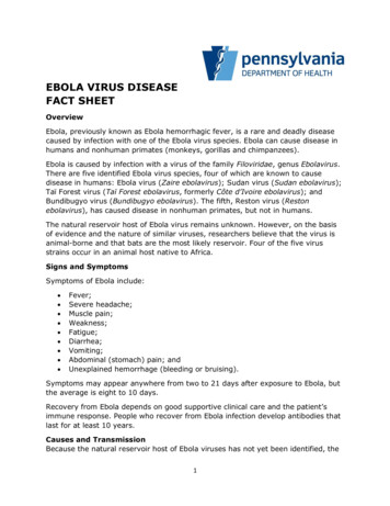
Ebola exposure in international responderswas considered intermediate risk if there was no exposure onto broken skin or mucous membranes or high risk if there was contact with these areas. In transcutaneous exposure (such as aneedle stick injury), risk was considered high if the sharp was a freshly used hollow-bore needle and high if the sharp object had been contaminated by contact with a confirmed EVDpatient or body fluids. The risk from a percutaneous injury was considered intermediate if thesharp was not known to have been directly contaminated [25].Factors associated with risk of exposure, experiencing a febrile/diarrhoeal illness in WestAfrica or within 1 mo of return, and being tested for EBOV during such illness were determined using logistic regression. Factors reaching p 0.1 in the univariate analysis wereincluded in a multivariable model. Tests for trend were calculated using the likelihood ratiotest to compare a linear model with a categorical model.ResultsParticipantsIt was not possible to estimate the number who received the survey via email, but online consent was received from 300 individuals, all of whom completed the survey; of these, 268(89.3%) returned an OFS and were included in the analysis. Fifty-three UK control participants consented and provided an OFS using the same procedures.Descriptive dataThe median age for those who returned a sample was slightly older than for those who did not(39 versus 33 y, respectively; p 0.07); all other characteristics were similar between thegroups. Of the 268 returnees who returned samples, 152 (57%) were female; the median agewas 36 y (interquartile range [IQR] 30–45). The majority had most recently worked in SierraLeone (253, 94%) and had worked in PPE (230, 86%) (Table 1). The median age of the controlswas 35 y (IQR 31–40), and 35 (66%) were female.Contact and exposurePossible exposure was ascertained through several different questions, and free-text responseswere encouraged to describe incidents more fully. Twenty-one (8%) individuals reported having had direct physical contact with patients with confirmed or suspected EVD whilst notwearing PPE. Fifty-five (21%) answered “yes” (47) or “unsure” (8) when asked whether theyhad had a significant exposure or a “near miss” event. Using the free-text descriptions, exposures were allocated a level of risk of transmission (Box 1). In all, 43 (16%) individuals hadpotential exposures: 27 (10%) very low risk, 10 (4%) low risk, five (2%) intermediate risk, andone high risk (Fig 1).Table 2 shows the factors associated with reporting any transmission risk compared toreporting no/unclear risk. The amount of time spent in PPE in the laboratory or the clinicalred zone was associated with increased odds of experiencing an EBOV transmission risk (p fortrend 0.02, excluding those not wearing PPE). Returnees were more likely to report an exposure risk if they had been performing a clinical role (29% of 123) compared to a laboratory role(3% of 90) (odds ratio 13.4 [95% CI 3.8–47.0] after adjusting for time in PPE) (Table 2). Afteradjusting for role and days in PPE, no other factors were associated with risk. The differentmethods of PPE removal were associated with the role, so were not independently associatedwith risk.Over one-third of returnees reported contact with recovered EVD patients (98/268, 37%).Most of these (57/98, 58%) were direct physical contact (handshaking or embracing) withPLOS Medicine https://doi.org/10.1371/journal.pmed.1002300 May 16, 20176 / 18
Ebola exposure in international respondersTable 1. Characteristics of 268 individuals who travelled to West Africa in response to the 2014–2016Ebola epidemic.CharacteristicNumber (percent) or median(IQR)DemographicsFemaleAge (years)152 (56.7%)36 (30–45)Country most recently worked inSierra Leone253 (94.4%)Liberia12 (4.5%)Guinea3 (1.1%)Roles undertaken (returnees could select more than y engagement/tracing18WASH staff11Finance3Engineer3Pharmacist2Social worker/burial team/information nist17Worked in PPE (laboratory or clinical)233 (86.9%)Time in country (days)30 (20–40)Previous experienceLaboratory work with filoviruses11Haemorrhagic fever outbreak7Both lab and outbreak work31One returnee reported each of these roles.IQR, interquartile range; PPE, personal protective equipment; WASH, water, sanitation, and 00.t001patients at the time of discharge from an Ebola treatment centre (ETC), which was encouraged. Others described living or working with recovered patients (9, 3%) or contact within afollow-up clinic setting (14, 5%), or they reported contact but did not provide a clear description (18, 7%). These contacts were not included as transmission risks.Self-reported illness and testingAmong 268 returnees who were never known to have had EVD, 57 (21%) experienced afebrile/diarrhoeal illness either in West Africa or within 1 mo of return. Table 3 shows the factors associated with illness and with being tested for EBOV if the returnee experienced diarrhoea and/or fever.Clinical staff were the most likely to have experienced an illness; 29% (35 of 123) comparedto 20% (11 of 55) of “other” staff and 12% (11 of 90) of laboratory staff (p 0.01) (Table 3).After adjusting for role, no other factors were associated with illness.PLOS Medicine https://doi.org/10.1371/journal.pmed.1002300 May 16, 20177 / 18
Ebola exposure in international respondersBox 1. Examples of risk classification, and associated free-textdescriptions from the survey, among 268 individuals who travelledto West Africa in response to the 2014–2016 Ebola epidemicNo or unclear risk: direct contact with local population with no known illness; tornouter glove, inner glove intact.“Children and some adults would approach you unexpectedly and grab your arm etc.Contact with locals, they did not look ill as were mainly walking on streets or playing”“Torn glove in isolator but remaining gloves intact and no fluid contact”Very low risk: living or working with someone with undiagnosed EVD but no directphysical contact; breaches in PPE with no visible contamination or broken skin exposure“Torn suit when caring for a sick woman who was fitting”“Facemask dislodged”“Person I was staying in same accommodation with became infected”Low risk: direct physical contact with patient who does not have vomiting, diarrhoea, orbleeding.“Contact with subsequently confirmed infected Medic. . .contact was symptomaticbut ‘dry’”“A local colleague working at our ETU [Ebola treatment unit] came to work whilesymptomatic for approximately 5 days without informing us, he tested positive anddied from EVD a week later”Intermediate risk: direct physical contact with a patient with diarrhoea, vomiting, orbleeding; direct contact with bodily fluids from a patient with EVD.“Cut hand on artesunate vial (through gloves) whilst in red-zone”“Being vomited on while wearing just gloves”“Touched mask with blood on gloves (could not breathe—had to pull mask fromface)”High risk: exposure of mucous membranes or broken skin to bodily fluids from an EVDpatient, or sharp injury from a needle/other used on an EVD patient (not freshly used ornot hollow-bore needle).“Sharp injury with a broken vial of medication inside the red zone with dirty and contaminated gloves”PLOS Medicine https://doi.org/10.1371/journal.pmed.1002300 May 16, 20178 / 18
Ebola exposure in international respondersFig 1. Risk of Ebola virus disease transmission in 268 individuals who travelled to West Africa inresponse to the 2014–2016 Ebola epidemic. PPE, personal protective 2300.g001Amongst 57 returnees who experienced a febrile/diarrhoeal illness, 30% (17 returnees)were tested for EBOV infection at the time. One of 21 (5%) who fell ill in country had anEBOV PCR, compared to 11 of 17 (65%) who fell ill on return (odds ratio 36.7 [95% CI 3.9–244.8]). Of those who were ill and were classified as having no or unclear risk (47 returnees),12 (25%) were tested, compared to five of the ten (50%) with some risk. The individual with ahigh-risk exposure did not describe an illness and was not tested. Of the five with intermediate-risk exposures, three were ill, and two of those were tested.Laboratory resultsThe frequency of NOD values for the 268 OFSs are shown in Fig 2, alongside those from 53controls. All the samples from UK controls and all but two of the samples from returnees werewell below the cutoff (maximum NOD 0.7). The two samples that were reactive on the first testhad similar results on re-testing. Plasma from these individuals was tested further, as describedbelow.Participant AParticipant A was a female laboratory worker who spent a total of 30 d in an Ebola laboratoryin West Africa. She did not describe a significant exposure or febrile illness, and was not testedwith PCR for EBOV infection at any point during her deployment or on return.The OFS was reactive on three tests, with NOD 3.55, 3.27, and 3.12. In plasma, the IgG capture assay was reactive twice (NOD 4.5 and 5.4), the DABA was reactive twice (NOD 2.6 and3.2), but the competitive assay was unreactive twice (NOD 0.44 and 0.49), and the in-houseindirect ELISA test at the Jenner Institute was unreactive. The EBOV PCR on plasma wasnegative.Participant BParticipant B was a female physician who spent 40 d in the clinical red zone, did not describe asignificant exposure or any illness, and was not PCR tested for EBOV infection during herdeployment or on return. The OFS showed a low level of reactivity on the IgG capture assay(NOD 1.05, 1.26, and 0.88). Similar low-level reactivity was seen in the IgG capture assay ofPLOS Medicine https://doi.org/10.1371/journal.pmed.1002300 May 16, 20179 / 18
Ebola exposure in international respondersTable 2. Factors associated with risk of Ebola virus disease transmission in 268 returnees who worked in West Africa during the 2014–2016 Ebolaoutbreak.FactorNumber (percent)Risk of EVD transmission, n(percent)Unadjusted OR(95% CI)Adjusted OR1(95% CI)p-ValueSexFemale152 (56.7)20 (13.2)1Male116 (43.3)23 (19.8)1.6 (0.8–3.1)Age group (years)19–3073 (27.2)8 (11.0)131–4099 (36.9)19 (19.2)1.9 (0.8–4.7)41–5045 (16.8)8 (17.8)1.8 (0.6–5.1) 5151 (19.0)8 (16.7)1.5 (0.5–4.3)Sierra Leone253 (94.4)41 (16.2)1Guinea/Liberia15 (5.6%)2 (13.3)0.8 (0.2–3.7)CountryFacility2Facility 172 (26.9)16 (22.2)1Facility 267 (25.0)7 (10.5)0.4 (0.2–1.1)Facility 353 (19.8)3 (5.7)0.2 (0.1–0.8)Facility 413 (4.9)6 (46.2)3.0 (0.9–10.2)Other63 (23.5)11 (17.5)0.7 (0.3–1.7)90 (33.6)3 (3.3)1Role3Laboratory work 0.011Clinical work123 (45.9)35 (28.5)11.5 (3.5–38.9)13.4 (3.8–47.0)Other role455 (20.5)5 (9.1)2.9 (0.7–12.7)8.7 (1.6–47.4)Days in PPE50.02; test fortrend, 0.0261–2577 (28.7)13 (16.9)1126–3583 (31.0)9 (10.8)0.6 (0.2–1.5)1.3 (0.5–3.6)36–6038 (14.2)9 (23.7)1.5 (0.6–4.0)2.3 (0.8–6.4) 6022 (8.2)9 (41.0)3.4 (1.2–9.6)4.7 (1.5–14.8)None48 (17.9)3 (6.3)0.3 (0.1–0.4)0.3 (0.1–1.4)Previous viral haemorrhagic fever workNo previous work247 (92.2)41 (16.6)1Previous work21 (7.8)2 (9.5)0.5 (0.1–2.3)PPE removal—chlorine spray7No spray98 (36.6)7 (7.1)1Chlorine spray132 (49.3)33 (25.0)4.3 (1.8–10.3)Not applicable38 (14.2)3 (7.9)1.1 (0.3–4.6)No assistance108 (40.3)13 (12.0)1Assistance122 (45.5)27 (22.1)2.1 (1.0–4.3)Not applicable38 (14.2)3 (7.9)0.6 (0.2–2.3)PPE removal—assistance71Only days in PPE and laboratory work were included in the final model.Facility is the Eb
their experience of illness. Oral fluid collection devices were sent and returned by post, and samples were tested using an EBOV IgG capture assay that detects IgG to Ebola glycopro-tein. Blood was collected from returnees with reactive samples for further testing. Unex-posed UK controls were also recruited.
![f][triazin-4-amino] Adenine C-Nucleoside for the Treatment of Ebola and .](/img/14/ad1031184.jpg)