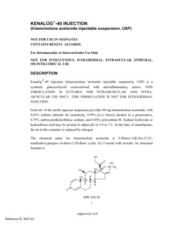
Transcription
European Journal of Trauma and Emergency EVIEW ARTICLETreatment options in extra‑articular distal radius fractures:a systematic review and meta‑analysisGuido W. Van Oijen1 · Esther M. M. Van Lieshout1Tjebbe Hagenaars1 · Michael H. J. Verhofstad1· Maarten R. L. Reijnders1 · Anand Appalsamy1 ·Received: 17 January 2021 / Accepted: 19 April 2021 The Author(s) 2021AbstractPurpose This systematic literature review aimed to make a detailed overview on the clinical and functional outcomes andto get insight into the possible superiority of a treatment method for extra-articular distal radius fractures.Methods Embase, Medline, Cochrane Library, Web of Science, and Google Scholar were searched for studies describingtreatment results. Five treatment modalities were compared: plaster cast immobilization, K-wire fixation, volar plating,external fixation, and intramedullary fixation.Results Out of 7,054 screened studies, 109 were included in the analysis. Overall complication rate ranged from 9% afterplaster cast treatment to 18.5% after K-wire fixation. For radiographic outcomes, only volar tilt in the plaster cast group waslower than in the other groups. Apart from better grip strength after volar plating, no clear functional differences were foundacross treatment groups.Conclusion Current literature does not provide uniform evidence to prove superiority of a particular treatment method whenlooking at complications, re-interventions, and long-term functional outcomes.Keywords Review · Distal radius fractures · Extra-articular · Treatment · Non-operative · OperativeIntroductionA large variety exists in distal radius fractures; from highenergy, comminuted, intra-articular fractures, to low-energy,simple extra-articular fractures. Because of the difference inbone quality, fracture characteristics, associated soft tissueinjury, and patient’s needs between these groups, a differenttreatment approach might be necessary [1–3].Fractures of the distal radius can be treated operatively ornon-operatively. Closed reduction and plaster cast immobilization was traditionally preferred for both intra- and extraarticular fractures. Devices for operative treatment, such asKirschner wires, plates, external fixators, and intramedullaryimplants, have been introduced over the last decades andtheir appropriate use yields good functional results [4–6].Especially the introduction of volar locking plates has led* Esther M. M. Van Lieshoute.vanlieshout@erasmusmc.nl1Trauma Research Unit, Department of Surgery, ErasmusMC, University Medical Center Rotterdam, P.O. Box 2040,3000 CA Rotterdam, The Netherlandsto a significant increase in operative treatment rates, alsofor extra-articular fractures, because of the improvementin stability and therefore the possibility for early mobilization [7]. On the other hand, complaints and complicationsfrom tendon irritation are assumed more common and theextensive dissection of soft tissue might cause fracture andwound healing problems [7]. Other operative treatments alsoprovide better stability than plaster casting, but have theirown cons [8–10].Current guidelines suggest closed reduction and plastercast immobilization as the primary treatment for extraarticular distal radius fractures because the treatment isnon-invasive and cheap [11, 12]. However, it is unclear ifa non-invasive method also means fewer complications andan acceptable loss of function. There is still no consensusregarding the superiority of one of these methods for treatingextra-articular distal radius fractures.The aim of this systematic literature review and metaanalysis was to make a detailed overview on the clinicaland functional outcomes and to get insight into the possiblesuperiority of a treatment method in adult patients with anextra-articular distal radius fracture.13Vol.:(0123456789)
G. W. Van Oijen et al.Materials and methodsA literature search was conducted June 19, 2018. Embase,Medline, The Cochrane Library, Web of Science, and GoogleScholar were searched to identify relevant clinical studies thatreport on the outcomes of extra-articular distal radius fractures.De-duplication of studies was performed as described before[13]. The exact search strategy for the different databases isshown in Online Appendix 1. No language limits were usedand any potentially eligible non-English language manuscriptswere translated if possible by native speaking colleagues orusing Google translate, if no native speaker was available.patients, mean age of patients, number of female patients,duration of follow-up, and type of fracture classification.The outcome data that were retrieved, consisted of; complications and re-interventions, radiographic outcomes(i.e. consolidation rate, volar/dorsal tilt, radial inclination,radial height, and ulnar variance), range of motion, gripstrength, and functional outcome scores (i.e. Disabilities ofthe Arm, Shoulder, and Hand (DASH) score, Quick-DASHscore, Patient Rated Wrist Evaluation (PRWE-)score, VisualAnalog Scale (VAS) for pain, Gartland and Werley score,and the Mayo Wrist Score). All outcome data were pooledfor four different follow-up periods: 0–3 months, 3–6months, 6–12 months, and 12 months or longer (longterm).Selection criteriaQuality assessmentThe studies were eligible for inclusion if they met the following criteria: (1) Patients who suffered and were treated for anextra-articular distal radius fracture; (2) age 18 years or older;(3) patients were either treated using a plaster cast, K-wire(s),plate fixation, external fixator, or an intramedullary device;(4) primary data for at least one outcome parameter had to beavailable.Studies without a clear description of fracture type or intervention, or studies that lacked sufficient data for analysis wereexcluded. In addition, studies with incomplete registration ofcomplications were excluded. Studies describing a not-commonly used device or technique for extra-articular fractureswere excluded (i.e. dorsal plates, Epibloc system, cannulatedscrew, above elbow cast, etc.). If a study consisted of one eligible and one non-eligible cohort, this study was included inour study, but only if data for the eligible group was provided.Two authors (GWVO and AA) independently screened thesearch results for potentially eligible studies by checking thetitle and abstract in phase one. Any disagreement has beenresolved by consensus or consultation of a third author (TH).After the exclusion of all irrelevant studies, the full text ofthe remaining studies was obtained, and the eligibility wasevaluated to complete selection in phase two. This again wasdone independently by two authors (GWVO and MRLR). Anydisagreement was resolved as described above. If the full-textstudy was not available, the corresponding author was contacted and asked.The study protocol has not been registered or publishedbefore.Risk of bias and methodological quality of the includedstudies were assessed using the Newcastle–Ottawa Quality Assessment Scale. This scale results in a score rangingfrom 0 to 9. Scores of 8 and 9 were defined as high-qualitystudies, scores of 5–7 were defined as medium quality, andscores of 1–4 were defined as low quality [14].Search strategyData analysisMeta-analysis for binary data was performed using MedCalcfor Windows. Heterogeneity testing was performed usingCochran’s Q and the I 2 statistic. Q is distributed as a chisquare statistic with k (number of studies) minus 1 degreesof freedom. Q has low power as a comprehensive test ofheterogeneity [15]. The I 2 statistic describes the percentageof variation across studies that is due to heterogeneity ratherthan chance [16, 17]. A random-effects model was used ifthe I2 value was larger than 40%. When the I 2 was lower than40% a fixed-effects model was used. Pooled estimates (i.e.,proportion) are reported with their 95% confidence intervals (CI). When there was no overlap in the 95% confidenceintervals, the difference was considered statistically significant. For the continuous data a pooled estimate (i.e., mean)was calculated in Microsoft Excel, using sample size as aweighting factor. Since most studies only provided the meanvalues but no standard deviation, 95% confidence interval,or standard error, a formal meta-analysis was not feasible forthe continuous data.Data extractionResultsIn phase three, the same two authors independently extractedthe following data from the included studies: author, yearof publication, study design, type of treatment, number ofThe primary search resulted in 14,398 hits and after deduplication 7054 studies remained. After screening thesestudies by title and abstract 1137 remained for full-text evaluation. A total of 109 studies, with 136 different cohorts13
Treatment options in extra-articular distal radius fractures: a systematic review and were marked eligible for the analysis (Fig. 1 and Supplementary Table S1). These 136 cohorts consisted of a totalof 6707 patients divided over the five different treatmentmodalities. The pooled average age and gender of patientsfor each treatment method are outlined in Table 1.Among the 109 studies were 21 randomized controlledtrials, 37 prospective studies, and 51 retrospective studies (Supplementary Table S1). Among the 136 cohorts,31 reported on outcomes after plaster cast immobilization [18–48], 37 on K-wire fixation [5, 18, 21, 41, 42, 45,47, 49–76], 41 on volar plate fixation [4–6, 54–56, 60, 61,72, 74, 77–107], 16 on external fixation [6, 30, 34, 36, 53,108–118], and 11 on intramedullary fixation [4, 6, 43, 81,92, 96, 119–123]. Most of the included studies were of lowor medium quality, 40% (N 44) and 50% (N 55), respectively. Ten of the included studies were of high quality, asoutlined in Supplementary Table 2.ComplicationsThe pooled overall complication rate ranged from 9%after plaster cast treatment to 19% after K-wire fixation(Table 2). Superficial infections were most prominent andhigher in the K-wire (4.7% [95% CI 2.5–7.6%]) and ExFixFig. 1 Flowchart of study selection13
G. W. Van Oijen et al.Table 1 Study subjects pertreatment modalityTreatmentCohorts(N)Population(N)Mean age(95% CI)Proportion female(95% CI)Plaster castK-wiresVolar plateExternal fixatorIMN313741161121851525224542832461.7 (53.5-69.9)57.9 (47.2-68.7)58.8 (51.7-65.9)50.6 (31.4-69.8)59.3 (50.0-68.6)78.5 (71.9–84.5)67.2 (56.1–77.4)73.2 (63.4–82.0)77.6 (63.4–89.1)77.6 (71.3–83.1)Data are shown as proportion with 95% CIgroups (10.1% [95% CI 4.5–17.6%]). Volar plating hadthe highest rate of carpal tunnel syndrome (3.5% [95% CI2.4–4.8]) which was higher than plaster cast immobilization and K-wire fixation (0.4 [95% CI 0.0–1.7] and 0.7[95% CI 0.2–1.4], respectively). In 6.1% of the K-wiregroup, the implant(s) failed (i.e., loosening or breakage ofmaterial). This was significantly higher than in the volarplate fixation group (0.8%). There was significantly lessre-dislocation in the volar plate group when comparedwith plaster cast (1.0% versus 9.3%). Pooled re-intervention rates ranged from 3.8% to 5.3%. Regarding these reinterventions, only one significant difference was found:the use of antibiotics was 4.2% in the K-wire group versus0.8% in the volar plating group. Data of all other complications and re-interventions had overlapping confidenceintervals across the groups and are described in Table 2and Table 3 and shown in Fig. 2 and Fig. 3.Radiographic outcomeRadiographic outcomes are shown in Fig. 4 and Supplemental Table 2. Because no non-union was reported forany of the treatment methods, consolidation rates werenot different. The pooled mean radial inclination, radialheight, and ulnar variance were similar for all five treatments after 12 months’ follow-up. Overall, volar platefixation shows consistent good outcome for all four radiological measurements. Especially the ulnar variance wasclearly better than the other four treatment modalitieswith 0.3 mm, 0.1 mm, 0.3 mm, and 0.3 mm at 0–3,3–6, 6–12, and 12 months after surgery, respectively.The palmar tilt in the plaster cast group was consistentlylower than in other groups in all follow-up periods. Theproportion of patients with a good or excellent Lidströmscore was reported for three treatments only: plaster castimmobilization, K-wire fixation and external fixation.Proportions were 72% [95% CI 45.3–92.2], 90% [95% CI81.3–95.2], and 88% [95% CI 59.9–99.9], respectively.13Functional clinical outcomeRange of motion and grip strength are outlined in Fig. 5and Supplemental Table 2. For several outcome parameters,none or a small number of studies provided data for analysis. Differences in range of motion and grip strength wereespecially found in the short-term follow-up. For example,volar plate fixation showed a relatively good grip strength(69% of the non-injured side) and pro-/supination (81.4and 80.0 degrees, respectively) at 0–3 months’ follow-up.Both pro- and supination were above the limit of disabilityfor the entire follow-up period. At long-term follow-up, noclear functional differences were found across the treatmentgroups.Patient‑reported outcomesA large variety of patient rated outcome measures were usedin literature. This resulted in small numbers of study subjectsand uncertain values. Three outcome measures that wereused most often, were chosen to be analyzed and are shownin Fig. 6: Disabilities of Arm, Shoulder, and Hand (DASH)score, Gartland and Werley score, and a Visual Analog Scale(VAS) for pain. Assessment of the DASH score showed lowpooled means for volar plating (13.0 points) and intramedullary fixation (15.0 points) at 0–3 months’ follow-up. Again,at long term, follow-up scores were similar across the treatment groups. For both the Gartland and Werley score and theVAS for pain, an improvement was seen for all treatmentsover time and no clear differences were found.DiscussionThis systematic review and meta-analysis aimed to evaluate complications, re-interventions, radiographic outcomes,functional outcomes, and patient-reported outcomes of themost commonly described treatment methods for extra-articular distal radius fractures. The data do not show a clearsuperiority or inferiority for any of the treatments evaluated.
Treatment options in extra-articular distal radius fractures: a systematic review and Table 2 Pooled complication rates per treatment modalityParameterAny complicationTreatmentPlaster castK-wiresVolar plateExternal fixatorIMNCRPSPlaster castK-wiresVolar plateExternal fixatorIMNCTSPlaster castK-wiresVolar plateExternal fixatorIMNInfectionPlaster castK-wiresVolar plateExternal fixatorIMNDeep infectionPlaster castK-wiresVolar plateExternal fixatorIMNImplant failurePlaster castK-wiresVolar plateExternal fixatorIMNParesthesia superficial Plaster castradial nerveK-wiresVolar plateExternal fixatorIMNTendon irritationPlaster castK-wiresVolar plateExternal fixatorIMNTendon rupturePlaster castK-wiresVolar plateExternal fixatorIMNStudies(N)Population(N)Q(p-value)I2(95% CI)Pooled proportion(95% 367481622919882.8 ( 0.001)148.4 ( 0.001)63.4 ( 0.001)24.0 (0.008)18.5 (0.005)85.4 ( 0.001)28.8 (0.119)21.4 (0.317)12.5 (0.188)5.4 (0.606)0.4 (0.999)8.5 (0.993)14.5 (0.884)0.8 (0.999)0.7 (0.994)0.8 (1.000)49.9 ( 0.001)7.7 (0.994)41.8 ( 0.001)0.7 (0.998)0.8 (1.000)2.6 (1.000)3.1 (1.000)4.3 (0.959)0.7 (0.998)NA.87.3 ( 0.001)17.1 (0.582)5.9 (0.753)0.7 (0.998)0.6 (0.997)17.8 (0.537)25.2 (0.090)18.7 (0.028)34.3 ( 0.001)0.8 (0.999)4.6 (0.999)16.9 (0.461)0.8 (0.999)3.3 (0.651)0.8 (0.998)13.0 (0.840)2.6 (1.000)0.8 (0.999)6.4 (0.381)90.3 (84–94)87.2 (82–91)68.4 (50–80)58.4 (19–79)67.6 (28–85)90.6 (84–94)27.1 (0–57)11.1 (0–46.4)27.9 (0–65.4)0 (0–58.8)0 (0–0)0 (0–0)0 (0–17)0 (0–0)0 (0–0)0 (0–0)59.9 (35.3–75.1)0 (0–0)73.7 (53.3–85.2)0 (0–0)0 (0–0)0 (0–0)0 (0–0)0 (0–0)0 (0–0)NA.79.4 (68.5–86.5)0 (0–42.3)0 (0–42.6)0 (0–0)0 (0–0)0 (0–44.4)32.6 (0–61.9)52.0 (1.4–76.6)79.6 (60.3–89.5)0 (0–0)0 (0–0)0.0 (0–49.7)0 (0–0)0 (0–62.9)0 (0–0)0 (0–23.9)0 (0–0)0 (0–0)6.1 (0–73.1)9.0 (2.0–20.3)18.5 (10.9–27.6)13.3 (9.6–17.4)18.1 (11.8–25.4)18.2 (9.5–29.0)2.4 (0.0–9.1)1.1 (0.5–2.0)2.2 (1.4–3.5)1.4 (0.4–3.4)1.9 (0.5–4.7)0.4 (0–1.7)*0.7 (0.2–1.4)*3.5 (2.4–4.8)*0.9 (0.1–3.1)1.4 (0.3–4.1)0.5 (0.1–1.7)*4.7 (2.5–7.6)*0.8 (0.3–1.6)*10.1 (4.5–17.6)*0.8 (0.1–3.1)*0.5 (0.1–1.7)0.6 (0.2–1.3)0.7 (0.2–1.4)0.9 (0.2–2.6)1.4 (0.3–3.9)NA.6.1 (2.5–11.1)*0.8 (0.3–1.5)*1.0 (0.2–2.9)1.4 (0.3–3.9)0.5 (0.0–1.9)*1.3 (0.6–2.4)*2.2 (1.0–3.9)3.4 (1.0–7.2)10.1 (2.9–21.1)*0.5 (0.1–1.8)0.7 (0.2–1.7)2.0 (1.2–3.3)0.9 (0.1–3.1)1.0 (0.1–3.7)0.5 (0.1–1.9)1.0 (0.4–2.0)0.8 (0.3–1.7)0.9 (0.1–3.1)1.2 (0.2–3.9)13
G. W. Van Oijen et al.Table lation(N)Q(p-value)I2(95% CI)Pooled proportion(95% CI)RedislocationPlaster cast10500103.0 ( 0.001)91.2 (86.1–94.5)9.3 (2.5–19.9)*K-wires2076336.8 (0.008)48.4 (13.3–69.3)2.3 (1.0–4.1)Volar plate1772612.8 (0.686)0 (0–39.0)1.0 (0.4–2.0)*External fixator102496.3 (0.705)0 (0–46.9)2.6 (1.0–5.3)IMN71988.0 (0.236)25.3 (0–67.34)2.6 (0.9–5.8)*Non-overlapping 95% CI.Table. 3 Pooled reintervention rates per treatment (p-value)I2(95% CI)Pooled proportion(95% CI)Any reinterventionNonoperativeK-wiresVolar plateExternal fixatorIMNNonoperativeK-wiresVolar plateExternal fixatorIMNNonoperativeK-wiresVolar plateExternal fixatorIMNNonoperativeK-wiresVolar plateExternal fixatorIMNNonoperativeK-wiresVolar plateExternal 81170214NA.49790119721457.9 ( 0.001)45.5 ( 0.001)46.2 ( 0.001)6.2 (0.521)11.3 (0.126)0.8 (0.998)42.5 ( 0.001)2.1 (1.000)36.3 ( 0.001)0.7 (0.998)0.78 (0.998)0.7 (1.000)2.3 (1.000)0.8 (0.999)0.6 (0.997)10.6 (0.157)9.2 (0.684)6.9 (0.976)2.0 (0.846)2.4 (0.932)NA.22.9 (0.062)54.5 ( 0.001)5.2 (0.524)0.7 (0.998)84.5 (73.1–91)71.4 (50.1–83.4)61.1 (35.8–76.4)0 (0–63.6)38.1 (0–72.7)0 (0–0)67.0 (43.4-80.8)0 (0–0)78.0 (58.3–88.4)0 (0–0)0 (0–0)0 (0–0)0 (0–0)0 (0–0)0 (0–0)34.0 (0–70.8)0 (0–43.6)0 (0–0)0 (0–39.0)0 (0–7.6)NA.38.9 (0–66.9)68.8 (49.3–80.8)0 (0–66.6)0 (0–0)4.6 (1.1–10.2)4.1 (1.4–8.2)5.1 (3.0–7.8)5.3 (2.8–9.1)3.8 (1.7–7.2)0.5 (0.1-1.9)*4.2 (1.7–7.8)*0.8 (0.3–1.7)*4.1 (0.4–11.5)0.8 (0.1–3.1)0.5 (0.1–1.9)0.6 (0.1–2.0)0.9 (0.3–1.9)0.9 (0.1–3.1)0.8 (0.1–3.1)1.9 (0.8–3.9)1.3 (0.4–2.8)0.6 (0.2–1.5)2.4 (0.7–5.9)1.2 (0.2–3.7)NA.2.5 (1.3–4.3)5.5 (3.0–8.8)5.2 (2.6–9.3)1.4 (0.3–3.9)AntibioticsIncision and DrainageRe-osteosynthesisNon-planned hardware removal*Non-overlapping 95% CI.The main treatment goals for extra-articular distal radiusfractures are to regain an adequate wrist function and/orto release pain. In order to study and objectivize this, amultitude of questionnaires and outcome scores have beenintroduced over the last decades. However, which outcomeparameters should be used for valid and accurate assessmentof wrist function are still unclear. Also, the variety of treatment options for distal radius fractures impedes the ability13to prove superiority or inferiority of the currently used treatment methods. Cochrane reviews and the American Academy of Orthopaedic Surgeons both labeled the evidence as“inconclusive” [124, 125]; [126]; [12]. This review focusedon extra-articular distal radius fractures only, where bothCochrane and the AAOS stated this inconclusiveness almosta decade ago for intra-articular fractures as well. All eligibleliterature has been included to show trends in techniques
Treatment options in extra-articular distal radius fractures: a systematic review and Fig. 2 Pooled complication rates per treatment modality. Data are shown as pooled proportion with 95% CIFig. 3 Pooled re-intervention rates per treatment modality. *Early,unplanned removal due to infection, loosening, failure, or other reasons. Data are shown as pooled proportion with 95% CI.used over time. Only volar plates have been subject to significant technological improvement, this probably explains thesteep increase of studies in the twenty-first century (Fig. 7).Surprisingly, unintended outcome has been reported morefrequently than intended outcome: 87 out of 136 cohorts inthis study reported at least one type of complication. Theoverall complication and subsequent re-intervention ratesare not significantly different for the five treatment modalities. However, not all studies reported consistently on allcomplications and re-intervention rates. Different definitionsof complications and a possible extra focus on specific complications in certain studies, likely induced broad ranges ofcomplication rates, and probably underreporting. For example, finger stiffness was described in only four studies witha rate up to 24% in a plaster cast immobilization cohort ofMardani et al. [18], where the other 105 studies did not evenmention it.No differences in functional and patient-reported outcomes at long-term follow-up ( 12 months) could bedetected. This was also mentioned by Costa et al. andArora et al. [5]; [77]. However, in the short term, differences in functional and patient-reported outcomes seemto exist. In all treatment groups, a lowering trend wasobserved after the 0–3 months’ follow-up in both the13
G. W. Van Oijen et al.Fig. 5 Range of motion and grip strength per treatment modality. aFlexion (degrees), b Extension (degrees), c Ulnar deviation (degrees),d Radial deviation (degrees), e Pronation (degrees), f Supination(degrees), g Grip strength (% of contralateral side). Data are shownas sample size weighted mean. The grey line represents the lowestvalues that will not cause any functional impairment (disability value[127])towards surgical treatment. These arguments advocate forboth short- and long-term cost-effectiveness comparisonof the various treatment methods.Radiological outcomes also seem to be in favor of volarplate fixation, as shown by a positive volar tilt, adequateradial inclination, and a negative or low ulnar variance.Radiographic parameters showed worse outcomes for plaster cast immobilization with concomitant high re-dislocationrates. Diaz-Garcia et al. published a systematic review in2011 to examine outcomes of unstable distal radius fracturesafter treatment with either volar locking plate, non-bridgingexternal fixation, bridging external fixation, percutaneousFig. 4 Radiographic outcomes per treatment modality. a Volar tilt (degrees), b Ulnar variance (mm), c Radial inclination (degrees) and d Radialheight (mm). Data are shown as sample size weighted mean.13 DASH score and the Gartland & Werley score, whichmeans less disability and better function. In line withthese findings, also an improvement in grip strength and adecrease in pain were seen in the same time period for alltreatment groups. However, volar plates and intramedullary fixation had a much faster recovery than other modalities. Also, range of motion was immediately close to, oreven above the limit of disability at 0–3 months’ follow-upfor both treatments (Fig. 5). Pronation and supination forall follow-up moments were consistently within the normal range of motion as established by, e.g. the Guides, tothe Evaluation of Permanent Impairment by the AmericanMedical Association [127]. This early recovery of patientstreated with volar plating and intramedullary fixationmight be an important factor in treatment choice. In current society, one might argue that long rehabilitation periods are no longer acceptable for most patients. Moreover,a strong lobby from the industry to sell implants and a highstrive to restore anatomy perfectly by surgeons may push
Treatment options in extra-articular distal radius fractures: a systematic review and 13
G. W. Van Oijen et al.Fig. 6 Patient-reported outcome measures per treatment modality. a DASH score b Gartland & Werley (% good or excellent) c Gartland & Werley Score, d: VAS score for pain. Data are shown as sample size weighted mean.Kirschner-wire fixation, or plaster cast immobilization. Theyalso found that plaster casting was associated with worseradiological outcomes than operative treatment [128]. Oursystematic review reveals that since 2011 nothing has reallychanged.The first thing that stood out during screening of potentialstudies, was the small number of studies that specificallyreported on different types of distal radius fractures (AO/OTA or other classification systems). Many studies reportedtreatment outcomes for distal radius fractures in general,without any fracture classification. Distinction between different fracture types is essential to provide adequate treatment. A simple extra-articular fracture requires a differentapproach than a comminuted intra-articular one.This systematic review had several limitations. First,most studies that were included were retrospective or prospective observational studies, with disappointing reporting13quality for continuous data. For a proper meta-analysis ofcontinuous data the mean, a measure of dispersion (eitherstandard deviation, standard error or confidence interval),and sample size are necessary. Many studies in this reviewfailed to provide these data. Moreover, the number of large,well-designed RCTs on extra-articular fractures is very low,and therefore it is very well possible to have a type 2 error.Second, selection bias in the source studies might haveinfluenced the results of the meta-analysis, since stable andsimple extra-articular fractures might have been treated nonoperatively, whereas the more dislocated and comminutedfractures might have been treated by volar plating or external fixator. However, current data did not allow for metaregression analyses. Therefore, it is not entirely certain ifthe current whether the findings are related to the treatmenttype, or can be explained by a difference in indication. Third,there was high heterogeneity in type of outcome parameters
Treatment options in extra-articular distal radius fractures: a systematic review and Fig. 7 Included studies pertreatment modality per timeperiod. Data are shown asproportion of the total includedstudies per treatment modalityin time periods of five years(N included studies)and time points of measurements reported. Goldhahn et al.mentioned already in 2014 that this heterogeneity is problematic in research on distal radius fractures and suggesteda set of core domains to assess outcomes as a possible solution [129]. We have not discovered any clear improvementin data consistency since this article has been published.However, the effect of this study can still be expected overthe next years due to its recent character. We agree withGoldhahn et al. that minimal requirements of outcome inthe domains patient history, physical examination and radiological findings should be established and journals shouldonly publish papers that meet these criteria. Only then thevalue of future systematic reviews in the field of orthopedictrauma will increase.able to draw sound conclusions on superiority of one of thetreatment methods.Supplementary Information The online version contains supplementary material available at https:// doi. org/ 10. 1007/ s00068- 021- 01679-z.Acknowledgment We thank Mr. W.M. Bramer, Biomedical information specialist at the Erasmus MC (Rotterdam, The Netherlands) forassistance with the literature search.Author contributions All authors contributed to the study conceptionand design. Material preparation, data collection and analysis were performed by Guido van Oijen, Esther van Lieshout, Maarten Reijnders,and Anand Appalsamy. The first draft of the manuscript was writtenby Guido van Oijen and all authors commented on previous versionsof the manuscript. All authors read and approved the final manuscript.Funding No external funding was received for this study.ConclusionCurrent literature does not provide enough evidence to support superiority of a particular treatment method for extraarticular distal radius fractures, when looking at complications, re-interventions, and long-term functional outcomes.A broad range of outcome parameters have been used, whichmakes the data partly impossible to compare. From a methodological point of view, the quality of data used in thissystematic review and subsequent conclusions that can bedrawn, appear to be rather weak. This paper should thereforeencourage future investigators to use more sound researchmethods. Consensus on outcome measures and completeness of reporting is necessary to conduct high-quality studieswith standardized outcome assessment. This is needed to beAvailability of data and material Not applicable.Code availability Not applicable.DeclarationsConflict of interest The authors declare that they have no competinginterests.Open Access This article is licensed under a Creative Commons Attribution 4.0 International License, which permits use, sharing, adaptation, distribution and reproduction in any medium or format, as longas you give appropriate credit to the original author(s) and the source,provide a link to the Creative Commons licence, and indicate if changeswere made. The images or other third party material in this article areincluded in the article’s Creative Commons licence, unless indicated13
G. W. Van Oijen et al.otherwise in a credit line to the material. If material is not included inthe article’s Creative Commons licence and your intended use is notpermitted by statutory regulation or exceeds the permitted use, you willneed to obtain permission directly from the copyright holder. To view acopy of this licence, visit http:// creat iveco mmons. org/ licen ses/ by/4. 0/.References1. Brogren E, Petranek M, Atroshi I. Incidence and characteristicsof distal radius fractures in a southern Swedish region. BMCMusculoskelet Disord. 2007;8:48.2. de Putter CE, Selles RW, Polinder S, Hart
Meta-analysis for binary data was performed using MedCalc for Windows. Heterogeneity testing was performed using Cochran's Q and the I2 statistic. Q is distributed as a chi-square statistic with k (number of studies) minus 1 degrees of freedom. Q has low power as a comprehensive test of heterogeneity [15]. The I2 statistic describes the .
