Transcription
(2022) 23:176Hou et al. Respiratory Open AccessRESEARCHReduced frequencies of Foxp3 GARP regulatory T cells in COPD patients areassociated with multi‑organ loss of tissuephenotypeJia Hou1*†, Xia Wang2†, Chunxia Su3†, Weirong Ma1, Xiwei Zheng1, Xiahui Ge4* and Xiangguo Duan5*AbstractBackground: Expression of glycoprotein A dominant repeat (GARP) has been reported to occur only in activatedhuman naturally occurring regulatory T cells (Tregs) and their clones, and not in activated effector T cells, indicatingthat GARP is a marker for bona fide Tregs. A different phenotype of chronic obstructive pulmonary disease (COPD)may have a different immunologic mechanism.Objective: To investigate whether the distribution of Tregs defined by GARP is related to the multi-organ loss of tissue phenotype in COPD.Methods: GARP expression on T cells from peripheral blood and bronchoalveolar lavage (BAL) collected frompatients with COPD was examined by flow cytometry. The correlation of GARP expression to clinical outcomes andclinical phenotype, including the body mass index, lung function and quantitative computed tomography (CT) scoring of emphysema, was analyzed.Results: Patients with more baseline emphysema had lower forced expiratory volume, body mass index (BMI), worsefunctional capacity, and more osteoporosis, thus, resembling the multiple organ loss of tissue (MOLT) phenotype.Peripheral Foxp3 GARP Tregs are reduced in COPD patients, and this reduction reversely correlates with quartilesof CT emphysema severity in COPD. Meanwhile, the frequencies of Foxp3 GARP Tregs, which are characteristic ofpro-inflammatory cytokine production, are significantly increased in COPD patients, and correlated with increasingquartiles of CT emphysema severity in COPD. Tregs in BAL show a similar pattern of variation in peripheral blood.Conclusion: Decreased GARP expression reflects more advanced disease in MOLT phenotype of COPD. Our resultshave potential implications for better understanding of the immunological nature of COPD and the pathogenicevents leading to lung damage.†Jia Hou, Xia Wang and Chunxia Su contributed equally to this work*Correspondence: houj@live.com; 15000760961@126.com;2455281549@qq.com1Department of Respiratory and Critical Care Medicine, General Hospitalof Ningxia Medical University, Ningxia, China4Department of Respiratory Medicine, Seventh People’s Hospitalof Shanghai University of TCM, Shanghai, China5College of Clinical Medicine, Ningxia Medical University, Ningxia, ChinaFull list of author information is available at the end of the article The Author(s) 2022. Open Access This article is licensed under a Creative Commons Attribution 4.0 International License, whichpermits use, sharing, adaptation, distribution and reproduction in any medium or format, as long as you give appropriate credit to theoriginal author(s) and the source, provide a link to the Creative Commons licence, and indicate if changes were made. The images orother third party material in this article are included in the article’s Creative Commons licence, unless indicated otherwise in a credit lineto the material. If material is not included in the article’s Creative Commons licence and your intended use is not permitted by statutoryregulation or exceeds the permitted use, you will need to obtain permission directly from the copyright holder. To view a copy of thislicence, visit http:// creat iveco mmons. org/ licen ses/ by/4. 0/. The Creative Commons Public Domain Dedication waiver (http:// creat iveco mmons. org/ publi cdoma in/ zero/1. 0/) applies to the data made available in this article, unless otherwise stated in a credit line to the data.
Hou et al. Respiratory Research(2022) 23:176Page 2 of 14Keywords: COPD, T-regulatory cells, GARP, Emphysema, MOLTIntroductionChronic obstructive pulmonary disease (COPD) isan inflammatory lung disease related to an abnormalimmune response that results in airflow limitation.Although cigarette smoke exposure is the most common risk factor for COPD, pulmonary and systemicinflammatory responses persist after smoking cessation in COPD patients [1], suggesting non-environmental factors are driving inflammation in susceptibleindividuals. Cumulating evidence show that cigarettesmoking can generate new self-antigens that may trigger the activation of abnormal "autoreactive" immuneresponses [2, 3]. Hence, the immunoregulatory function may determine the susceptibility to, and severityof, COPD.Naturally occurring C D4 CD25 regulatory T cells(nTregs) are a distinct subpopulation of lymphocytesthat play a critical role in controlling immune responsesand promoting and maintaining self-tolerance. nTregsmust depend on the forked-head helix transcriptionfactor (Foxp3) for differentiation and development aswell as maintenance of function, and the identificationof Foxp3 facilitated murine nTreg identification [4].Foxp3 has been regarded as a specific molecular markerfor Treg cells. However, human effector T cells havetheir Foxp3 expression significantly upregulated uponactivation, and such cells do not have immunomodulatory capacity [5]. Therefore, the use of Foxp3 as aspecific marker for human nTregs is increasingly questioned [6]. Some other common surface markers usedto identify nTregs such as CD25, cytotoxic T-lymphocyte antigen 4 (CTLA-4), and glucocorticoid-inducedtumor necrosis factor receptor (GITR) encounteredthe same problem, i.e., these markers are constitutively expressed, not only in nTregs, but also in activated effector T cells [7–9]. Likewise, both nTregs andactivated effector T cells show decreased expressionof CD127. The search for surface markers specific forhuman nTregs has been an important topic in this field[5, 10, 11]. It is uncertain whether molecular markersthat are specifically expressed in nTreg cells exist.The breakthrough in this field has been the discovery of the glycoprotein A dominant repeat (GARP orLRRC32). Using DNA microarray technology, Unutmazet al. [12] discovered for the first time that activatedhuman Treg cells express a transmembrane protein,GARP, which is not expressed by activated or restingeffector T cells. GARP is not induced to be expressedon activated effector T cells, which have high levels ofFoxp3 expression induced by TGF-β stimulation butlack suppressive function. GARP mRNA is barelydetectable in freshly isolated C D4 T cells, but GARPmRNA levels rise rapidly in Treg following T cell receptor (TCR)-mediated activation, whereas they do notoccur in non-Treg cells [13–15]. In addition, high GARPexpression distinguishes Treg cells from interleukin 17(IL-17)-secreting cells [12]. Forced expression of GARPin non-Treg cells using transgenic techniques results inthe acquisition of some or all the phenotype and function of Tregs (including Foxp3 expression). Knockdownof GARP in nTreg cells results in a significant decreasein suppressive activity, while Foxp3 expression does notchange. Thus, GARP is a highly specific and functionally relevant marker molecule expressed by activatednTreg cells and has been regarded as a potential targetfor immunotherapy [16–19].In this study, we aimed at investigating whetherthere was abnormal distribution of bona fide activated Foxp3 regulatory T cells identified by GARP in theblood and BAL of patients with COPD, and whetherthis distribution was associated with clinical phenotypeof COPD. The findings should provide new insights intothe underlying mechanisms of persistent inflammationand adaptive immunology in COPD.Materials and methodsPatient enrolment and study designForty-three patients with COPD were recruited atthe General Hospital of Ningxia Medical University,China. Fourteen never-smokers and 12 smokers ( 20pack-year history) with normal lung function werealso recruited as controls. COPD diagnosis and severity classification were performed according to the criteria of the Global Initiative for Chronic ObstructiveLung Disease (GOLD) [20]. Patients having a history ofasthma and/or allergic rhinitis, or suffering from infections, malignancy, autoimmune disorders, and otherimmune-related diseases were excluded. Patients onantibiotics, systemic steroids, chemotherapeutics andimmunosuppressors within the four weeks before theenrollment, were also excluded. None of the COPDpatients had experienced respiratory infection or acuteexacerbation over the last four weeks prior to the study.The study was approved by the Ethics Committeeand Medical Faculty of Ningxia Medical University(approval no. 2020–678) and conducted in accordance with the Declaration of Helsinki regarding human
Hou et al. Respiratory Research(2022) 23:176medical research. All the subjects provided writteninformed consent prior to study participation.Computed tomography (CT) scan quantificationof emphysemaAll COPD patients underwent at least one high-resolution chest CT scan using multidetector-row scanners(Siemens Healthcare GmbH, Germany). Detailed CTprotocols have been described elsewhere [21]. Quantitative analysis of emphysema is based on the proportion ofCT voxels below 950 Hounsfield units (low attenuationarea, LAA) using the software of 3DSlicer (http:// www. slicer. org).Cell collection and processingPeripheral blood samples from each subject were collected in EDTA treated tubes and were processed toisolate peripheral blood mononuclear cells (PBMCs) forflow cytometry procedures. Blood samples were layeredonto Ficoll-Paque Plus (Amersham Biosciences), thencentrifuged (400 g for 20 min at 22 C), and PBMCswere harvested. PBMCs were washed twice in PBS solution at 300 g for 5 min at 4 C, then were resuspendedand viable counts obtained.Sequential BAL preparation was performed and processed as previously described [22]. To avoid the acuteeffects of smoking, smokers were asked to stop smokingat least 12 h prior to the BAL procedure. Using a wedgedflexible bronchoscope (Olympus BF-290, Tokyo, Japan)under topical lidocaine, four separate 50 mL aliquots ofsterile 0.9% saline were infused into one segment of theright middle lobe and gently suctioned. Since the lavagefluid from the first 50 mL aliquot is known to containcells and material from the central airways rather thanthe peripheral bronchial and alveolar, the lavage fluidfrom the first aliquot was discarded. The leftover lavage fluid was filtered and resuspended at 106 cells/ml inRPMI-1640 medium at 4 C after being washed twice inphosphate-buffered saline. All BAL and peripheral bloodsamples were processed immediately after sampling.Flow Cytometry Analysis Software (FACS) analysisFreshly isolated PBMCs were stained and incubatedwith relevant antibodies for 30 min at 4 in a darkroom. Cells were washed twice before being analyzedby a BD LSRII flow cytometer (BD Biosciences, SanJose, CA, USA). For intracellular cytokine staining, cellswere activated by phorbol myristate acetate (PMA) andionomycin (Sigma) for 5 h, followed by fixation andpermeabilization using a commercial kit (e-Bioscience,San Diego, California, USA), according to the manufacturer’s instructions. Both interferon-γ (IFN-γ) and IL-17antibodies were used for intracellular cytokine staining.Page 3 of 14Foxp3 expression was performed via the fixation andpermeabilization procedure. The following monoclonalantibodies (mAbs) were used for extracellular staining:CD4, CD25, CD127 and GARP. The following mAbs wereused for intracellular staining: IL-17, IFN-γ and Foxp3.For GARP transduction, cells were activated with CD3/CD28 beads (Invitrogen). All antibodies were purchasedfrom BD Biosciences or Pharmigen (San Jose, CA, USA)and e-Bioscience (San Diego, CA, USA). Data acquiredby flow cytometry were analyzed using FlowJo software(Tree Star).T‑cell proliferation assaysCD4 CD25 responder cells (5 104) from healthy controls were stained with carboxyfluorescein succinimidylester (CFSE, Molecular Probes), and were coculturedwith 5 104 CD25 GARP Tregs from healthy controlor COPD patients sorted by the Aria sorter in anti-CD3coated (5 μg/mL OKT3 mAb; eBioscience) plates in thepresence of soluble anti-CD28 (5 μg/mL; eBioscience) for72 h. Proliferation of the cells was assessed on day fouror day five by analyzing the dilution of CFSE in the cellswith flow cytometry.Statistical analysisGroup data were expressed as the mean and S.E.M oras the median and interquartile range, where appropriate. For data distributed normally, one-way analysis ofvariance (ANOVA) with post-hoc multiple comparisonsusing the Tukey’s method was performed to determinesignificance of differences between groups. For data notdistributed normally, comparisons between groups weremade using one-way Kruskal–Wallis tests, and a p-valueof 0.05 was considered significant. If this test indicated significance, the Dunn’s multiple comparison testwas used for post-hoc analysis for comparisons betweentwo groups. Correlation was assessed by calculatingSpearman’s rank correlation coefficient. Here, a p-valueof 0.05 was considered significant. All significance levelswere based on two-tailed tests. Statistical analyses wereperformed using Prism (GraphPad Prism 7.0).ResultsDemographic characteristics of study subjectsInitially, 66 stable COPD patients were recruited butonly those with full CT profile (n 43) were includedin the current analysis. The demographic characteristics of COPD patients enrolled in this study are shownin Table 1. All the patients with COPD were stratified byCT scan quantification of emphysema quartiles (Fig. 1and Additional file 1: Fig. S1). Table 2 illustrates the mainbaseline clinical features of COPD patients included inthe study, stratified by CT emphysema quartiles.
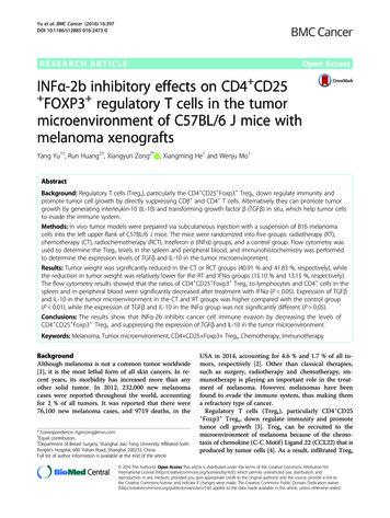
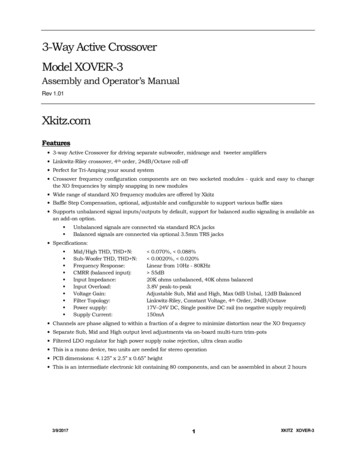
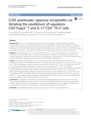
Hou et al. Respiratory Research(2022) 23:176Page 4 of 14Table 1 Demographics and spirometry of all participantsNever smokersSmokersCOPD14124365 567 666 1028 629 625 8Pack-years0Current smoker %037 1844 2398 850 22Subjects (n)DemographicsAge (year)Male sex %64BMI (kg m 2)Smoking status75837223PhysiologyFEV1% predictedFEV1/FVC %94 585 482 348 16Data are presented as median (IQR) for smoking status, mean SD for all othersFEV1, forced expiratory volume in 1 s; FVC, forced vital capacityIn line with a previous report [23], patients with moresevere emphysema were more likely to have lower BMI,worse oxygen saturation, F EV1, symptom and healthstatus, and were more likely to be a heavy ex-smoker(Table 2). They were also more likely to report osteoporosis and less likely to report having diabetes or cardiovascular disease. Most subjects in all quartiles were receivinginhaled corticosteroid, whereas a minority of subjects( 2%) were receiving oral corticosteroid (Table 1). Retrospective analysis showed that patients with more emphysema had a higher frequency of acute exacerbations,hospitalizations associated with accelerated lung function loss over the past two years.Selective expression of GARP on suppressive activatedTreg CellsAs described previously [12], GARP is specificallyhighly expressed in the C D25 or Foxp3 populations(Fig. 2A). Treg cells selectively express GARP onlyCOPD Q2(LAA 10.4%)TGF‑β can induce Foxp3 expression but not GARPexpressionIt has been shown that TGF-β induces Foxp3 expression in both murine and human T cells [8, 9], but unlikemurine cells, TGF-induced human C D25 Foxp3 cells do not confer a regulatory phenotype. Consistentwith previous studies [12], we further determined thatTGF-β induce Foxp3 expression but not GARP expression (Fig. 4), suggesting the transcription factor Foxp3is not essential for the induction of GARP and GARPcan truly distinguish activated nTreg cells from inducedTreg cells and activated T cells that have no immunosuppressive function.COPD Q3LAA 25.0%COPD (Q4)LAA 44.7%CT RA950COPD Q1(LAA 7.3%)after TCR stimulation (Fig. 2B). To further investigatethe potential to use GARP as a marker for activatedTreg cells with suppressive capacity, C D25 GARP cells were sorted with cytometry after TCR stimulation to determine their functional activity. As Fig. 2C,D shows, C D25 GARP cells can more stronglyinhibit the proliferation of responder cells comparedto C D25 GARP cells. Remarkably, a much higheramount of pro-inflammatory cytokines, includingIL-17 and IFN-γ were detected in C D25 GARP cellsin response to PMA stimulation (Fig. 2E–G), whichidentified this subpopulation as the previously reportedpro-inflammatory cytokines secreting Tregs [24]. Compared with C D25 GARP cells, the C D25 GARP population showed a higher expression of TIGIT,CTLA-4, HELIOS, and HLA-DR (Fig. 3A–F) suggestingthat these cells are a proliferating, activated fraction ofTregs. Taken together, GARP not only defines activatedregulatory T cells with highest inhibitory capacity butalso discriminate C D25 Foxp3 T cells that secretepro-inflammatory cytokines.Fig. 1 Central coronal chest CT emphysema quantification. The quantification of emphysema, based on the RA950, is demonstrated in therepresentative coronal CT scan from four individuals with varying severity of emphysema. From this classification, normal lung tissue is denotedblue, and emphysema is denoted purple. Patients with more advanced emphysema have more purple areas. RA950: relative area of the CT densityhistogram voxels 950 HU
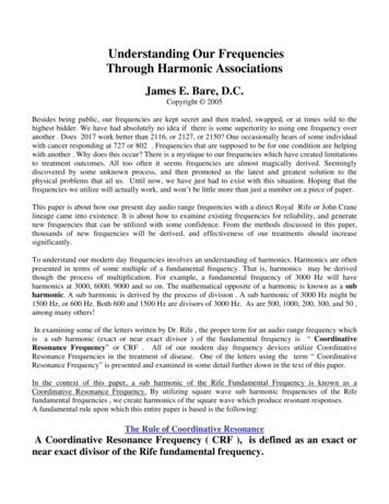
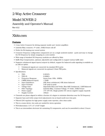
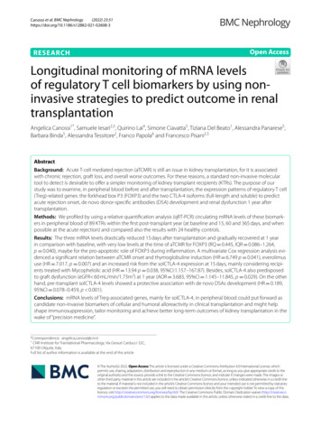
Hou et al. Respiratory Research(2022) 23:176Page 5 of 14Table 2 Baseline characteristic of patients stratified by quartiles of CT emphysema severitySubjects (n)Q1LAA 7.5%Q2LAA 7.5–15%Q3LAA 15–25%Q4LAA 25%9111310p-value(ANOVA)DemographicsAge years64 965 867 766 8Male sex %68707876BMI (kg m 2)26 727 525 623 6BMI 21 kg m 2%14121930NS 0.001 0.001 0.001Smoking statusPack-yearsCurrent smoker %41 193340 213043 221545 1914 0.01 0.001Medications %Inhaled steroids15255652 0.001Oral corticosteroids0000NS59 1352 1446 1444 16 0.0195.8 2.996.2 3.992.8 3.491.4 2.3 0.001PhysiologyFEV1% predFEV1/FVC %Oxygen saturation %Comorbidities (%)55 951 1041 943 6 0.01Cardiovascular diseases15201713 0.01Osteoporosis18203542 0.01Diabetes91056 0.01Longitudinal outcomesCOPD hospitalizations PPPY*0.40.81.63.4 0.001COPD exacerbations PPPY*1.22.63.43.8 0.001FEV1 decline mL·year 128 2632 3035 3336 32 0.01LAA: low attenuation area; BMI: body mass index; FEV1: forced expiratory volume in the first second; FVC: forced vital capacity; PPPY: per patient per year*Average number of events per year(See figure on next page.)Fig. 2 GARP specifically defines suppressive activated Tregs. A Gating strategy and representative flow cytometry plots of GARP expression areshown. GARP is selectively expressed in CD4 CD25 and C D4 Fop3 cells after TCR stimulation. CD25 GARP and CD25 GARP subpopulationscan be sorted for in vitro suppressive function assay. B Induction of GARP on C D4 Fop3 cells after TCR stimulation (anti-CD3 and anti-CD28 beads).C In vitro suppressive function assay of the CD25 GARP and CD25 GARP subsets. The evaluation of the suppressive function of each subset wasperformed by CFSE staining of the responder cells. Percentages of dividing responder cells per well were indicated. Data are representative of fiveseparate experiments. D The percentages of CD25 GARP and CD25 GARP cells prohibiting proliferation in each group are shown in a summarygraph. EFlow cytometry of the secretion of IFN-γ vs. IL-17 by GARP and GARP subsets after stimulation with PMA ionomycin for 5 h. Summarygraph showing the percentages of IFN-ɣ and IL-17 secreting cells in each subset, respectively (F, G). One-way ANOVA with post hoc pairwisemultiple comparisons using the Tukey’s method. For D, F and G, data are from n 5 independent experiments using T cells from different healthydonors (n 5 subjects)Decreased frequencies of circulating F oxp3 GARP Tregsassociated with multi‑organ loss of tissue phonotypeWe analyzed circulating Foxp3 GARP cells in nonsmoker, smokers, and COPD subjects at different stagesof disease. Cytometric assays revealed that healthy smokers had a higher frequency of F oxp3 GARP Tregs compared with non-smokers and COPD subjects (Fig. 5A,B). In COPD patients, the frequency of F oxp3 GARP Treg subsets progressively decreased from Q1 to Q4,whereas the frequency of F oxp3 GARP cells increasedfrom Q1 to Q4 (Fig. 5A, B). Not surprisingly, the frequency of Foxp3 GARP Tregs were inversely correlated with the LAA (r 0.63, P 0.001) whereas thefrequency of Foxp3 GARP cells correlated with LAA ofCOPD patients (r 0.72, P 0.001) (Fig. 5C). In addition to the emphysema quartiles, we also analyzed circulating Foxp3 GARP Tregs in COPD patients stagedusing the Global Initiative on Obstructive Lung Disease
Hou et al. Respiratory Research(2022) 23:176Page 6 of 14FSCFSCACD4SSCBPost ac va onBefore ac va on13.64%12.60%CGARPGARP0.01%3.52%Foxp3Foxp3DP 0.01100Proliferation (%)EP 0.01806040200derponRes- 5 GCD2derponRes ARPndespo ReARPr 5 GCD2FGP 0.001P 0.001P 0.00180IL-17 secreting cells(%)IFN- γ secreting cells(%)P 0.0016040200--5CD2PGAR- 5CD2PGAR 5CD2Fig. 2 (See legend on previous page.)PGARP 0.001P 0.001543210-5CD2-RPGA-P GAR5CD2 P GAR5CD2
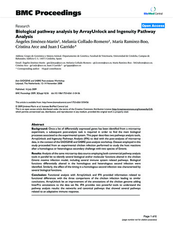
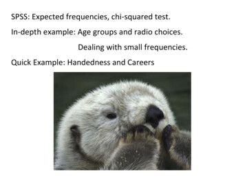
Hou et al. Respiratory Research(2022) 23:176Page 7 of 14ABCP 0.001P 0.001P 0.00180 CTLA-4 %2060402000-- PP GAR GARARPp3 Gp3p3FoxFoxFoxEP 0.001Fox20--RPGAF-RP A3 GoxpFo RP GAxp3-P GARp3FoxF P GARp3FoxP 0.001HLA-DR %403oxp-RPP 0.00180600GAP 0.001P 0.00180-p3-HELIOS %P 0.001P 0.0018040FP 0.00110060TIGIT %DP 0.0016040200--- PP GAR GARARPp3p3FoxFox3GpFoxFig. 3 Functional markers relevant to nTregs are significantly expressed in Foxp3 GARP T cells. A Gating approach for CD4 T cell subpopulationsbased on Foxp3 and GARP expression. B Representative flow cytometry plots showing the expression of classical Tregs markers in three C D4 T cellsubpopulations from healthy donors. C Statistical graphs depicting the expression of those markers in each subpopulation of C D4 T cells
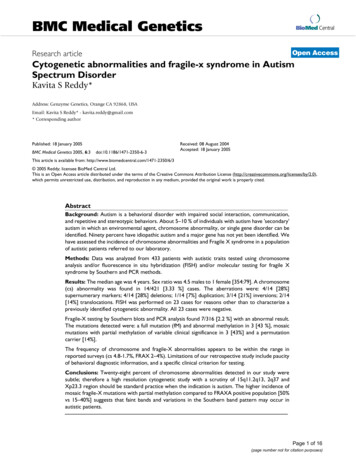
Hou et al. Respiratory Research(2022) 23:176Page 8 of 14disease, and these markers progressively decrease as thedisease progresses.Progressive changes of Treg cell function during COPDdevelopmentThe altered GARP expression prompted us to performin vitro experiments to further evaluate the immunefunction of nTregs in COPD patients. Consistent with theimmune phenotype results, the frequency of Foxp3 Il17-secreting cells were increased in COPD patients andassociated with the severity of emphysema (Fig. 6A, B).However, the immuno-suppressive capacity of F oxp3 Tregs tends to decrease progressively with increasingemphysema severity (Fig. 6C–E). Together, these finding suggest that the compromised immunomodulatorycapacity and enhanced pro-inflammatory capacity areinvolved in lung tissue destruction in COPD.Is GARP Expression a More Accurate Marker to Detect TregsDuring Chronic Inflammation in COPD?Fig. 4 TGF-β treatment leads to different induction of Foxp3 andGARP in TN cells. TN cells were activated via TCR (CD3CD28 beads)with or without 20 ng/mL TGF-β. Foxp3 and GARP expression weremeasured after two days of activation(GOLD) criteria and compared them with both nonsmoker and smoker controls. No significant reductionin the frequency of Foxp3 GARP subsets was foundin COPD patients at different GOLD grades. Furthermore, there was no statistically significant difference in Foxp3 GARP cells of COPD patients as they progressedfrom GOLD II to IV (Additional file 1: Fig. S2).Next, we measured markers associated with suppressive (CTLA-4 and TIGIT) and proliferative function (Ki67) on Tregs. In line with the expression trend of GARP,the proportion of CTLA-4, TIGIT and progressivelydecreased from Q1 to Q4 in COPD patients. However,the patients with COPD at Q1 and Q2 expressed higherlevels of Ki-67 than the patients at Q3 and Q4 (Fig. 5D).Taken together, the results indicate that the expressionlevels of functional markers on the surface of Treg cellsare normal or mildly increased in the early stages of theReliably discriminating Tregs from activated T cells ischallenging, because Tregs markers are potentially modified during T-cell activation or differentiation, especially under chronic inflammation [11]. That promptedus to look into whether GARP could be a better markerfor identifying bona fide Tregs in the chronic immuneactivation of COPD. Using the classic definition, thepercentage of Tregs in peripheral blood of different populations was examined. Treg identification using CD4 Foxp3 CD127 (Fig. 5E, F) revealed no differences inthe frequency of circulating Tregs in patients with COPDcompared to smokers and never-smokers, confirmingprior research [25, 26]. Combined with in vitro functional assays, this striking discordance between the frequency of FOXP3 CD127 T cells and F OXP3 GARP T cells suggest that GARP expression can better identifyTregs during systemic inflammation in COPD.The similar pattern of GARP variation in BALTo clarify the GARP expression of pulmonary Tregs,BAL samples were obtained from seven never-smokers, eight smokers and twenty COPD patients havingclinical investigational bronchoscopy. Interestingly,(See figure on next page.)Fig. 5 GARP expression on CD4 Foxp3 T cell are progressively decreased in different severity of emphysema. A Representative flow cytometryplots of GARP expression are shown. B The proportions of Foxp3 GARP and Foxp3 GARP subsets among CD4 T cells in all subjects. C Negativestatistical correlation between frequency of circulating FoxP3 GARP and LAA% (C, Left). Positive statistical correlation between frequency ofcirculating FoxP3 GARP cells and LAA% (C, Right). r 0.63, p 0.001 and r 0.72, p 0.001 by Pearson’s correlation. D Expression of Treg cellfunction-specific markers (CTLA-4 and TIGIT) and activation markers (Ki-67) in peripheral F oxP3 GARP cells. One-way ANOVA with post-hocpairwise multiple comparisons using Tukey’s method. *p 0.05; **p 0.01; ***p 0.001; ****p 0.0001. E Representative staining of CD4 T cellsfor GARP, CD127, and FOXP3 expression. CD4 T cells were stained for CD127 and FOXP3 expression as resting T cells and for GARP and FOXP3expression following 1 day of TCR activation. F Analysis of Foxp3 CD127 T cells from all subjects
Hou et al. Respiratory Research(2022) 23:176Page 9 of 14AB***************25% of Foxp3 GARPin CD4 T cells642NSSQ1Q2Q320151050Q4COPDC403030LAA %LAA 5% of Foxp3 GARP- in CD4 T cells***% of TIGIT on Foxp3 GARP T cells% of CTLA-4 on Foxp3 GARP T cellsQ310********************NSQ2r 0.72P 0.001% of Foxp3 GARP in CD4 T cells50Q120100SCOPDr -0.63P 0.00150NSQ3Q4100****% of Ki67 on Foxp3 GARP T cells% of Foxp3 GARP in CD4 T Q1Q2Q3Q4*60Res ng0NSSQ1Q24.5%13.6%% of Foxp3 CD27in CD4 T cellsCD1276.3%6420NSSQ1Q2Q3COPDFig. 5 (See legend on previous page.)Q3COPD8Post-ac va on**20FE**40COPDCOPD*Q4Q4
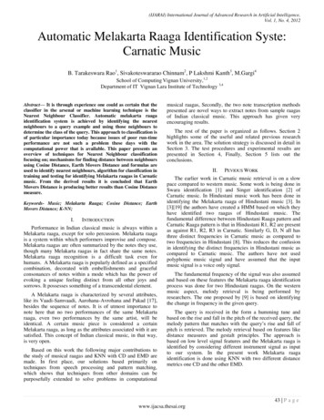
Hou et al. Respiratory Research(2022) 23:176Page 10 of 14AB*****% of Foxp3 IL17 on CD4 T Proliferation (%)100*****806040200NSSQ1Q2Q3COPDQ4DFig. 6 Impaired suppressive function and enhanced IL-17 production of Tregs from COPD patients. A Representative intracellular staining ofFoxp3 vs. IL-17 in non-smoker (n 6), smoker (n 6), and COPD patients (n 6) at a different quartile after 5 h of stimulation with PMA/ionomycinand GolgiStop. B The proportions of Foxp3 IL-17 subsets among CD4 T cells in all subjects. One-way Kruskal–Wallis test with post-hoc multiplecomparisons using the Dunn’s method. **p 0.01; ***p 0.001; C CD25 GARP Tregs and CD25 GARP responder T cells (Tresp) were isolated byFACS. For suppression and proliferation assays, CFSE-labeled Tresp cells were co-cultured with Treg cells with plate-bound anti-CD3 and solubleanti-CD28 for 96 h, alone or in 1:4 ratios. Tresp cells were isolated from non-smoker and Tregs were isolated from non-smoker, smoker and COPDpatients in different quartiles. D Suppressive function of Tregs decreased with the CT scan quantification quartile. The flow cytometry plot showsdilution of CFSE labeled responder cells. The percentage of dividing cells is shown. E Suppressive function assay revealed that C D25 GARP Tregcells from COPD patients have a reduced suppressive capacity and associated with the severity of emphysema. Data are from n 8 independentexperiments (at least n 3 subjects in 2 technical replicates). One-way ANOVA with post hoc pairwise multiple comparisons using Tukey’s test**p 0.01; ***p 0.001we found strong GARP expressions of Tregs in BALwithout activation of CD3CD28 beads. Foxp3 GARP Tregs in BAL shows a similar pattern of variation inperipheral blood, that is, frequencies of Foxp3 GARP Tregs decreased whereas F oxp3 GARP T cellsincreased in COPD patients and associated withthe severity of multi-organ loss of tissue phonotype(Fig. 7A, B).
Hou et al. Respiratory ResearchPage 11 of ) 23:176Foxp3*B*****6420NSSQ1-2Q3-4COPD******40% of Foxp3 GARPin CD4 T cells% of Foxp3 GARP in CD4 T cells8***3020100NSSQ1-2Q3-4COPDFig. 7 Decreased Foxp3 GARP Tregs in BAL of COPD patients. A Representative intracellular staining of Foxp3 vs. GARP in BAL fluid fromnon-smoker (n 7), smoker (n 6) and COPD patients (n 15) at a different quartile. B The proportions of Foxp3 GARP Tregs and Fop3 GARP Tcell among C D4 T cells in all subjects. One-way ANOVA with post-hoc pairwise multiple comparisons using the Tukey’s test *p 0.05; **p 0.01;***p 0.001 DiscussionAs the most common chronic inflammatory respiratorydisease, COPD is characterized by airflow limitation thatis not fully reversible. Cellular and molecular mechanisms involved in this disease remain ill-defined. nTregsplay a critical role in controlling immune responses andmaintaining immune homeosta
Chronic obstructive pulmonary disease (COPD) is an inammatory lung disease related to an abnormal immune response that results in airow limitation. Although cigarette smoke exposure is the most com-mon risk factor for COPD, pulmonary and systemic inammatory responses persist after smoking cessa-tion in COPD patients [], suggesting non-environ1 -