Transcription
HindawiVeterinary Medicine InternationalVolume 2018, Article ID 8515812, 7 pageshttps://doi.org/10.1155/2018/8515812Research ArticleChicken Coccidiosis in Central Java, Indonesia: A Recent UpdatePenny Humaidah Hamid ,1 Yuli Purwandari Kristianingrum,2April Hari Wardhana,3 Sigit Prastowo,4 and Liliana Machado Ribeiro da Silva51Department of Parasitology, Faculty of Veterinary Medicine, Universitas Gadjah Mada, Yogyakarta, IndonesiaDepartment of Pathology, Faculty of Veterinary Medicine, Universitas Gadjah Mada, Yogyakarta, Indonesia3Indonesian Research Center for Veterinary Sciences, Bogor, Indonesia4Department of Animal Science, Universitas Sebelas Maret, Surakarta, Indonesia5Institute of Parasitology, Justus Liebig University Giessen, Giessen, Germany2Correspondence should be addressed to Penny Humaidah Hamid; penny hamid@ugm.ac.idReceived 10 November 2017; Revised 29 December 2017; Accepted 11 January 2018; Published 8 February 2018Academic Editor: Francesca ManciantiCopyright 2018 Penny Humaidah Hamid et al. This is an open access article distributed under the Creative Commons AttributionLicense, which permits unrestricted use, distribution, and reproduction in any medium, provided the original work is properly cited.Avian coccidiosis is a huge problem worldwide. Heavily infected animals that show severe clinical signs and coccidiostat resistanceare causing important economic losses. The present study aimed to update the recent cases of coccidiosis in Central Java, Indonesia,and to show the importance of the disease in the region. A total of 699 samples were obtained from different chicken breed. DifferentEimeria species were detected in 175 individuals (25.04%). Three different groups of chicken breed were considered: local chicken(autochthonous chickens of Sentul and Jawa), commercial broiler, and layer. Broiler chickens showed the highest prevalence ofinfection (34%), followed by layer (26.26%) and local chickens (10.45%). Mild to severe clinical signs of avian coccidiosis wereobserved in 42% of the infected animals, while 58% of the infected animals showed no clinical signs other than low feed conversionrates. Seven different Eimeria species were identified: E. tenella was the most prevalent (43.3%), followed by E. maxima (26.3%),E. necatrix (15.7%), E. acervulina (8%), E. praecox (3.1%), E. mitis (2.2%), and E. brunetti (1.3%). Coinfections with several Eimeriaspecies were diagnosed. With this study we found massive usage of coccidiostat in the region even though its usage cannot guaranteecoccidiosis-free chicken production.1. IntroductionPoultry industry is the most predominant meat productionin Indonesia reaching 2,030,880 tons in 2016 [1]. Nowadays,chicken meat is consumed not only as fresh meat, but alsoas derived products, for instance, processed food products(e.g., nugget, meatballs), frozen products, and canned meat.The increasing consumption of meat in this region is proportional to the increasing purchasing power in Asian countriesobserved nowadays. Further, chicken meat is cheaper thancattle or lamb meats and it is a common option to completehuman demand for protein of animal origin. In fact, production of chicken increased in Asia by 68.83%, featuringthe highest growth when compared to other regions such asAustralia, New Zealand, Africa, and Europe [2]. In Indonesia,chicken populations are dominantly commercial broiler andlayer, followed by different kinds of autochthonous/localchicken breed [3]. Different management systems are common in Indonesia, ranging from very sophisticated intensivefarms to traditional production kept by small producers invillages.Increasing production of poultry meat in Indonesia ismandatory to fulfill accelerated demands as a consequenceof the increment of the population. To guarantee a highand constant production level of chicken products, diseasescontrol programs are needed. One of the most well-knowndiseases worldwide is avian coccidiosis [4]. The losses causedby coccidiosis are due to not only mortality but also poorweight gain and feed conversion rate together with the costs oftreatment [5]. Avian coccidiosis is caused by Eimeria species,which are highly species-specific. Each of the seven speciesknown to infect chicken has a special predilection site in theintestine to complete its life cycle [6]. General clinical signs,being reduced weight gains and egg yields, together with
2anorexia and poor feed utilization, pale combs, and dehydration, associated with mucus and/or bloody diarrhoea, arecommon. Besides, coccidiosis is a predisposing factor fornecrotic enteritis, whose incidence has been rising due to thestop of antibiotic usage in some countries [7, 8]. Mechanismof how Eimeria promote Clostridium spp. massive growth isnot exactly understood. As reviewed elsewhere [7], severalfactors contribute to clostridial pathogenicity in intestineof Eimeria infected chicken. Coccidiosis led to chickenimmunodepression, with consequent increased proliferation,adhesion, and toxin production of Clostridium spp. [7].There are seven species causing chicken coccidiosis: E. tenella(haemorrhagic typhlitis) and E. necatrix (haemorrhage ofthe small intestine) considered highly pathogenic, E. brunetti(necrotising enteritis) that is slightly less pathogenic, E. maxima and E. acervulina that cause mild to severe catarrhalicenteritis, and E. mitis and E. praecox considered as lowpathogenic species [9]. In fact, previous experiments using E.praecox evince different virulence between strains and evenexceed E. acervulina which is known as more pathogenicspecies [10]. The phenomenon enhances variability of clinicalmanifestation in chicken, which is influenced by total doseingested, strains virulence, and also susceptibility of thehost when infection occurred [10, 11]. Eimeria spp. life cycleis direct with short prepatency periods of 4–6 days and,consequently, the disease spreads fast between hosts. Shortlyafter ingestion of sporulated oocysts, invading sporozoitespenetrate epithelial intestinal cells and endogenous development proceeds with 2–4 merogonies and gamogony. Observations of the animals as well as necropsies to determine thelocation and type of the lesions (lesion scores) in each flockare important to confirm diagnosis and identify the speciesvirulence upon outbreak.It has been known that avian coccidiosis is a serious threatin Indonesian poultry industry. Infections by Eimeria spp. inchickens reared in traditional farming management systemreached 39.3% [12] with E. tenella as the most prevalentspecies. Technical services of farm industries of commerciallayer and broiler chickens reported cases from many areasin Java, Kalimantan, and Sumatra islands. Nowadays andcontrary to what is expected, coccidiosis cases are occurring not only in traditional management systems where notreatments or control measures are being performed, butalso in intensive and semi-intensive farms where treatmentsand control measures are considered. For example, chemoprophylaxis with anticoccidials in feed or drinking water isperformed in broilers throughout the whole fattening perioduntil premarketing withdrawal and in laying hens until thestart of egg laying to avoid residues of anticoccidial drugsin the eggs [6]. Furthermore, Eimeria spp. resistance toseveral anticoccidial drugs has been reported [13], with theemergence of drug-resistant strains of all Eimeria species inchickens [6]. Additionally, persistent Eimeria spp. infectionsin Indonesia may also be supported by the high environmental humidity which might contribute significantly to thecompletion of the parasite life cycle [14]. Given that theoocysts remain in the environment after being shed [15]and that wet floors favour oocysts sporulation, infection ismaintained.Veterinary Medicine InternationalIn this study, we provide recent information of coccidiosiscases in Central Java, Indonesia. The update of chickencoccidiosis situation is pivotal for consideration of controlstrategies as both preventive and cure programs in the area,in order to fulfil the high demands for chicken products.2. Materials and MethodsEthical clearance regarding the experiment was issued byethics committee, LPPT Gadjah Mada University, number00087/04/LPPT/VII/2017.Random sampling was performed in flocks of traditionaland semi-intensive management systems allocated in CentralJava and Special Region of Yogyakarta in the followingprovinces: Kulon Progo, Gunung Kidul, Bantul, Yogyakarta,Magelang, and Boyolali, 7 47.44 south latitude and 110 8.24eastern longitude ordinates.In total 47 flocks were visited. Sample collection was carried out from April to June 2017. Chicken breeds included inthis study were autochthonous chickens from Sentul and Jawabreed, commercial layers, and broilers. The Jawa chicken (Figures 1(d) and 1(e)) is characterized by mottled feather almostin whole body part and yellow skin and beaks. While Sentulchicken (Figures 1(b) and 1(c)) has more homogeneous greycolor, its skin and beaks are also grey-colored and the colorbecomes darker in older chicken. Sentul chicken has an average of adult body weight in male of 2600 207 g and femaleof 1408 123 g; this chicken is able to produce 17 1 eggsper laying period. Jawa chicken, commonly known as “ayamkampung” chicken, has an adult body weight in males of1600 107 g, females 1450 150 g, and it is able to produce 13 2 eggs per laying period. Jawa and Sentul chicken are farmedfor meat production. These two chicken breeds are differentfrom Bekisar (hybrid from jungle fowl) which is usuallyreared by citizens in Java Island due to its fascinating soundand mostly for contest or hobby purposes. It is difficult toobtain Bekisar fertile eggs, so certain breeders are producingfertile eggs by traditional mating of Gallus varius (as parentstock), which is now increasingly rare except in certain island,and or artificial insemination with female of Jawa chicken.Faecal samples from chickens presenting severe or mildclinical signs of coccidiosis, as well as from animals withoutclinical signs, were randomly collected. Flotation techniquewith saturated NaCl [16] was performed to determine thepresence of Eimeria spp. oocysts. Faecal samples containingoocysts were mixed with potassium dichromate (2% finalconcentration) and allowed to sporulate at room temperature. Eimeria species present in each sample were identifiedaccording to the morphology of the sporulated oocysts.Moreover, some chickens presenting severe clinical signs ofcoccidiosis, such as bloody diarrhea, were collected for further investigations at the laboratory. Necropsy of chickenswith severe clinical signs was performed and lesion score wasdetermined. Additionally, histopathology sections were performed and analysed after haematoxylin and eosin staining.3. Results and DiscussionOf the total 699 samples obtained from different chickenbreed, Eimeria spp. oocysts were detected in 175 of the
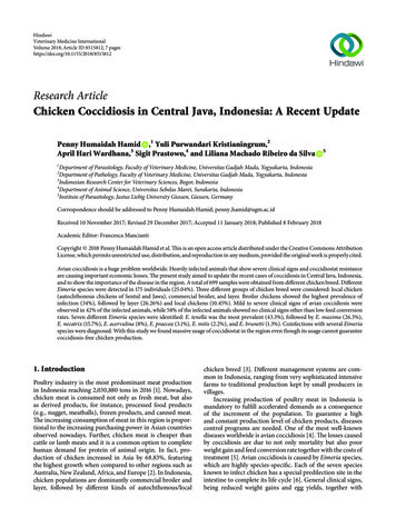
Veterinary Medicine International3(a)(b)(d)(c)(e)Figure 1: Conditions of flocks with coccidiosis in a semi-intensive farming system in Central Java. (a) Semi-intensive farming of broilers(19 days old), with almost all chicken positive for E. tenella infection, showing unspecific clinical signs. Sentul breed in (b) 5-day-old and (c)21-day-old cases. Jawa breed in (d) 5-day-old and (e) 21-day-old cases.chicken (25.04%). Broiler chicken showed the highest prevalence with 34% of the animals infected, followed by layer(26.26%) and local chicken (10.45%) as presented in Table 1.Clinical signs were observed in 42% of the infected chickenranging from mild (lethargy and light diarrhoea) to severeclinical signs such as bloody diarrhoea. Nonetheless, 58%of the infected animals (positive for presence of oocysts byflotation technique) did not present any clinical signs usuallydescribed for coccidiosis. Local chicken of Sentul and Jawa(Figure 1) mostly from villages showed less prevalence ofEimeria spp. oocysts (Table 1).Seven Eimeria species were identified during this study. E.tenella was the most prevalent species with 43.3% prevalence,followed by E. maxima with 26.3%, E. necatrix with 15.7%,
4Veterinary Medicine InternationalTable 1: Prevalence of chicken coccidiosis in Central Java by chickentype.Total number of samplesNumber of positive samplesNumber of negative samplesPrevalence of chicken coccidiosis (%)Chicken 026.2610.4560% infection4020E. tenellaE. praecoxE. necatrixE. mitisE. maximaE. brunettiE. acervulina0mixed species infection (%)number of positive (%)Figure 2: Eimeria species identification in positive faecal samples.E. acervulina with 8%, E. praecox with 3.1%, E. mitis with2.2%, and E. brunetti with 1.3% (Figure 2). Several Eimeriaspp. with less pathogenic characteristics were found in coinfection (Figure 2). All these seven Eimeria spp. are evidentlyspread worldwide [17]. Eimeria species have been found withdifferent prevalence during regional surveys in France [18],Jordan [19], Romania [20], Brazil [21], or China [22]. Ourfindings are in agreement with previous reports that show E.tenella as the predominant species found in Asia [23, 24].Animals infected with less pathogenic Eimeria species, E.maxima, E. acervulina, E. praecox, and E. mitis, showed noclinical signs. Eimeria tenella was the most frequent speciesfound in this study in comparison to other pathogenic Eimeria, that is, E. necatrix and E. brunetti. Although consideredto be the most pathogenic species, clinical symptoms ofsome chicken infected with E. tenella were not quite clear.Mortality was never observed massively in a short period oftime but it consistently occurred during fattening periods. Itwas observed that chicken with slight bloody diarrhoea stillpresented normal appetite and food intake although bodyweight conversion was diminished when compared to broilerchicken at the same age. Pathologic findings such as caecaballooning were only observed when chickens were necropsied. Necropsied chickens infected with E. tenella showedclinical manifestation of bloody diarrhoea and ballooningcaeca with various parasitic stages (Figure 3). Some chickenshowed grooves in caeca lumen (Figure 3) categorized ashaving score 2 in E. tenella infection. Score lesions could notbe identified in all necropsied chicken in accordance withspecies-specific pathogenicity since mixed infections werecommonly observed. Intestines parts with no bloody diarrhoea did not show erythrocytes extravasations out of bloodvessels but massive lengthened villi were observed in grosssection (Figure 3).This study also showed that coccidiosis affects all chickenexamined: layer, broiler, and local chicken, Sentul and Jawa.Nevertheless, local chickens had less prevalence in comparison with broiler and layer. We collected faecal samples fromchicken reared free in the villages where they have enoughspace for exercises and more natural feed supply. In contrastto broiler and layer examined, farmers usually use coccidiostat in feed during layer and growing/fattening period of localchicken. However, no evidence existed so far in resistanceand susceptibility of these local chickens to avian coccidiosis.These current data exhibited local chicken performancein facing field infection and may show ability in eliminatinginfection naturally. That resistance to avian coccidiosis isinherited and genes-associated was reported elsewhere [25,26]. So far, reports in Indonesia related to parasite-resistancebreed was checked in thin-tail sheep which is less susceptibleto Fasciola hepatica and F. gigantica infections [27]. In thiscontext, further study of resistance to avian coccidiosis inlocal chicken breed will be useful and informative for breeding selection. Local chickens in this study (Jawa and Sentul)had lower body weight at the same age compared to broiler.However, some people choose to consume meat from localchicken due to the taste preference, therefore having higherprice. Price per kg body weight of a local chicken cannot becompared to broiler; thus it is the reason why some farmersprefer to grow local chicken than broiler although its growthrate is slower. Additional information of coccidiosis-resistantphenotype in this study will benefit local chicken farmingand may give insight into breeding strategy of local chickenbreeds.In the flocks of broiler sampled in this study, coccidiostatwas supplemented in the feed and applied during almostthe whole life of chicken. Exemplary of this observation, allchickens in Figure 1 were randomly sampled and all presentedballooning caeca although coccidiostat as feed additive wasbeing applied. The flock of Figure 1 consisted of 5,000 chickens with semi-intensive farming system. This incidence mayreflect a case of coccidiostat resistance in the field. Nowadays,avian eimeriosis resistance to various commercial anticoccidial drugs is reported and reviewed worldwide [22, 28–30].This might be a concern since no scientific reports are published in Indonesia so far. Combination or rotation of coccidiostat has to be performed in order to prolong drug efficaciesand prevent resistance to specific active compound. Insteadof chemical compounds, natural feed additives can be considered to enhance chicken performance during critical phasesof fattening period. Research in natural herbs shows evident
Veterinary Medicine International5(a)(b)(c)(d)Figure 3: Macroscopic lesions and histopathologic sections of chicken intestine with coccidiosis in Central Java. (a) Macroscopic lesion ofthe caecum showing ballooning condition (arrows) with bloody lumen. (b) Different pinhead size structures were massively formed in lumenof caecum (arrow). (c) Haemorrhagic and inflammatory cells infiltration (arrow) in the infected caecum. (d) Macrogamonts of Eimeria spp.in the caecum (arrows). The species is identified as E. tenella based on the predilection site (caecum), morphology of oocyst, and sporulationtime of collected oocysts from faeces.improvement of immune response to avian Eimeria infections [29, 31–33]. Moreover, combination of live attenuatedvaccines with anticoccidial drugs may enhance immunesystem for infection. This approach is considered effective inimproving chicken endurance in accordance with applicationof coccidiostats [34]. In addition, a wise decision in choosingvaccination strategy is mandatory through update of information and vaccine products in the market with all its prosand cons [34].Several farms with infected chicken showed clearly thatlitter management may contribute to parasite persistence andhard elimination. The humidity of litter made from rice husk(Figure 1) was observed to be high. Moisture of litter togetherwith its surface temperature in Indonesia tropical climate ispresumed to be very close to the optimum conditions forsporulation, as reviewed elsewhere [34]. However, total oocyst counts in the litter need to be performed to evaluatecontribution of the litter in Eimeria persistency within a flock,but that was not performed in this study. Some observedfarms for broiler were also having very short period oftransition times before entering new brooding period ofbroiler for new rearing batch giving a very limited time toclean and rest the parasites cycles. Overall results in this studyimply evaluation of management systems of flocks, including litter, in-out processes, vaccination, and/or combinedvaccine-coccidiostat application during fattening.4. ConclusionChicken coccidiosis is a persistent problem in Central Java.Most of the animals were asymptomatic but showing low feedconversion rates. In addition, local chicken breed, that is, Sentul and Jawa, showed less prevalence than broiler and layer.This study also pointed out that massive usage of coccidiostatin feed cannot guarantee chicken free from coccidiosis.Conflicts of InterestThe authors declare that they have no conflicts of interest.Authors’ ContributionsPenny Humaidah Hamid, Yuli Purwandari Kristianingrum,April Hari Wardhana, and Sigit Prastowo designed and performed the research. Penny Humaidah Hamid, Sigit Prastowo, and Liliana Machado Ribeiro da Silva wrote the manuscript. All authors approved the manuscript.AcknowledgmentsThe research was funded by Ministry of Agriculture, KP4SGrant no. 76.76/PL.040/H.1/04/2017.K.
6References[1] Ministry of Agriculture Republic of Indonesia, Livestock andAnimal Health Statistic, Direktorat Jenderal Peternakan danKesehatan Hewan Kementerian Pertanian RI, Jakarta, Indonesia, 2016.[2] FAO, Livestock poultry production, 2014, http://faostat.fao.org/.[3] Ministry of Agriculture Republic of Indonesia, Produksi danpopulasi peternakan di Indonesia, 2016, https://www.populasi ayam kampung dan ras&aqs chrome.69i57.8442j0j8&sourceid chrome&ie UTF-8.[4] M. W. Shirley and H. S. Lillehoj, “The long view: a selective review of 40 years of coccidiosis research,” Avian Pathology, vol.41, no. 2, pp. 111–121, 2012.[5] R. B. Williams, “A compartmentalised model for the estimationof the cost of coccidiosis to the world’s chicken productionindustry,” International Journal for Parasitology, vol. 29, no. 8,pp. 1209–1229, 1999.[6] P. Deplazes, J. Eckert, A. Mathis, G. von Samson-Himmelstjerna, and H. Zahner, Parasitology in Veterinary Medicine, Wageningen Academic Publisher, Wageningen, Netherlands, 2006.[7] R. B. Williams, “Intercurrent coccidiosis and necrotic enteritisof chickens: rational, integrated disease management by maintenance of gut integrity,” Avian Pathology, vol. 34, no. 3, pp. 159–180, 2005.[8] F. Van Immerseel, J. De Buck, F. Pasmans, G. Huyghebaert, F.Haesebrouck, and R. Ducatelle, “Clostridium perfringens inpoultry: an emerging threat for animal and public health,” AvianPathology, vol. 33, no. 6, pp. 537–549, 2004.[9] H. D. Chapman, “Milestones in avian coccidiosis research: areview,” Poultry Science, vol. 93, no. 3, pp. 501–511, 2014.[10] R. B. Williams, R. N. Marshall, M. Pages, M. Dardi, and E. delCacho, “Pathogenesis of Eimeria praecox in chickens: virulenceof field strains compared with laboratory strains of E. praecoxand Eimeria acervulina,” Avian Pathology, vol. 38, no. 5, pp. 359–366, 2009.[11] P. L. Long, B. J. Millard, L. P. Joyner, and C. C. Norton, “A guideto laboratory techniques used in the study and diagnosis ofavian coccidiosis,” Folia Veterinaria Latina, vol. 6, pp. 201–217,1976.[12] A. H. Salfina and S. Partoutomo, “A study on the infection rateof coccidia and distribution of coccidiosis of village chickens inSouth and East Kalimantan,” Indonesian Journal of Animal andVeterinary Sciences, vol. 1, pp. 37–40, 1995.[13] H. D. Chapman, T. K. Jeffers, and R. B. Williams, “Forty yearsof monensin for the control of coccidiosis in poultry,” PoultryScience, vol. 89, no. 9, pp. 1788–1801, 2010.[14] S. Ahad, S. Tanveer, and T. A. Malik, “Seasonal impact onthe prevalence of coccidian infection in broiler chicks acrosspoultry farms in the Kashmir valley,” Journal of ParasiticDiseases, vol. 39, no. 4, pp. 736–740, 2015.[15] G. G. Bushkin, E. Motari, A. Carpentieri et al., “Evidence for astructural role for acid-fast lipids in oocyst walls of cryptosporidium, Toxoplasma, and Eimeria,” mBio, vol. 4, no. 5, ArticleID e00387-13, 2013.[16] B. Foreyt, Veterinary Parasitology Reference Manual, Iowa StateUniversity Press, Ames, Iowa, Iowa, USA, 2001.[17] E. L. Clark, S. E. Macdonald, V. Thenmozhi et al., “CrypticEimeria genotypes are common across the southern but notnorthern hemisphere,” International Journal for Parasitology,vol. 46, no. 9, pp. 537–544, 2016.Veterinary Medicine International[18] R. B. Williams, A. C. Bushell, J. M. Répérant et al., “A survey ofEimeria species in commercially-reared chickens in Franceduring 1994,” Avian Pathology, vol. 25, no. 1, pp. 113–130, 1996.[19] M. Q. Al-Natour, M. M. Suleiman, and M. N. Abo-Shehada,“Flock-level prevalence of Eimeria species among broiler chicksin northern Jordan,” Preventive Veterinary Medicine, vol. 53, no.4, pp. 305–310, 2002.[20] A. Györke, L. Pop, and V. Cozma, “Prevalence and distributionof Eimeria species in broiler chicken farms of different capacities,” Parasite, vol. 20, no. 1, article 52, 2013.[21] J. C. Moraes, M. França, A. A. Sartor et al., “Prevalence of Eimeria spp. in Broilers by Multiplex PCR in the Southern Region ofBrazil on Two Hundred and Fifty Farms,” Avian Diseases, vol.59, no. 2, pp. 277–281, 2015.[22] L. H. Lan, B. B. Sun, B. X. Zuo, X. Q. Chen, and A. F. Du, “Prevalence and drug resistance of avian Eimeria species in broilerchicken farms of Zhejiang province, China,” Poultry Science, vol.96, no. 7, pp. 2104–2109, 2017.[23] B. H. Lee, W. H. Kim, J. Jeong et al., “Prevalence and cross-immunity of Eimeria species on Korean chicken farms,” Journal ofVeterinary Medical Science, vol. 72, no. 8, pp. 985–989, 2010.[24] M. M. Awais, M. Akhtar, Z. Iqbal, F. Muhammad, and M. I.Anwar, “Seasonal prevalence of coccidiosis in industrial broilerchickens in Faisalabad, Punjab, Pakistan,” Tropical AnimalHealth and Production, vol. 44, no. 2, pp. 323–328, 2012.[25] H. S. Lillehoj, Y. Hong, and C. Kim, “Quantitative genetic andfunctional genomics approaches to investigating parasite disease resistance and protective immune mechanisms in aviancoccidiosis,” Developments in Biologicals, vol. 132, pp. 67–75,2008.[26] Y. H. Hong, E.-S. Kim, H. S. Lillehoj, E. P. Lillehoj, and K.-D.Song, “Association of resistance to avian coccidiosis with singlenucleotide polymorphisms in the zyxin gene,” Poultry Science,vol. 88, no. 3, pp. 511–518, 2009.[27] J. A. Roberts, E. Estuningsih, S. Widjayanti, E. Wiedosari, S. Partoutomo, and T. W. Spithill, “Resistance of Indonesian thin tailsheep against Fasciola gigantica and F. hepatica,” VeterinaryParasitology, vol. 68, no. 1-2, pp. 69–78, 1997.[28] H. W. Peek and W. J. M. Landman, “Resistance to anticoccidialdrugs of Dutch avian Eimeria spp. field isolates originating from1996, 1999 and 2001,” Avian Pathology, vol. 32, no. 4, pp. 391–401,2003.[29] R. B. Williams, “Tracing the emergence of drug-resistance incoccidia (Eimeria spp.) of commercial broiler flocks medicatedwith decoquinate for the first time in the United Kingdom,”Veterinary Parasitology, vol. 135, no. 1, pp. 1–14, 2006.[30] L. Tan, Y. Li, X. Yang et al., “Genetic diversity and drug sensitivity studies on Eimeria tenella field isolates from Hubei Province of China,” Parasites and Vectors, vol. 10, no. 1, article 137,2017.[31] S. Ahad, S. Tanveer, I. A. Nawchoo, and T. A. Malik, “Anticoccidial activity of Artemisia vestita (Anthemideae, Asteraceae) a traditional herb growing in the Western Himalayas, Kashmir,India,” Microbial Pathogenesis, vol. 104, pp. 289–295, 2017.[32] T. A. Malik, A. N. Kamili, M. Z. Chishti, S. Tanveer, S. Ahad,and R. K. Johri, “In vivo anticoccidial activity of berberine oxolo(5,6-a)quinolizinium]—an isoquinoline alkaloid present in the rootbark of Berberis lycium,” Phytomedicine, vol. 21, no. 5, pp.663–669, 2014.
Veterinary Medicine International[33] K. P. Kheirabadi, J. K. Katadj, S. Bahadoran, J. A. T. da Silva, A.D. Samani, and M. C. Bashi, “Comparison of the anticoccidialeffect of granulated extract of Artemisia sieberi with monensinin experimental coccidiosis in broiler chickens,” ExperimentalParasitology emphasizes, vol. 141, no. 1, pp. 129–133, 2014.[34] R. B. Williams, “Anticoccidial vaccines forbroiler chickens: pathways to success,” Avian Pathology, vol. 31, no. 4, pp. 317–353,2002.7
ZoologyInternational Journal ofHindawiwww.hindawi.comCase Reports inVeterinary MedicineVolume 2018Hindawiwww.hindawi.comVeterinary MedicineInternationalScientificaVolume 2018Hindawiwww.hindawi.comVolume 2018Hindawiwww.hindawi.comVolume 2018The ScientificWorld JournalHindawi Publishing lume 20182013Journal ofVeterinary MedicineInternational Journal dawi.comVolume 2018International Journal ofVolume 2018International Journal ofEcologyAgronomySubmit your manuscripts al Journal ofCell BiologyVolume 2018Journal ofParasitology ResearchHindawiwww.hindawi.comApplied &EnvironmentalSoil ScienceInternational Journal .comVolume 2018Volume 2018Hindawiwww.hindawi.comVolume 2018BiotechnologyResearch InternationalHindawiwww.hindawi.comVolume 2018Hindawiwww.hindawi.comBiochemistryResearch InternationalVolume 2018Hindawiwww.hindawi.comVolume 2018Volume 2018BioMedResearch InternationalHindawiwww.hindawi.comVolume 2018Advances inVirolog yHindawiwww.hindawi.comPsycheVolume 2018Hindawiwww.hindawi.comArchaeaVolume 2018Hindawiwww.hindawi.comVolume 2018
VeterinaryMedicineInternational (a) (b) (c) (d) (e) F ofbroilers