Transcription
Kim et al. BMC Complementary and Alternative Medicine (2016) 16:116DOI 10.1186/s12906-016-1095-zRESEARCH ARTICLEOpen AccessAnti-wrinkle effects of Seungma-GalgeunTang as evidenced by the inhibition ofmatrix metalloproteinase-I production andthe promotion of type-1 procollagensynthesisMin Kyoung Kim1,3, Chae Young Bang2, Gwang Jun Yun1, Hyang-Yu Kim3, Young Pyo Jang1,3*and Se Young Choung2,3*AbstractBackground: Seungma-Galgeun-Tang (SMGGT), a traditional herbal medicinal formula, has been used to treatvarious skin problems such as inflammation and rashes in Korean traditional medicine. In order to clarify thescientific evidence for the biological efficacy of SMGGT on the prevention of skin aging and in particular wrinkleformation, molecular anti-wrinkle parameters were evaluated in cultured human dermal fibroblasts.Methods: Standard SMGGT was prepared from KFDA-certified herbal medicines and the chemical fingerprint ofSMGGT was verified by HPLC-ESI-MS to insure the quality of SMGGT. To evaluate the inhibitory effects of SMGGT onthe synthesis of matrix metalloproteinase-1 (MMP-1) and type-1 procollagen, the content of MMP-1 and type-1procollagen synthesizing enzymes in cultured human dermal fibroblasts were measured using an ELISA kit andWestern Blot, respectively.Results: The treatment of SMGGT water extract significantly inhibited the production of MMP-1 and promotedtype-1 procollagen synthesis concentration dependently.Conclusions: These results suggest that SMGGT has the potential to prevent wrinkle formation by down-regulatingMMP-1 and up-regulating type-1 procollagen in human dermal fibroblasts.Keywords: Seungma-Galgeun-Tang, Anti-wrinkle, Matrix metalloproteinase-1, Type-1 procollagenBackgroundSeungma-Galgeun-Tang (SMGGT) is a traditional herbalmedicine in China and Korea that is prepared by decoction of four medicinal herbs including Cimicifugae Rhizoma, Puerariae Radix, Paeoniae Radix, and GlycyrrhizaeRadix in water. This medicine has traditionally beenused to treat epidemic diseases such as smallpox andmeasles, as well as urticaria by dispelling the superficial* Correspondence: ypjang@khu.ac.kr; sychoung@khu.ac.kr1Division of Pharmacognosy, College of Pharmacy, Kyung Hee University,Hoegi-dongDongdaemun-gu, Seoul 130-701, South Korea2Department of Preventive Pharmacy and Toxicology, College of Pharmacy,Kyung Hee University, Hoegi-dongDongdaemun-gu, Seoul 130-701, SouthKoreaFull list of author information is available at the end of the articlemuscles, promoting eruption, tonifying the blood, andclearing away heat according to oriental medicinal theory and detoxification of inflammatory diseases [1]. Various studies have shown that SMGGT has anti-allergiceffects [2], and inhibits cytopathic effects of human respiratory syncytial virus in cell lines from the human respiratory tract [3] and enterovirus 71 infection in ahuman foreskin fibroblast cell line [4]. Recently, a newclinical application for an old herbal preparation hasbeen proposed by several Korean traditional clinics forprevention of wrinkle formation (unpublished clinicalreport).Wrinkle formation is one of the primary characteristics of skin aging, and is a complex process that involves 2016 Kim et al. Open Access This article is distributed under the terms of the Creative Commons Attribution 4.0International License (http://creativecommons.org/licenses/by/4.0/), which permits unrestricted use, distribution, andreproduction in any medium, provided you give appropriate credit to the original author(s) and the source, provide a link tothe Creative Commons license, and indicate if changes were made. The Creative Commons Public Domain Dedication o/1.0/) applies to the data made available in this article, unless otherwise stated.
Kim et al. BMC Complementary and Alternative Medicine (2016) 16:116Page 2 of 9age-dependent decline of skin cell function. The majorcause of winkles is loss of structural protein (type-1 collagen) in the dermal layer of the skin. Collagen makesup 70–80 % of the dry weight of skin and contributes tothe stability and structural integrity of tissues. The progressive decline of type I collagen synthesis in the dermiscontributes to the formation of wrinkles and aging skin[5]. Ultraviolet (UV) radiation is also known to induceskin damage, and chronic exposure has been shown toinduce clinical and histological damage [6]. UVB was reported to induce transcription factors such as activatorprotein-1 (AP-1) and nuclear factor kappa-light-chainenhancer of activated B cells (NF-κB) in the epidermis[7]. These factors impair the synthesis of collagen andinduce the expression of matrix metaloproteinases(MMPs) [7–9]. Therefore, the evaluation of the inhibitory effects of specific materials on MMP-1 expressioncould be used to identify promising compounds thatmay inhibit the degradation of collagen [10–12]. Collagen directly influences skin tension, and maintenance ofcollagen structure is related to the intrinsic aging andphoto-aging processes of the skin [13, 14].In this study, we investigated the anti-wrinkle potencyof SMGGT by evaluating its efficacy in reducing MMP-1expression and promoting type-1 procollagen synthesisin human dermal fibroblasts.(Ansan, South Korea). High purity nitrogen gas was provided by Shinyang Oxygen Co. (Seoul, South Korea). Lascorbic acid was purchased from Sigma-Aldrich (StLouis, MO, USA). An MMP-1 immunoassay ELISA kitwas purchased from Calbiochem Inc. (Darmstadt,Germany), and a type-1 procollagen immunoassayELISA kit was purchased from Takara Bio Inc. (Otsu,Japan). Protease and phosphatase inhibitor cocktailswere purchased from Roche (Mannheim, Germany).Western Blot was performed using following antibodies:anti-MMP-1 from Santa Cruz Biotechnology, Inc. (SantaCruz, CA, USA), type-1 procollagen from Abnova Corporation (Taipei, Taiwan), and β-actin from Santa CruzBiotechnology, Inc.HPLC-ESI-MS analysisSeungma-Galgeun-Tang (SMGGT) is composed of Cimicifuga heracleifolia Kom (Ranunculaceae, a root-likestem), Pueraria lobata Ohwi (Leguminosae, root), Paeonialactiflora Pallas (Paeoniaceae, root), and Glycyrrhiza uralensis Fischer et DC (Leguminosae, root and stolon) whichwere mixed in order at the ratio of 2:1:1:1 and the totalmixture weight was 125 g. KFDA-certified herbal medicines were purchased from a local herbal market in SouthKorea and their authenticity was re-confirmed by Professor Dong Il Kim, College of Korean Medicine, Dong-gukUniversity, Ilsan, Korea. The dried plants were depositedas voucher specimen in the herbarium of the College ofKorean Medicine, Dong-guk University (Ilsan, Korea) withvoucher specimen numbers assigned as DUMCKM2015044 (Cimicifugae Rhizoma), DUMCKM2015-001 (Puerariae Radix), DUMCKM2015-087 (Paeoniae Radix), andDUMCKM2015-014 (Glycyrrhizae Radix). SMGGT wasextracted with distilled water at 100 C for 4 h using asoxhlet extractor. The extract was passed through filterpaper (Hyundai Micro Co., Ltd., Korea) and the filtratewas freeze-dried (yield 8 g) and stored at 4 C.A total of 30 mg of SMGGT extract was dissolved in1 mL of water and filtered through a 0.45 μm syringe filter (Millipore, Bedford, MA, USA) before being subjected to HPLC. The HPLC system consisted of aDionex model P680 HPLC pump, ASI100 autosamplerand UVD340U PDA detector operated by Dionex Chromeleon software. The Waters μBondapak C18 (Milford,MA, USA) column (300 3.9 mm i.d.; 5 μm) was selected for analysis. The UV/Vis detection wavelengthwas set to 315 nm. The mobile phase was comprised ofacidified acetonitrile with acetic acid (0.1 %, solvent A)and acidified water with acetic acid (0.1 %, solvent B).All solvents were filtered through a 0.45 mm filter. Thegradient program was 0 min, 2 % of solvent A; 120 min,20 % of solvent A; 160 min, 100 % of solvent A at a flowrate of 0.8 mL per min. The injection volume was 10 μL.An AccuTOF single-reflectron time-of-flight massspectrometer equipped with an ESI source (Electrosprayionization, JEOL, Peabody, MA, USA) was operated withMassCenter system version 1.3.7b (JEOL). In the positiveion mode, typical values were set as follows: orifice 1 80 V and ring lens and orifice 2 10 and 5 V, respectively. The ion guide potential and detector voltage wereset to 2000 V and 2300 V, respectively. ESI parameterswere set as follows: needle electrode 2000 V, desolvating chamber temperature 250 C, and orifice 1temperature 80 C, respectively. Nitrogen gas was usedas nebulizer and flow rate was 1 L/min, respectively. Nitrogen gas was also used as desolvating and flow ratewas 3 L/min, respectively. Mass scale calibration was accomplished with a YOKUDELNA calibration kit (JEOL,Tokyo, Japan) for accurate mass measurements and calculations of the elemental composition. MS acquisitionwas set with a scan range of m/z 100 to 2000.Chemicals and reagentsHuman skin fibroblast cell cultureAcetonitrile (HPLC grade) and glacial acetic acid (99.0 %purity) were obtained from Duksan Pure Chemicals Co.Primary human foreskin dermal fibroblasts were established from biopsies of healthy male donor of 22 yearsMethodsPreparation of SMGGT water extract
Kim et al. BMC Complementary and Alternative Medicine (2016) 16:116Page 3 of 9old and in accordance with Institutional Review Board(IRB) approved by the Kyung Hee University Hospital(Seoul, Korea) (IRB approval no. 2012-01-006). The hospital obtained written informed consent from the donor,giving permission to collect their tissue and use for research purposes. The research adhered to the tenets ofthe Declaration of Helsinki. Fibroblasts were maintainedin Dulbecco’s modified eagle medium (DMEM) supplemented with 10 % fetal bovine serum, 2 mM glutamine,penicillin (100 U/mL), and streptomycin (100 mg/mL) ina 37 C humidified incubator containing 5 % CO2. Thefibroblasts were cultured until 90 % confluency and thensubcultivated. Cells cultured after five passages wereused for the experiments.For UVB-irradiated group, cells were washed with PBSand exposed to UVB (40 mJ/cm2) light. After irradiation,cells were incubated in serum-free DMEM. Cells inUVB-non-irradiated group were washed with PBS andfurther incubated in serum-free DMEM. After 24 h ofincubation, the cell-free supernatants from those twogroups were collected and used to assess MMP-1 degradation level. The effective inhibition of MMP-1 wasevaluated using a colorimetric method (Abcam, Cambridge, MA, USA) by ELISA microplate reader [17]. LAscorbic acid was used as a positive control and the results were normalized with cell numbers that wereconfluent.Cell viability assayThis assay measured the metabolic reduction of MTT toformazan (blue) by mitochondrial dehydrogenase, whichis active only in living cells [15]. Human dermal fibroblast cells were pre-incubated in 24-well plates at adensity of 104 cells per well for 24 h. On the second day,cells were exposed to various concentrations (0, 100,200, 400, and 500 μg/mL) of SMGGT water extract for48 h. After 48 h, the media was removed and washedwith phosphate-buffered saline (PBS, pH 7.4) and grownin 0.5 mg/mL MTT (prepared in PBS, filtered with a0.2 mm membrane) at 37 C. 4 h later, the MTT reagentwas removed, formazan crystals were dissolved in dimethyl sulfoxide (DMSO) solution, and absorptionvalues were read at 540 nm using an ELISA microplatereader (Bio-Tek instruments Inc., Winooski, VT, USA).UVB irradiationThe UVB light source was a sun lamp. To investigatethe effects of UVB, human dermal fibroblasts cells wereplated at a density of 104 cells per well in a 24-well plate.The cells were cultured in DMEM media for 24 h. After24 h, the media was replaced by 0.5 mL of phosphatebuffered saline (PBS pH 7.4), and the cells were subsequently exposed to UVB (40 mJ/cm2) light. Following irradiation, the cells were washed with PBS and culturedfor 1 day in serum-free DMEM media with or withoutvarious concentration of SMGGT water extract (0, 100,200, and 400 μg/mL) or L-ascorbic acid [16].MMP-1 inhibition assay in human dermal fibroblastsThe experimental group was divided into two, one isUVB-non-irradiated cells and the other is UVBirradiated (40 mJ/cm2) cells. Human dermal fibroblasts(5 104 cells) were pre-incubated in 24-well plates for24 h. The cells were confluent and cultured with serumfree DMEM or serum-free DMEM containing variousconcentration (100, 200, and 400 μg/mL) of SMGGTwater extract or L-ascorbic acid (200 μg/mL) for 48 h.Type-1 procollagen synthesis assayHuman dermal fibroblasts (5 104 cells) were seededonto 24-well plates for 24 h until they were confluentand then incubated with serum-free DMEM or serumfree DMEM containing various concentration (100, 200,and 400 μg/mL) of SMGGT or L-ascorbic acid (200 μg/mL) for 48 h. The study group was divided to UVB-nonirradiated and UVB-irradiated group. After incubation,the cell-free supernatants were collected from each welland the collagen contents were determined using a procollagen type-1 C-peptide assay kit (Takara Bio Inc.,Otsu, Japan) with ELISA microplate reader. L-Ascorbicacid was used as a positive control and the results werenormalized with cell numbers that were confluent.Western Blot analysisConfluent cultured fibroblasts were pre-treated withvarious concentration of SMGGT water extract for 1 hand then exposed with UVB (40 mJ/cm2) light. After irradiation, cells were incubated with serum-free DMEMcontaining SMGGT and further incubated for 24 h. Thecells were washed twice with cold PBS and lysed in lysisbuffer (20 mM Tris–HCl (pH 7.4), 0.32 mM sucrose,protease inhibitor, 1 mM PMSF, 0.5 M EDTA (pH 8.0),1 mM NaF, and 1 mM Na3VO4). Thirteen microgramsof protein per lane were separated by 8 % SDSpolyacrylamide gel electrophoresis. Proteins were transferred onto PVDF membranes in transfer buffer (25 mMTris–HCl (pH 7.4), 192 mM glycine and 20 % v/vmethanol). The transferred membranes were incubatedfor 2 h in blocking solution (5 % dried milk in Trisbuffered saline containing 0.1 % Tween-20) at roomtemperature. Blots were incubated with the appropriateprimary antibodies at a dilution of 1:1000, and then further incubated with horseradish peroxidase conjugatedsecondary antibody at a dilution of 1:5000. Bound antibodies were detected using enhanced chemiluminescence plus kits (Amersham International, LittleChalfont, UK).
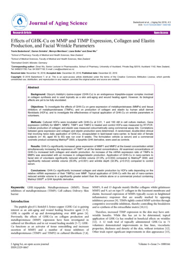
Kim et al. BMC Complementary and Alternative Medicine (2016) 16:116Statistical analysisResults are presented as means S.E.M. Statistically significant differences between groups were determinedwith one way analysis of variance (ANOVA) using statistical package for social sciences (SPSS) software. Multiple comparisons were performed using Turkey’smultiple-comparisons test. P-values 0.05 were considered to be statistically significant.ResultsEstablishment of a standard HPLC fingerprint of SMGGTThere have been several reports on the HPLC profiles ofsingle herbs included in SMGGT such as Puerarialobata Ohwi and Cimicifuga heracleifolia Kom [18, 19],but there were no HPLC profiling studies to date thathave looked at the SMGGT preparation. In order to establish a standard chromatogram for SMGGT, an HPLCstudy was performed. The representative chromatogramof SMGGT is shown in Fig. 1. Identification of majorPage 4 of 9peaks on the chromatogram was accomplished usingHPLC-ESI-MS.Identification of major components of SMGGT by HPLCESI-MSThe retention time, observed mass, mass difference,fragment ions, and proposed compounds of six peaksare listed in Table 1. The major components of SMGGTwere identified by comparing both UV and MS spectrato their spectroscopic data in the literature [20–22]. Thepeak 1 and peak 4 showed identical protonated ion ofm/z 417 in the positive ion mode but the fragment ionpeaks were differentiated as the neutral loss of 120 Da inpeak 1 (m/z 297, [M H-C4H9O4] ) attributed to thecharacteristic cleavage of C-glycosides and the neutralloss of 162 Da in peak 4 (m/z 255, [M H-C6H10O5] )attributed to the characteristic cleavage of O-glycosides.Based on the molecular ions and their fragmentationpatterns, peak 1 and peak 4 were identified as puerarinFig. 1 HPLC chromatogram of the SMGGT and UV-Visible absorption spectra of the six major peaks
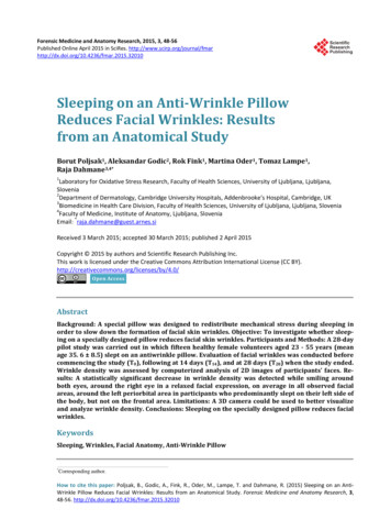
Kim et al. BMC Complementary and Alternative Medicine (2016) 16:116Page 5 of 9Table 1 The observed and calculated mass numbers of HPLC peaks of SMGGTPeak No. RT (min) Theoretical mass [M H] Observed Mass [M H] Mass difference (mmu) Fragment ions IdentificationReference151.93417.11854417.11338 16 5.94429.11301327.079943'-methoxypuerarin [20]464.31417.11854417.11534 3.20255.05128daidzin[21]567.74195.06573195.05935 6.38177.05273149.05859isoferulic acid[22]673.83419.13416419.13338 ) and daidzin (daidzein-7-Oglucoside), respectively. Likewise, the peak 3 showedprotonated ion at m/z 447 and the neutral loss of120 Da same as in peak 1. Based on the retentiontime, MS spectrum, and 30 Da difference compare tothe protonated puerarin ion at m/z 417, peak 3 wasidentified as 3’-methoxylpuerarin. The peak 2 represented protonated ion at m/z 481 and fragmented ionat m/z 179 due to the loss of single glucose and benzoic acid which specified it to be paeoniflorin. Thepeak 6 showed protonated ion at m/z 419 and fragmented ion at m/z 257 attributed to the loss of a glucose molecule and it was identified as liquiritin(liquiritigenin-4'-O-glucoside). Six major phytochemicals in SMGGT were identified as follows: isoferulicacid (from Cimicifugae Rhizoma), puerarin, 3'-methoxypuerarin, daidzin (from Puerariae Radix), paeoniflorin (from Paeoniae Radix), and liquiritin (fromGlycyrrhizae Radix). The identities of KFDA- certifiedherbal medicines were reconfirmed by HPLC-ESI-MSto ensure the quality of the herbal medicines contained in SMGGT.Cell viability(7.25 ng/mL) was slightly increased compared withvehicle-treated control from non-irradiated fibroblasts(6.65 ng/mL) and both values were decreased dose dependently as treated SMGGT concentration increased(Fig. 3a). The non-UVB irradiated cells, SMGGT significantly decreased MMP-1 levels in a dose-dependent manner by 2.3 %, 16.3 %, and 20.5 % at concentrations of 100,200, and 400 μg/mL percentile on the vehicle-treatedgroup. The positive control, 200 μg/mL of L-ascorbic acid,decreased MMP-1 by 4.0 % which is similar to the 100 μg/mL of SMGGT. In a way, the UVB irradiated cells alsodose-dependently decreased MMP-1 levels by 5.6 %,15.2 %, and 17.1 % at concentrations of 100, 200, and400 μg/mL, respectively (p 0.001). The L-ascorbic aciddecreased MMP-1 by 7.8 % similar to the 100 μg/mL ofSMGGT.Promotion of type-1 procollagen synthesisTo evaluate the effects of SMGGT on type-1 procollagensynthesis, cells were cultured with SMGGT or L-ascorbicacid for 48 h and then the culture medium was collected.UVB irradiation significantly decreased the levels of newlysynthesized type-1 procollagen compared with those ofUVB-non-irradiated cells. As shown in Fig. 3b, SMGGTThe cells were treated with various concentrations (0,100, 200, 400, and 500 μg/mL) of SMGGT water extractand for 48 h. The SMGGT extract did not induce anycytotoxicity up to 400 μg/mL concentrations but showedslight reduction in cell viability (89.4 %) at 500 μg/mL(Fig. 2). The SMGGT water extract did not show any effects on cell proliferation (data not shown). Therefore,three different concentrations (100, 200 and 400 μg/mL)of SMGGT were used in this study.Inhibitory effects on MMP-1 productionTo examine inhibitory effects of SMGGT on MMP-1, cellswere cultured with SMGGT or L-ascorbic acid (positivecontrol) for 48 h and then the cell-free supernatants werecollected to quantify the level of MMP-1. The study wasdivided into two groups that non-UVB irradiated fibroblasts and UVB irradiated fibroblasts. The level of MMP-1in vehicle-treated control from irradiated fibroblastsFig. 2 Cell viability test of SMGGT in human skin dermal fibroblastcells. Human dermal fibroblasts were cultured in DMEM until 80 %confluence. Cells were treated with various concentrations ofSMGGT water extract for 48 h, after which the MTT assay wasperformed. Data are expressed as a percentage of the control(without SMGGT). Each evaluation was performed in triplicate
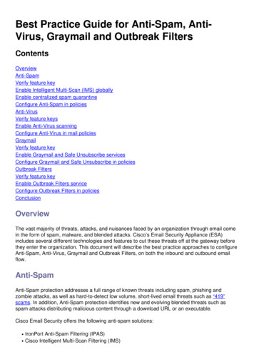
Kim et al. BMC Complementary and Alternative Medicine (2016) 16:116Page 6 of 9Fig. 3 Effects of SMGGT on MMP-1 production (a) and type-1 procollagen synthesis (b) in UVB-non-irradiation or UVB-irradiation (40 mJ/cm2)group of human dermal fibroblasts. Cells were treated with various concentrations of SMGGT water extract or L-ascorbic acid for 48 h, and thencell supernatants were collected for ELISA kit. Statistical significance for the results were given as *p 0.05; **p 0.01 and ***p 0.001. L-Ascorbicacid was used as a positive control and the results were normalized with cell numbers that were confluentsignificantly increased type-1 procollagen synthesis in adose-dependent manner by 2.2 %, 19.7 % and 34.8 % (percentile versus vehicle-treated group) at concentrations of100, 200, and 400 μg/mL in UVB-non-irradiated groups,respectively (p 0.001). The UVB-irradiated groups alsoshowed a dose-dependent synthesis promotion of type-1procollagen by the treatment with SMGGT; 19.7 %,33.8 %, and 39.4 % increase at concentrations of 100, 200,and 400 μg/mL, respectively, and these effects were muchpotent as positive control, 200 μg/mL of L-ascorbic acid,which increased the level by 23.6 % versus vehicle-treatedgroup. As a result, we concluded that the inhibition ofMMP-1 production and promotion of type-1 procollagensynthesis by SMGGT were superior to the effects of L-ascorbic acid and these effects were more prominent whenUVB was exposed. Similar pattern was observed withother herbal extract in previous report [23]. Therefore, wemeasured the changes of MMP-1 and type-1 procollagenexpression by the treatment of SMGGT water extract inboth UVB-non-irradiated and UVB-irradiated group toconfirm the effects in molecular level.MMP-1 and Type-1 procollagen expressionTo determine the anti-wrinkle effects of SMGGT waterextract, the expression of MMP-1 (Fig. 4a) and type-1procollagen (Fig. 4B) were evaluated in human dermal fibroblasts using Western Blot analysis. After UVirradiation, the cells were incubated with 0, 100, 200,and 400 μg/mL of SMGGT for 1 day. As shown inFig. 4a, SMGGT dramatically inhibited UVB-inducedMMP-1 expression; the MMP-1 expression level decreased to 30.7 % at 200 μg/mL and 30.9 % at 400 μg/mL compared with the UVB-irradiation control group.Figure 4b showed that SMGGT significantly promotedthe expression of Type-1 procollagen in UVB-irradiatedhuman skin fibroblast cells in a dose-dependent manner;the type-1 procollagen expression level increased to24.7 % at 100 μg/mL, 31.6 % at 200 μg/mL and 32.7 % at400 μg/mL.DiscussionSeungma-Galgeun-Tang (SMGGT) is a traditional herbalmedicine in China and Korea that is prepared by boiling
Kim et al. BMC Complementary and Alternative Medicine (2016) 16:116Page 7 of 9Fig. 4 Effects of SMGGT water extract on UVB-induced expression of MMP-1 (a) and type-1 procollagen (b) in human skin dermal fibroblast cells.After UVB-irradiation (40 mJ/cm2), cells were cultured with or without various concentrations of SMGGT for 24 h. Each evaluation was performedin triplicate. Data are expressed as means S.E.M. #p 0.05 and ##p 0.01 vs. vehicle control; *p 0.05 and **p 0.01 vs. UVB-irradiation onlyfour medicinal herbs, Cimicifugae Rhizoma, PuerariaeRadix, Paeoniae Radix, and Glycyrrhizae Radix in water.This medicine has traditionally been used to treat epidemic diseases such as smallpox and measles, as well asurticaria by dispelling the superficial muscles, promotingeruption, tonifying the blood, and clearing away heat viadetoxification in inflammatory diseases [1]. Through arecent survey on Korean traditional physicians, a newclinical application has been proposed for this preparation in several Korean traditional medicine hospitals asa preventative agent for excess wrinkle formation. Furthermore, Puerariae Radix was reported to increasemRNA expression for type-1 collagen in humanosteoblast-like SaOS-2 cells [24] and Cimicifugae Rhizoma has been known to inhibit matrix proteinases onosteoarthritis [25]. The present study aimed to investigate the effects of SMGGT extract, which includes Puerariae Radix and Cimicifugae Rhizoma, on the anti-agingpotential in human dermal fibroblasts and to providemolecular evidences for its anti-wrinkle efficacy inclinique.The skin aging process can be classified into intrinsicaging, a natural course determined by internal geneticfactors, and extrinsic aging, caused by external factorssuch as sun exposure, gravity, and smoking. Extrinsicaging caused by sunlight has been termed photo-aging[26]. UVB irradiation (290–320 nm) is the primary factor contributing to biological reactions in the skin, inducing inflammatory responses, apoptosis, and subsequentskin damage [27]. Interestingly, Seungma-Galgeun-Tang(SMGGT) had been reported to have anti-inflammatoryeffects in the skin [28]. In this study, we demonstratedthe anti-aging activity of SMGGT water extract in human dermal fibroblast cells compared with those of L-ascorbic acid as a positive control. It has been reportedthat L-ascorbic acid, one of vitamin-C, is a potent antiphotoaging substance which is able to enhance collagensynthesis and inhibit MMP-1 expression [29]. UV irradiation induces photo-damage and causes premature skinaging [30]. Among the factors responsible for mediatingUV-induced skin aging are matrix metalloproteinases(MMPs), which are up-regulated in dermal fibroblasts byUV irradiation [31]. UVB irradiation induces the expression of MMPs in human dermal fibroblasts which leadsto the breakdown of collagen and other extracellularmatrix proteins and causes pre-mature aging (photoaging) of human skin [32]. Therefore, the inhibition ofthis increase in MMP expression has been reported toimprove UV-induced photo-aging in terms of protectionfrom collagen degradation [30]. As shown in Figs. 3 and4, SMGGT water extract treatment significantly inhibited the production of MMP-1 and promoted the production of the type-1 procollagen in human dermalfibroblast cells, which provides strong support for theclinical efficacy for SMGGT. This was reconfirmed bythe quantitation of MMP-1 and type-1 procollagen protein expression in UVB-irradiated human fibroblasts.The dose-dependent inhibition of MMP-1 expressionand promotion of type-1 procollagen expression by thetreatment with SMGGT demonstrates the clinical efficacy of this preparation for skin aging caused by extrinsic stresses including sunlight.
Kim et al. BMC Complementary and Alternative Medicine (2016) 16:116Page 8 of 9The molecular mechanism underlying the protectiveeffects of SMGGT on UVB-irradiated skin aging werenot revealed in this pilot study. Since collagen isknown to be synthesized from dermal fibroblasts asprecursor molecules called procollagen which is regulated by transforming growth factor β2 (TGF- β2)and activator protein-1 (AP-1), a transcription factorconsisting of c-Jun and c-Fos promoting collagenbreakdown by up regulating matrix metalloproteinases(MMPs) [33, 34], the potential molecular targets ofSMGGT would be some of these important cytokinesand transcription factor.Authors’ contributionsCYB, GJY, MKK, HYK, SYC and YPJ participated in the design of this study. Theexperiments and writing the manuscript were done by CYB, HYK, and MKK.YPJ supervised the work and reviewed the draft and final manuscripts andinterpretation of results. All authors read and approved the final manuscriptfor submission.ConclusionsIn present study, a new application of the traditionalherbal preparation SMGGT was tested by analyzing itseffects on two major contributors to skin aging, MMP-1and type-1 procollagen. The protective effect of SMGGTon UVB-irradiated skin aging in human fibroblast cellssupports the clinical efficacy of SMGGT and suggeststhat this herbal preparation could be a potential candidate for an active ingredient in cosmeceutical productsthat prevent and cure wrinkle formation.Ethics approval and consent to participatePrimary human foreskin dermal fibroblasts were established from biopsies of healthy male donor of 22 yearsold and in accordance with Institutional Review Board(IRB) approved by the Kyung Hee University Hospital(Seoul, Korea) (IRB approval no. 2012-01-006). The hospital obtained written informed consent from the donor,giving permission to collect their tissue and use for research purposes. The research adhered to the tenets ofthe Declaration of Helsinki.Consent for publicationNot applicable.Availability of data and materialsThe datasets supporting the conclusions of this articleare included within the article.AbbreviationsANOVA: one way analysis of variance; AP-1: activator protein-1; DMEM: Dulbecco’smodified eagle medium; DMSO: dimethyl sulfoxide; EDTA: ethylenediaminetetraacetate; ELISA: enzyme-linked immunosorbent assay; HPLC-ESI-MS: highperformance liquid chromatography-electro spray ionization-mass spectrometry;KFDA: Korea Food and Drug Administration; MMP-1: matrix metalloproteinase-1;MTT: um bromide; NF-κB: nuclearfactor kappa-light-chain-enhancer of activated B cells;PMSF: phenylmethanesulfonyl fluoride; SMGGT: Seungma-Galgeun-Tang;SPSS: statistical package for social sciences; UVB: ultraviolet light B.Competing interestsThe authors declare that they have no competing interests.AcknowledgementsWe would like to thank Editage (www.editage.co.kr) for editing andreviewing this manuscript for English language.FundingThe funding needed for the design of the study and collection, analysis, andinterpretation of data and in writing the manuscript was supported by agrant from t
prevention of wrinkle formation (unpublished clinical report). Wrinkle formation is one of the primary characteris-tics of skin aging, and is a complex process that involves * Correspondence: ypjang@khu.ac.kr; sychoung@khu.ac.kr 1Division of Pharmacognosy, College of Pharmacy, Kyung Hee University, Hoegi-dongDongdaemun-gu, Seoul 130-701, South .