Transcription
Published online: 2019-10-01THIEME166Filling AbilityUsing Two Carrier-Based TechniquesOriginalArticleMancino et al.Microscopic and Chemical Assessments of theFilling Ability in Oval-Shaped Root Canals Using TwoDifferent Carrier-Based Filling TechniquesDavide Mancino1,2Naji Kharouf1,2Joseph Hemmerlé1,21 Department of Endodontics, Faculty of Dental Medicine,Strasbourg University, Strasbourg, France2 UMR-S 1121 Inserm, Biomaterials and Bioengineering, Strasbourg,FranceYoussef Haïkel1,2Address for correspondence Davide Mancino, DDS, PhD,1 rue place de l’Hôpital, Departement of Endodontics,faculty of Dental Medicine, 67000 Strasbourg, France(e-mail: endodontiefrancaise@outlook.com).Eur J Dent 2019;13:166–171AbstractKeywords carrier-basedtechniques filling ability GuttaCore oval-shaped canals ThermafilObjectives The aim of this study was to assess the filling ability in oval-shaped canalsusing two different carrier-based filling techniques.Materials and Methods Twenty-four human mandibular premolars with one ovalcanal were selected. Canals were shaped using WaveOne Gold Primary and ProGlider.Samples were divided into two groups and filled as follows: Thermafil and GuttaCore.The proportions of gutta-percha-filled areas (GPFAs), sealer-filled areas (SFAs), andvoid areas (VA), at 2 and 5 mm, were analyzed using optical numeric microscope, scanning electron microscope, and energy-dispersive X-ray.Statistical Analysis Data were compared by Kruskal–Wallis one-way analysis of variance on ranks, with statistical significance set at α 0.05.Results At 2 and 5 mm distances from the apex, this study discloses no statisticallydifferent filling ability for the two techniques. Concerning each sample treated usingboth filling systems, the presence of tags was visualized. At working length (WL)-5,and WL-2, the maximum tag penetration depth for the GuttaCore group into the dentinal tubules was, respectively, 96 μm and 48 μm, whereas the values in the thermafilgroup were 109 μm and 55 μm, respectively.Conclusions Our results clearly show that Thermafil and GuttaCore can fill ovalshaped canals in appropriate way. Furthermore, we can state that the GuttaCore obturator allows to preserve the same filling ability than Thermafil obturator, in view ofthe fact that there was no difference, in terms of GPFA, SFA, and VA between the twodifferent carrier-based obturation techniques.IntroductionA successful root canal treatment depends on appropriateaccess cavity preparation, suitable shaping, proper cleaning, and tridimensional filling of the root canal system.1-9Although literature highlights the importance of shapingand cleaning step during the endodontic therapy, there is noevidence other than clinical feeling to support or deny anyparticular filling technique.10-14 Thus, the role played by thefilling step is inevitably underestimated. Often the choice ofDOI https://doi.org/10.1055/s-0039-1695661ISSN 1305-7456.a filling technique is based on factors such as speed, simplicity, or economics, rather than on the endodontic anatomyor on other quantitative parameters.15 Without the slightest doubt, a tridimensional shaping and cleaning of the rootcanal system are essential requirements to perform a faultless filling.13,15The root canal filling should be regarded as the finalstep of the disinfection. No cleaning technique allows toeradicate 100% of bacteria present in an infected root canalsystem.16-23 2019 Dental InvestigationSociety
Filling Ability Using Two Carrier-Based TechniquesThe task of canal filling must allow to entomb the remaining bacteria, to ensure the periapical healing as long as possible.24 To provide a tridimensional seal, the root canal filling involves the use of gutta-percha in combination witha root canal sealer. The sealer is used to fill the voids andgaps remaining between the gutta-percha material and theroot canal walls, like a gasket, especially in oval- or irregular-shaped root canals. On the other hand, the sealer represents the weak link in the chain of root canal filling for threemain reasons: first of all, for its setting reaction with subsequent shrinkage, then for its intrinsic porosities and at last forits possible reabsorption over time.15,19 These factors are likelythe cause of void areas’ (VA) formation that, of course, constitute a relevant problem in terms of sealing. Hence, the sealer thickness should be as smaller as possible. In this respectwarm gutta-percha obturation techniques, and in particularThermafil system (Dentsply Sirona), have been developed toperform tridimensional root canal filling with a smaller sealerthickness.11,12 However, the presence of its plastic carrier couldbe a real problem. First, the clinicians, who lack of the basicprinciples for use Thermafil obturator, could perform a rootcanal filling with a naked plastic carrier in the last millimeterof the canal.25,26 Second, the preparation of the postspace, ifnecessary, in a canal filled with a Thermafil obturator couldbe very difficult and could cause a postoperative perforationas result of a lack of attention or experience on the part ofthe clinician. Third, the retreatment management in a toothfilled with a Thermafil obturator could be very difficult for ageneral practitioner. For these reasons, we decided to test twodifferent techniques of carrier-based obturation: thermafiland GuttaCore (Dentsply Sirona). GuttaCore is a carrier-basedobturator and it has emerged as an effective filling system.Materials and MethodsThis study was revised and approved by the Ethics Committee of Medical, Odontology school, and Strasbourg UniversityHospital (protocol no. 2018–89).A total of 87 mandibular first premolars, freshly extracted for orthodontic reasons, with a total length between20 and 22 mm, were selected. Informed consent wasobtained from all patients, and all the procedures wereperformed according to the Helsinki Declaration of 1975.After debridement of the root surface, specimens wereimmersed in a 1% sodium hypochlorite (NaOCl) solution at4 C for 24 hours and then stored in saline solution.Periapical radiographs were taken, placing the premolarsaccording to their buccolingual and mesiodistal diameter topreselect the teeth with a long/short diameter ratio 1.5 at5 mm from the apex. Preselected teeth were then scannedwith cone-beam computed tomography (CBCT) to select theteeth with the following morphological parameters: Presence of one canal A real oval canal at 5 mm from the apex with a long/short diameter ratio 1.5 at 5 mm from the apex Canal with a total length of 10 2 mm from canal orificeto apical foramenMancino et al. 10 to 30 primary root curvature in buccolingual andmesiodistal view according to the Schneider method3 Main curvature radius 4 mm4 Therefore, only 28 mandibular first premolars ensuredinclusion criteria, 4 were discarded because of severecalcification of the root canal, leaving a total of 24 teeth The tooth length was standardized to 18 mm by removing the coronal portion of the teeth Teeth having canals with apical diameters larger thansize 20 were discarded.Shaping ProcedureSpecimens were mounted on Protrain (Simit Dental srl, Mantova, Italia) to perform endodontic therapy simulating clinical situation.After access cavity preparation, all root canals wereshaped, keeping the pulp chamber constantly flooded with6% sodium hypochlorite. An experienced endodontist shapedeach premolar. Canal scoutings were performed with a #10K file at a working length (WL) 0.5 mm. Length determination was obtained using an electronic apex locator (Root ZX; JMorita Co, Kyoto, Japan). Then, the apex was dried and sealedwith a cyanoacrylate glue to simulate a closed system.Mechanical glide path was performed using ProGlider(Dentsply Sirona) and an endodontic engine (X-Smart-IQmotor, Dentsply Sirona) at 300 rpm and 4 Ncm at the full WL.Each tooth was shaped at the WL, using WaveOne GoldPrimary (Dentsply Sirona) (0.25 tip, 0.07 taper) and anX-Smart-IQ motor (300 rpm, 4 Ncm).New instruments were used to shape each premolar, andinstruments were removed from the canal and cleaned afterevery two pecking motions until the WL was reached.Recapitulation was performed between each instrument,taking a #10 K file to WL 0.5 mm.After the shaping procedure, to assure a tridimensionalcleaning of the root canal system, an aqueous 17% solutionof ethylenediaminetetraacetic acid was flooded into the pulpchamber and was then activated using a dynamic activationby EndoActivator (Dentsply Sirona) for 120 seconds in eachcanal. After rinsing with physiological saline, a 6% solutionof NaOCl flooded into the pulp chamber was activated usingEndoActivator for 120 seconds in each canal. Again, eachcanal was rinsed using physiological saline and then driedusing sterile paper points.The prepared teeth were randomly divided in two equalgroups (n 12 per group): thermafil group (TG) and GuttaCore group (GG).Thermafil System GroupAfter application of a standardized drop (0.230 0.005 g) ofAH Plus sealer (Dentsply Sirona) with a coated sterile paperpoint at the entrance of each canal, a second sterile paperpoint, taken to WL, was used to remove any excess of sealerand smoothly distribute the sealer in the whole canal system.We placed the silicone stopper at the established WL on theprimary WaveOne Gold Thermafil.A Thermafil obturator (Dentsply Sirona) was heated in aThermaPrep Plus oven until the first “beep” signal, indicatingEuropean Journal of Dentistry Vol. 13No. 2/2019167
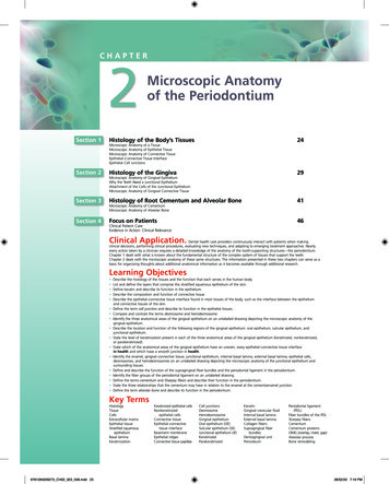
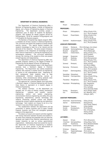
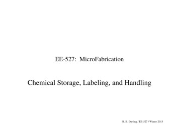
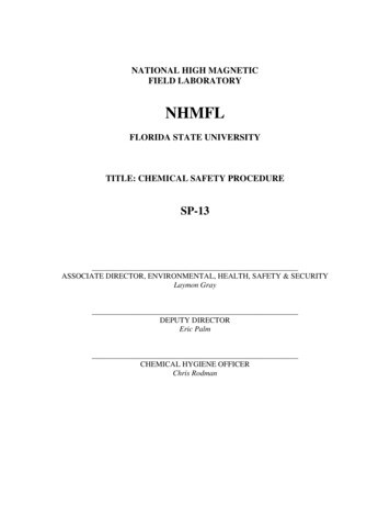
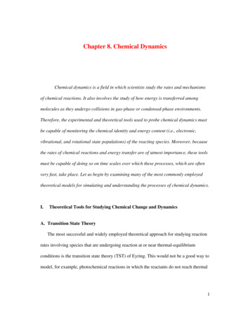
168Filling Ability Using Two Carrier-Based TechniquesMancino et al.it is ready for use. The obturator was then directly insertedinto the canal orifice with gentle apical pressure, withouttwisting or forcing, to the WL.GuttaCore Pink Obturator GroupAfter application of a standardized drop (0.230 0.005 g) ofAH Plus sealer (Dentsply Sirona) with a coated sterile paperpoint at the entrance of each canal, a second sterile paperpoint, taken to WL, was used to remove any excess of sealerand smoothly distribute the sealer in the whole canal system.With the help of the calibration rings on the WaveOne obturator, we placed the silicone stopper at the established WL.A WaveOne Gold obturator (Dentsply Sirona) was heatedin a GuttaCore Oven (Dentsply Sirona) until the first “beep”signal, indicating it is ready for use. The obturator was thendirectly inserted into the canal orifice with gentle apicalpressure, without twisting or forcing, to the WL.Sectioning and Surface MeasurementsAfter embedding in Epon 812 resin (Euromedex; Souffelweyersheim, France), two sections were made perpendicular tothe longitudinal axis of the root tooth at 2 and 5 mm from theapex with a wire saw (Walter EBNER; Le Locle, Switzerland).Thereafter, 1200, 2400, and 4000 P-grade (number of abrasive grains per cm2) abrasive papers were used successively topolish the surface. All samples were etched with a 20% (w/w)citric acid solution for 2 minutes (Sigma-Aldrich, Darmstadt,Germany), thoroughly rinsed with distilled water, and finallydehydrated in a graded series of ethanol solutions.compared by Kruskal–Wallis one-way analysis of variance onranks, with statistical significance set at α 0.05.ResultsOptical Microscope ObservationsFor both groups, the superficies of gutta-percha-filled areas(GPFAs), sealer-filled areas (SFAs), and voids, that is, unfilledareas (VA) in the canal at WL-5 and WL-2 were observed andmeasured. All measurements are expressed in mean standard error on the mean (n 12).At WL-5, concerning TG group, the VA was 0.22 0.001%,the SAF was 0.74 0.003%, and the GPFA was 99.04 0.02%.Concerning the GG, the VA was 0.20 0.001%, the SAF was0.61 0.003%, and the GPFA was 99.19 0.003%.At WL-2, concerning TG group, the VA was 0.13 0.004%,the SAF was 1.54 0.01%, and the GPFA was 98.33 0.04%.Concerning the GG, the VA was 0.15 0.001%, the SAF was1.09 0.004%, and the GPFA was 98.76 0.004%.Scanning Electron Microscope ResultsSEM analyses ( Figs. 1 and 2) confirmed the results obtainedby optical microscopy. Furthermore, concerning each sample treated using both filling systems, the presence of tagswas visualized ( Figs. 3 and 4). At WL-5 and WL-2, themaximum tag penetration depth for the GuttaCore into theOptical Numeric Microscope AnalysisSamples were first observed using an optical numeric microscope (KEYENCE; Osaka, Japan) and then analyzed usingthe VHX-5000 Communication software (KEYENCE; Osaka,Japan) to measure the quantity of voids and sealer filledareas.Scanning Electron Microscope AnalysisSpecimens were prepared for scanning electron microscopy(SEM) and sputter-coated with a gold-palladium alloy (20/80weight%) by using a HUMMER JR sputtering device (Technics;California, United States). The coated samples were observedwith a Quanta 250 FEG SEM (FEI Company, Eindhoven, theNetherlands) operating with an accelerating voltage of theelectrons of 10 kV.Fig. 1 Scanning electron micrograph of sectioned root at 5 mm fromthe apex filled with Thermafil obturator.Energy Dispersive X-Ray AnalysisThe specimens also underwent chemical analysis to analyzethe GuttaCore material and the related tags. Energy dispersive X-ray (EDX) microanalysis was performed with an EDAXOctane Plus spectrometer (AMETEK Materials Analysis Division, 91 McKee Dr, Mahwah, New Jersey, United States).Statistical AnalysisCollected data were analyzed with SigmaPlot release 11.2(Systat Software, Inc., San Jose, California, United States). Theproportions of voids and sealer areas corresponding to oneof two treatments GuttaCore (GC) or Thermafill (TH) wereEuropean Journal of Dentistry Vol. 13No. 2/2019Fig. 2 Scanning electron micrograph of sectioned root at 5 mm fromthe apex filled with GuttaCore obturator.
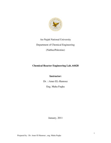
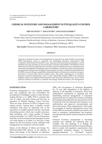
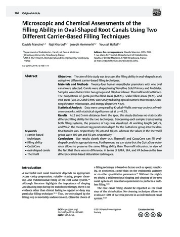
Filling Ability Using Two Carrier-Based TechniquesMancino et al.Chemical Analysis FindingsA spectrum taken from a tag at WL-5 and WL-2 for the twogroups was likened at a spectrum of Thermafil ( Fig. 5) andGuttaCore gutta-percha ( Fig. 6) and at a spectrum of Ahplus sealer showing their chemical analysis. This allowed usto affirm that the TG and GG tags consist of gutta-percha.DiscussionFig. 3 Scanning electron micrograph showing the penetration of thegutta-percha in the dentinal tubules on extracted tooth filled with aThermafil obturator.Fig. 4 Scanning electron micrograph showing the penetration of thegutta-percha in the dentinal tubules on extracted tooth filled with aGuttaCore obturator.dentinal tubules was, respectively, 96 and 48 μm, whereasfor the Thermafil the maximum tag penetration depth was,respectively, 109 μm and 55 μm. Furthermore, the shape ofthe tags for GG and TG was different; rectangular for GC andcircular for TG.To perform tridimensional filling, the essential requirementsare a previous tridimensional shaping and tridimensional cleaning. Obviously, this task is much more difficult inthe case of oval-shaped root canals. To fill suitably an ovalshaped root canal, the void and the sealer areas should be assmall as possible, and this is possible at the condition thatgutta-percha is thermoplastic.The present study was designed to quantify the GPFA, SFA,and VA on the teeth filled using two different techniques ofcarrier-based obturation, Thermafil and GuttaCore, with thegoal to provide a measure of quality. The Thermafil obturatorconsists of two parts: the plastic carrier, derivative of polysulfone; and the gutta-percha, whereas GuttaCore consists ofa carrier manufactured from a cross-linked, thermoset elastomer of gutta-percha coated in gutta-percha. The presenceof the plastic carrier in a Thermafil obturator could makea retreatment and/or the postspace preparation a difficulttask.25,26 What’s more, the clinicians who lack the basic principles for use Thermafil obturator could perform a root canalfilling with a naked plastic carrier in the last millimeter ofthe canal.Indeed, the GuttaCore carrier was designed to facilitateremoval during retreatment and/or postspace preparation byfast, simply, and safe trephining through the core.27-29Moreover, from a clinical point of view, our internal data(preclinical hands-on on different filling techniques, Strasbourg University) showed that the learning curve to master the GuttaCore filling is faster than the learning curveto master the Thermafil technique, with less possibility ofFig. 5 Energy-dispersive X-ray spectrum of the Thermafil material. Major peak can be seen indicating C and smaller peaks indicating O, Zn,and Br.European Journal of Dentistry Vol. 13No. 2/2019169
170Filling Ability Using Two Carrier-Based TechniquesMancino et al.Fig. 6 Energy-dispersive X-ray spectrum of the GuttaCore material. Major peaks can be seen indicating C, Zn and O, and smaller peak indicating Si.Fig. 7 Scanning electron micrograph showing the external surfaceof GuttaCore with the imprint of the dentinal tubules after previousdecalcification and removal of dental tissues.extrusion. In other words, the GuttaCore technique is lesstechnique sensitive and less difficult to master comparedwith the Thermafil technique.To reduce the possible errors, we decided to analyze eachspecimen using different instruments. At first, we choose thepremolars by using radiographic and CBCT control images todemonstrate the good sampling homogeneity.Afterward, specimens were observed using an opticalnumeric microscope at a magnification of 500. They werealso observed by using SEM at magnifications up to 30,000,using direct observations and not replicates. Finally, we useda last control by using EDX analyses to know exactly the composition of the different filled areas, including tags.At 5 and 2 mm distance from the apex, each specimen ofboth groups showed plasticized gutta-percha. No statisticaldifference was found for the GPFA, VA, and SAF values corresponding to the two tested thermo-plasticizing techniques.Consequently, the null hypothesis, that there are no differences, at 5 and 2 mm from the apex, in the percentagesof GPAF, VA, and SAF in oval-shaped root canals, filled bythe Thermafil obturator and GuttaCore obturator using AHplus sealer was accepted. SEM micrographs, at 2 and 5 mmEuropean Journal of Dentistry Vol. 13No. 2/2019distances from the apex, disclosed the presence of tags forthe GG, filling dentinal tubules. Moreover, SEM observationsrevealed the presence of tags for both groups. Gutta-perchatags ( Fig. 7) could be able to block physically potentiallyremaining bacteria, so entombing them.Chemical microanalyses allowed us to conclude that thetags of both groups were composed of gutta-percha material.Of course, the present findings depend on the employedgutta-percha product qualities. Thermafil and GuttaCoregutta-percha is breakable at the solid state, but it changeson gluey, swollen proprieties when heated. The flowabilityof GC gutta-percha is higher than that of the standard gutta-percha of gutta-percha cones.30At 2 and 5 mm distances from the apex, the present resultsreveal no statistically different filling ability for the two tested carrier-based techniques.Moreover, at 2 and 5 mm distances from the apex, for eachspecimens treated using the two different filling techniques,the presence of gutta-percha tags was visualized.Our results clearly show that the GuttaCore obturationcan fill oval-shaped canals in an excellent way. Hence, wecan state that GuttaCore system would be a filling techniqueless sensitive and less difficult to master compared withthe Thermafil technique, preserving the same filling ability,in view of the fact that there was no difference, in terms ofGPFA, SFA, and VA between the two different carrier-basedobturation techniques.FundingNone.Conflict of InterestNone declared.References1 Krasner P, Rankow HJ. Anatomy of the pulp-chamber floor.J Endod 2004;30(1):5–162 Boveda C, Kishen A. Contracted endodontic cavities: the foundation for less invasive alternatives in the management of apical periodontitis. Endod Topics 2015;33:169–186
Filling Ability Using Two Carrier-Based Techniques3 Eaton JA, Clement DJ, Lloyd A, Marchesan MA. Micro-computed tomographic evaluation of the influence of root canal system landmarks on access outline forms and canal curvaturesin mandibular molars. J Endod 2015;41(11):1888–18914 Alovisi M, Pasqualini D, Musso E, et al. Influence of contractedendodontic access on root canal geometry: an. in vitro. study.J Endod 2018;44(4):614–6205 Mancino D, Kharouf N. Root canal treatment of dilaceratedsecond maxillary premolars: planning the shaping procedure.J Clin Exp Dent 2018;10(6):e624–e6276 Zehnder M. Root canal irrigants. J Endod 2006;32(5):389–3987 Gu LS, Kim JR, Ling J, Choi KK, Pashley DH, Tay FR. Reviewof contemporary irrigant agitation techniques and devices.J Endod 2009;35(6):791–8048 Kanisavaran ZM. Chlorhexidine gluconate in endodontics: anupdate review. Int Dent J 2008;58(5):247–2579 Mohammadi Z, Abbott PV. The properties and applications ofchlorhexidine in endodontics. Int Endod J 2009;42(4):288–30210 Wu M, van der Sluis LW, Wesselink PR. A preliminary studyof the percentage of gutta-percha-filled area in the apicalcanal filled with vertically compacted warm gutta-percha. IntEndod J 2002;35(6):527–53511 De-Deus G, Gurgel-Filho ED, Magalhães KM, Coutinho-FilhoT. A laboratory analysis of gutta-percha-filled area obtainedusing Thermafil, System B and lateral condensation. Int EndodJ 2006;39(5):378–38312 De-Deus G, Maniglia-Ferreira CM, Gurgel-Filho ED, PaciornikS, Machado AC, Coutinho-Filho T. Comparison of the percentage of gutta-percha-filled area obtained by Thermafil and System B. Aust Endod J 2007;33(2):55–6113 Ørstavik D. Materials used for root canal obturation: Technical,biological and clinical testing. Endod Topics 2005;12:25–3814 Peng L, Ye L, Tan H, Zhou X. Outcome of root canal obturationby warm gutta-percha versus cold lateral condensation: ameta-analysis. J Endod 2007;33(2):106–10915 Whitworth J. Methods of filling root canals: principles andpractices. Endod Topics 2005;12:2–2416 Carver K, Nusstein J, Reader A, Beck M. In vivo antibacterialefficacy of ultrasound after hand and rotary instrumentationin human mandibular molars. J Endod 2007;33(9):1038–1043Mancino et al.17 Bukiet F, Couderc G, Camps J, et al. Wetting properties and critical micellar concentration of benzalkonium chloride mixed insodium hypochlorite. J Endod 2012;38(11):1525–152918 Park E, Shen YA, Haapasalo M. Irrigation of the apical rootcanal. Endod Topics 2012;27:54–7319 Wang Z, Shen Y, Haapasalo M. Effectiveness of endodontic disinfecting solutions against young and old Enterococcus faecalis biofilms in dentin canals. J Endod 2012;38(10):1376–137920 Ordinola-Zapata R, Bramante CM, Cavenago B, et al. Antimicrobial effect of endodontic solutions used as final irrigants on adentine biofilm model. Int Endod J 2012;45(2):162–16821 Haapasalo M, Endal U, Zandi H, Coil JM. Eradication of endodontic infection by instrumentation and irrigation solutions.Endod Topics 2005;10:77–10222 Haapasalo M, Shen YA. Current therapeutic options for endodontic biofilms. Endod Topics 2012;22:79–9823 Haapasalo M, Qian W, Shen YA. Irrigation: beyond the smearlayer. Endod Topics 2012;27:35–5324 Sundqvist G, Figdor D, Endodontic Treatment of Apical Periodontitis. In: Ørstavik D, Pitt Ford TR, eds. Essential Endodontology: Prevention and Treatment of Apical Periodontitis.Oxford: Blackwell; 199825 Wilcox LR. Thermafil retreatment with and without chloroform solvent. J Endod 1993;19(11):563–56626 Wong AW, Zhang S, Li SK, Zhang C, Chu CH. Clinical studies oncore-carrier obturation: a systematic review and meta-analysis. BMC Oral Health 2017;17(1):16727 Beasley RT, Williamson AE, Justman BC, Qian F. Time requiredto remove guttacore, thermafil plus, and thermoplasticizedgutta-percha from moderately curved root canals with protaper files. J Endod 2013;39(1):125–12828 Rödig T, Wagner J, Wiegand A, Rizk M. Efficacy of the ProTaper retreatment system in removing Thermafil, GuttaCore orvertically compacted gutta-percha from curved root canalsassessed by micro-CT. Int Endod J 2018;51(7):808–81529 Schroeder AA, Ford NL, Coil JM. Micro-computed tomographyanalysis of post space preparation in root canals filled withcarrier-based thermoplasticized gutta-percha. Int Endod J2017;50(3):293–30230 Greco K, Carmignani E, Cantatore G. The Thermafil root canalobturation system. G Ital Endod 2011;25:97–109European Journal of Dentistry Vol. 13No. 2/2019171
A WaveOne Gold obturator (Dentsply Sirona) was heated in a GuttaCore Oven (Dentsply Sirona) until the first "beep" signal, indicating it is ready for use. The obturator was then directly inserted into the canal orifice with gentle apical pressure, without twisting or forcing, to the WL. Sectioning and Surface Measurements