Transcription
Glomus tumor of small intestine: case report and review of literatureYan TanWuhan UniversityXinyi WangWuhan UniversityGuifang YangWuhan UniversityLan LiuWuhan UniversityJun FangWuhan UniversityYing ChangWuhan UniversityQiu ZhaoWuhan UniversityYafei Zhang ( zhangyafei1127@163.com )Wuhan University https://orcid.org/0000-0003-3329-8029Case ReportKeywords: Glomus tumor, Small intestine, Case reportPosted Date: March 16th, 2021DOI: https://doi.org/10.21203/rs.3.rs-318644/v1License: This work is licensed under a Creative Commons Attribution 4.0 International License. Read Full LicensePage 1/7
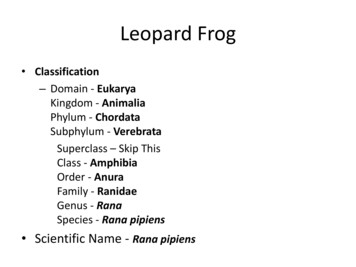
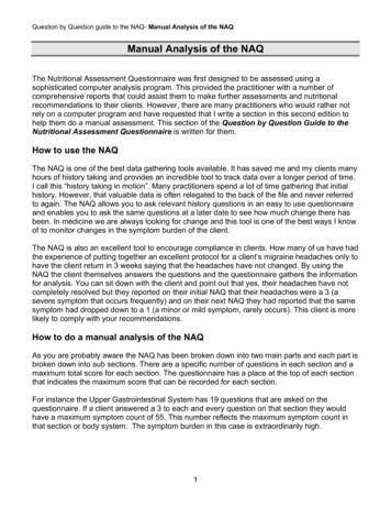
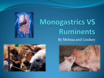
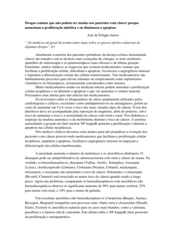
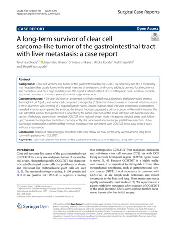
AbstractBackgroundGlomus tumors are exceedingly rare tumors arising from the normal glomus body. Only a very few cases of glomus tumors located in the small intestinewere reported to date. Here, we present a case of glomus tumor of the jejunum in a 30-year-old female patient, and discuss its clinical, imaging, andpathologic features.Case presentationA 30-year-old female referred to our hospital with the chief complain of melena and fatigue for 3 weeks. Oral single-balloon enteroscopy discovered a2.0*2.0cm tumor without epithelial lining in the jejunum. The patient then underwent partial enterectomy. HE stain illustrated that the tumor wasinterspersed with congestive capillaries of various size, and admixed with smooth muscle bundles. Immunohistochemical study showed that the tumorcells were strongly positive for SMA and collagen type Ⅳ. The Ki-67 proliferation index was less than 1% and mitotic activity is very low (about 1/50 HPF).The case was finally diagnosed as benign glomus tumor.ConclusionsGlomus tumors arising from the small intestine are extremely rare. The final diagnosis is made by histological and immunohistochemical examination.Although there is no standardized management pathway for these patients, early diagnosis and treatment are important for a good prognosis.BackgroundGlomus tumors (GTs) account for approximately 2% of all soft tissue neoplasms [1]. However, rare visceral GTs do occur. GTs within the gastrointestinaltract have been reported to usually occur at the gastric, especially gastric antrum. The vast majority of gastric GTs follow a benign clinical course, withouthistologic or clinical evidence of malignancy [2]. GTs rarely involve small intestine, with only 8 cases reported in the English literature, among which 4 casespresent malignant features [3–5]. Here, we report a patient with glomus tumor of the jejunum and review the literature on the clinicopathologic features,diagnosis, and differential diagnosis of small intestinal GTs.Case PresentationA 30-year-old female referred to our hospital with the chief complain of melena and fatigue for 3 weeks. There were no significant positive signs other thananemic appearance. Routine blood examination showed that her hemoglobin level was 6.7g/dL. Contrast-enhanced computed tomography (CT) revealedan obvious arterially enhancing mass in the small intestine and continuous enhancement in the delayed phase of enhancement (Fig. 1). Capsuleendoscopy indicated a possible mucosal eminence in the jejunum (Fig. 2a). Because the photographs captured by capsule endoscopy were limited, oralsingle-balloon enteroscopy was further applied to determine the mass. After insertion of about 250cm past pylorus, a 2.0*2.0cm tumor without epitheliallining was discovered (Fig. 2b). The patient was then transferred to the Department of Gastrointestinal Surgery and underwent a partial enterectomy.At surgery, a 3.5cm long segment of jejunum was removed. Gross pathology showed a 2.0*1.3*1.3cm gray-red polypoid protuberance in the small intestinalmucosal surface, with a 0.7cm width pedicle (Fig. 3a). HE stain illustrated that the tumor was well limited by a thin fibrous capsule, interspersed withcongestive capillaries of various size, and admixed with smooth muscle bundles (Fig. 3b and 3c). Muscularis propria and serosa didn’t show any damage.Immunohistochemical study showed that the tumor cells were strongly positive for SMA and collagen type Ⅳ (Fig. 3d and 3e), and negative for CD31, CD34,CD117, Desmin, DOG-1, CgA, Syn, S100, EMA, STAT6, β-catenin, Caldesmon and Calponin. Ki-67 proliferation index was less than 1% (Fig. 3f) and mitoticactivity is about 1/50 HPF.According to the CT results, endoscopic images and pathologic findings, the case was finally diagnosed as benign GT. Postoperatively, the gastrointestinalhemorrhage was completely resolved. And no recurrence or metastasis was observed during a follow-up period of 6 months.DiscussionGTs arising from the small intestine are extremely rare. So far, only 8 cases have been described in the English literatures [3–5]. Among them, two caseslocated at the duodenum, one case at jejunum, and five cases at the ileum. The clinicopathologic features of small intestinal GTs are summarized inTable 1. The case reported in our study is a 30-year-old female. Together with this case, the sex distribution is basically balanced (5 males and 4 females).Small intestinal GTs usually present non-specific symptoms such as abdominal pain and intestinal bleeding (melena or hematochezia). Larger tumors cancause intestinal stenosis and obstruction.Page 2/7
Table 1Clinicopathological characteristics of documented small intestinal GTsReferenceAge/SexLocationSize in meChen et icresectionSMA( ),vimentin( ),caldesmon( ),CD34( ), Ki-67 (80% ),CD117(-), desmin(-), Dog1(-), S100(-), leukocytecommon antigen m6.0*5.0SerosaLaparoscopicresectionSMA( ), Ki-67(70%),CD34( ), Nestin( ),EMA(-), CD117(-), Dog-1(-),S100(-), aLaparoscopicresectionKi-67 ( 5% )BenignFollow-up29/FIleum12.8*10.2*13.1Whole layerSurgicalresectionSMA( ), collagen typeIV( ), caldesmon( ),calponin( ), CD117(-),CD34(-), S100(-),cytokeratin(-), -)MalignantFollow-upKnackstedtet al. 2007[7]65/MDuodenumNot , CD117(-), CD56(-)BenignFollow-upShelton etal. 2007 [8]48/FAmpulla3.0Minimalinvasion ofcommonbile ductandpancreaticductWhippleNot availableMalignantUnknownGeraghtyet al. 1991[9]60/MIleum0.6SerosaSurgicalresectionSMA( ), desmin(-),chromogranin(-), euronspectificendase(-)UnknownDie of unrelatedcausesHamiltonet al. ctionNot availableUnknownUnknown2020 [3]Ma et al.2020 [4]Campanaet al.2014 [5]Abu-Zaidet al.2013 [6]Due to the deep location and non-specific symptoms, small intestinal tumors including GTs are usually hard to differentiate and diagnose. Abdominalcontrast-enhanced CT may be useful to distinguish GTs from other small intestinal tumors. According to previous description (mainly on gastric GTs),hemangioma-like globular enhancement with central fill-in and persistent homogeneous enhancement were both visualized in GTs [11]. As for the reportedcase, the tumor on contrast-enhanced CT showed homogeneous high attenuation in the arterial, venous, and delayed phases. GTs in gastric usually displaya submucosal pattern of growth. In the case reported by Knackstedt C et al., the GT located in duodenal bulb was polyp-like [7]. Shelton JH et al. reported aGT of the ampulla, in which a protruding mass was seen [8]. However, until now, there is no enteroscopic image presented in the jejunum or ileum GTs. Inthis paper, for the first time, we acquired the endoscopic imaging of small intestinal GT by balloon-assisted enteroscopy. Intriguingly, the enteroscopicimaging showed a fibrous capsule-covered mass, without any epithelial coverage, and on the surface of the tumor, no definite hemorrhage, erosions orulcers were observed. Only some minor erosions on the area around the tumor edge were seen, which may cause the persistent melena.Most GTs depend on postoperative pathological diagnosis. Under the microscope, the tumor consists of a large number of smooth muscle bundles anddilated capillaries. Immunohistochemically, tumor cells were stained positive for SMA and collagen type Ⅳ. Negative CD117 and DOG-1 expressions help inexcluding the diagnosis of gastrointestinal stromal tumors, while negative CgA and Syn expressions help in excluding neuroendocrine neoplasms [12].Criteria for malignant GTs proposed by Folpe et al. include tumors with a deep location and a size of more than 2 cm, or atypical mitotic figures, ormoderate to high nuclear grade and 5 mitotic figures/50 HPF [13]. According to the criteria, the present case was considered as low possibility ofmalignancy. And 6 months follow-up showed no indication of recurrence or metastasis after a partial enterectomy, which supporting the benign diagnosis.All the small intestinal GTs recorded in the literature have received surgical resection, but no patients received regional lymphadenectomy. After surgery,also no patients received adjuvant radiotherapy or chemotherapy. However, 2 patients developed metastases in the 8 reported cases. Therefore,standardized systemic treatment needs to be further studied. For gastric GTs, endoscopic or laparoscopic resection is recommended. Also due to the fact ofextremely rare cases of malignant GTs in gastric, no standardized neoadjuvant or adjuvant treatment was suggested. Radiotherapy or chemotherapy hasbeen utilized for the treatment of malignant GTs of head and neck. However, in the reported cases, tumor progression was not reliably altered [14].ConclusionPage 3/7
In conclusion, we have reported a case of GT of small intestine in a 30-year-old female patient. The final diagnosis is made by histological andimmunohistochemical examination. Due to the exceedingly rare occurrence, there is no standardized management pathway for these patients. As there is apotential for malignancy, early diagnosis and treatment are important for a good prognosis.AbbreviationsGTs: glomus tumors; CT: computed tomographyDeclarationsAcknowledgementsNot applicable.Authors’ contributionsAll authors contributed in the writing of the manuscript and read and approved the final manuscript.FundingNone.Availability of data and materialsThe data used and/or analyzed during the current study are available from the corresponding author on reasonable request.Ethics approval and consent to participatePatient provided informed consent; the present report were approved by the Ethics Committee of the Zhongnan Hospital of Wuhan University.Consent for publicationWritten informed consent was obtained from the patients for the publication of this case report and any accompanying images. A copy of the consent formis available for review by the Editor of this journal.Competing interestsThe authors declare that they have no competing interests.Author details1Department of Gastroenterology, Zhongnan Hospital of Wuhan University, Wuhan, China. 2Clinical Center and Key Lab of Intestinal and ColorectalDiseases of Hubei Province, Wuhan, China. 3Department of Pathology, Zhongnan Hospital of Wuhan University, Wuhan, China.References1. Gombos Z, Zhang PJ. Glomus tumor. Arch Pathol Lab Med. 2008;132:1448-52.2. Mago S, Pasumarthi A, Miller DR, Saade R, Tadros M. The Two Challenges in Management of Gastric Glomus Tumors. Cureus. 2020;12:e9251.3. Chen JH, Lin L, Liu KL, et al. Malignant glomus tumor of the intestinal ileum with multiorgan metastases: A case report and review of literature. World JGastroenterol. 2020;26:770-6.4. Ma YH, Li P, Jiang GZ, Jin RJ, Li WC. Gastrointestinal glomus tumors: a clinicopathological analysis of fifteen cases. Zhonghua Bing Li Xue Za Zhi.2020;49:22-7. Chinese.5. Campana JP, Goransky J, Mullen EG, Palavecino EM. Intestinal benign glomus tumor: description and review of the literature. Dig Dis Sci.2014;59:2594-6.6. Abu-Zaid A, Azzam A, Amin T, Mohammed S. Malignant glomus tumor (glomangiosarcoma) of intestinal ileum: a rare case report. Case Rep Pathol.2013;2013:305321.7. Knackstedt C, Wasmuth H, Donner A, Trautwein C, Winograd R. Diagnosis of an unusual tumor in the duodenum. Endoscopy. 2007;39 Suppl 1:E94.8. Shelton JH, Lamont JP, Zieske A, Mallat DB. Glomus tumor of the ampulla. Gastrointest Endosc. 2007;66:395-6; discussion 396.9. Geraghty JM, Everitt NJ, Blundell JW. Glomus tumour of the small bowel. Histopathology. 1991;19(3):287-9.10. Hamilton CW, Shelburne JD, Bossen EH, Lowe JE. A glomus tumor of the jejunum masquerading as a carcinoid tumor. Hum Pathol. 1982;13:859-61.11. Hur BY, Kim SH, Choi JY, et al. Gastroduodenal glomus tumors: differentiation from other subepithelial lesions based on dynamic contrast-enhancedCT findings. AJR Am J Roentgenol. 2011;197:1351-9.12. Grande C, Haller DG. Gastrointestinal stromal tumors and neuroendocrine tumors. Semin Oncol Nurs. 2009;25:48-60.Page 4/7
13. Folpe AL, Fanburg-Smith JC, Miettinen M, Weiss SW. Atypical and malignant glomus tumors: analysis of 52 cases, with a proposal for thereclassification of glomus tumors. Am J Surg Pathol. 2001;25(1):1-12.14. Wolter NE, Adil E, Irace AL, et al. Malignant glomus tumors of the head and neck in children and adults: Evaluation and management. Laryngoscope.2017;127:2873-82.FiguresFigure 1Features of the GT on contrast-enhanced CT. a, b CT images during arterial phase (coronal and horizontal planes) showed an obvious enhanced nodule inthe small intestine (arrow head); c, d CT images during delayed phase (coronal and horizontal planes)Page 5/7
Figure 2Endoscopic features of the GT. a Image of capsule endoscopy; b Image of oral single-balloon enteroscopyFigure 3Page 6/7
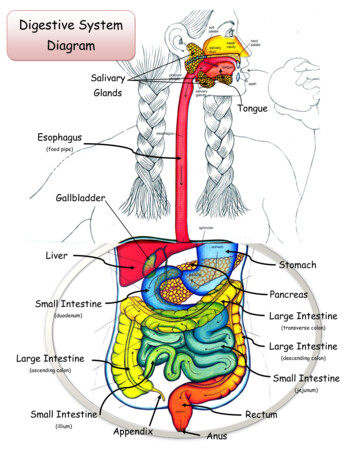
Gross findings, histological and immunohistochemical characteristics of the GT. a Gross image of resected mass; b H&E, 40; c H&E, 100; d SMAimmunostaining, 200; e Collagen type IV immunostaining, 200; f Ki-67 immunostaining, 400.Page 7/7
In this paper, for the rst time, we acquired the endoscopic imaging of small intestinal GT by balloon-assisted enteroscopy. Intriguingly, the enteroscopic imaging showed a brous capsule-covered mass, without any epithelial coverage, and on the surface of the tumor, no denite hemorrhage, erosions or ulcers were observed.