Transcription
(2020) 6:260Okada et al. surg case rephttps://doi.org/10.1186/s40792-020-01028-zOpen AccessCASE REPORTA long‑term survivor of clear cellsarcoma‑like tumor of the gastrointestinal tractwith liver metastasis: a case reportTakuhisa Okada1,2* , Yasumitsu Hirano1, Shintaro Ishikawa1, Hiroka Kondo1, Toshimasa Ishii1and Shigeki Yamaguchi1AbstractBackground: Clear cell sarcoma-like tumor of the gastrointestinal tract (CCSLTGT) is extremely rare. It is a mesenchymal neoplasm that usually forms in the small intestine of adolescents and young adults, is prone to local recurrenceand metastasis, and has a high mortality rate. We report a patient with CCSLTGT with lymph node- and liver metastases, who continues to survive 6 years after initial surgical resection.Case presentation: A 38-year-old woman presented with lightheadedness. Laboratory analysis revealed anemia(hemoglobin, 6.7 g/dL), and enhanced computed tomography (CT) demonstrated a mass in the small intestine, about6 cm in diameter, with swelling of 2 regional lymph nodes. Double-balloon small intestine endoscopic examinationrevealed a tumor accompanied by an ulcer; the biopsy findings suggested a primary cancer of the small intestine. Shewas admitted, and we then performed a laparotomy for partial resection of the small intestine with lymph node dissection. Pathologic examination revealed CCSLTGT with regional lymph node metastases. About 3 years later, followup CT revealed a single liver metastasis. Consequently, she underwent a laparoscopic partial liver resection. Histopathologic examination confirmed that the liver metastasis was consistent with CCSLTGT. It has now been 3 yearswithout a recurrence.Conclusion: Repeated radical surgical resection with close follow-up may be the only way to achieve long-termsurvival in patients with CCLSTGT.Keywords: Clear cell sarcoma-like tumor of the gastrointestinal tract, Liver metastasis, Long-term survivalIntroductionClear cell sarcoma-like tumor of the gastrointestinal tract(CCSLTGT) is a very rare malignant tumor of mesenchymal origin. Histopathologically, CCSLTGT has characteristic spindle-shaped tumor cells that proliferate in sheets,and osteoclast-like multinucleated giant cells are seen[1, 2]. On immunohistologic staining, S-100 protein andSOX10 are positive but HMB-45 is negative, a finding*Correspondence: t.okd@gunma‑u.ac.jp1Department of Gastroenterological Surgery, Saitama Medical UniversityInternational Medical Center, 1397‑1, Yamane, Hidaka‑City, Saitama‑Pref350‑1298, JapanFull list of author information is available at the end of the articlethat distinguishes CCSLTGT from malignant melanomaand soft-tissue clear cell sarcoma (CCS). As with CCS,Ewing sarcoma breakpoint region 1 (EWSR1) gene fusionis noted [3, 4]. Because CCSLTGT is a highly malignant tumor, it is important to distinguish it from othermesenchymal neoplasms, such as gastrointestinal stromal tumors (GIST). Local recurrence is common withCCSLTGT, as are lymph node metastases and distantmetastases to the liver and lung. These metastases occurrapidly and usually result in death [2]. We report herein apatient with liver metastasis after resection of CCSLTGTof the small intestine. She is alive, without further recurrence, 6 years after her initial surgery. The Author(s). 2020 Open Access This article is licensed under a Creative Commons Attribution 4.0 International License, whichpermits use, sharing, adaptation, distribution and reproduction in any medium or format, as long as you give appropriate credit to theoriginal author(s) and the source, provide a link to the Creative Commons licence, and indicate if changes were made. The images orother third party material in this article are included in the article’s Creative Commons licence, unless indicated otherwise in a credit lineto the material. If material is not included in the article’s Creative Commons licence and your intended use is not permitted by statutoryregulation or exceeds the permitted use, you will need to obtain permission directly from the copyright holder. To view a copy of thislicence, visit http://creat iveco mmons .org/licen ses/by/4.0/.
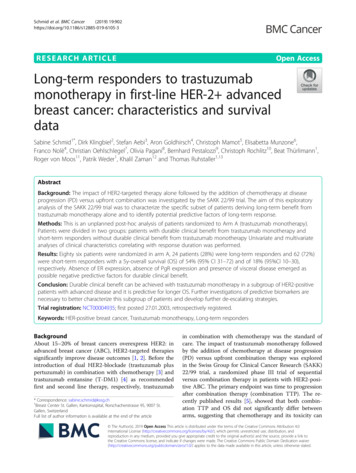
Okada et al. surg case rep(2020) 6:260Case reportA 38-year-old Japanese woman presented with lightheadedness. Her medical history was significant for malignantlymphoma at the age of 18 years. Laboratory analysisrevealed anemia (hemoglobin, 6.7 g/dL), and enhancedcomputed tomography (CT) revealed a mass in thesmall intestine measuring about 6 cm in diameter, withswelling of 2 regional lymph nodes (Fig. 1). The patientunderwent a double-balloon small intestine endoscopy,which revealed a submucosal tumor accompanied by anulcer (Fig. 2). Examination of the biopsy specimen withimmunostaining was negative for malignant lymphomaand GIST, and a very poorly differentiated cancer of thesmall intestine was suspected. The use of positron emission tomography–CT (PET–CT) confirmed an abnormalaccumulation of F-18 fluorodeoxyglucose in the tumorand lymph nodes (Fig. 3). We conferred the diagnosis ofa primary malignant tumor of the small intestine withlymph node metastasis. She was admitted and underwent a laparotomy for partial resection of the small intestine with lymph node dissection. The tumor was locatedin the jejunum and was an ulcerative lesion, measuring5.8 3.9 cm. Gross examination revealed a white, solidtumor with a clear boundary (Fig. 4a, b). The tumor waslocated between the muscularis mucosa and the muscularis propria of the small intestine (Fig. 4c, d). There wasa fibrous septum separating the sheet-like epithelial cellsPage 2 of 9from a network of eosinophilic cells and an oval-shapedbundle of spindle-like tumor cells. The tumor demonstrated a pseudorosette pattern around several bloodvessels and displayed a pseudopapillary growth pattern.Throughout the neoplasm, the tumor cells were associated with numerous osteoclast-type giant cells. Onimmunohistochemical examination (Table 1), the tumorcells stained positive for the anticytokeratin antibodies AE1 and AE3 (on part of the surface layer) and forS-100, and partially positive for the neural cell adhesionmolecule (CD56) and synaptophysin. The cells stainednegative for anticytokeratin (CAM 5.2), lymphocytecommon antigen (CD45), desmin, α-smooth muscle actin(ɑ-SMA), c-kit, CD34, HMB-45, melan-A, chromogranin A, CD1a, and CD21. The osteoclast-type giant cellswere positive for CD68. Fluorescence in situ hybridization (FISH) using an EWSR1 probe revealed a split signal in the tumor cells: seemingly an EWSR1 gene fusion(Fig. 5). Taken together, these results yielded a diagnosisof CCSLTGT. Metastases were found in 3 of 20 lymphnodes.About 3 years later, follow-up CT revealed a singleliver metastasis (Fig. 6a). The results of gadoxetic acidenhanced magnetic resonance imaging (EOB-MRI) andPET–CT were also suggestive of liver metastasis (Fig. 6b–d). Consequently, the patient underwent a laparoscopicpartial liver resection. Histopathologic examinationFig. 1 CT findings. a Horizontal view. Primary tumor (the white arrow). b, c Horizontal view. Lymph nodule (the arrowhead). d Sagittal view. Primarytumor (the white arrow) and lymph nodule (the arrowhead)
Okada et al. surg case rep(2020) 6:260Page 3 of 9Fig. 3 PET–CT confirmed an abnormal accumulation of F-18fluorodeoxyglucose in the tumor and lymph nodes. a Primary tumor’saccumulation. b Lymph nodule’s accumulationFig. 2 Findings of double-balloon endoscopy. a, b Primary tumorlooked like submucosal tumor accompanied by an ulcerrevealed that the liver metastasis was consistent withCCSLTGT (Fig. 7a–c). It has now been 3 years since thisprocedure, and follow-up CT every 3 to 6 months hasshown no evidence of recurrence.DiscussionThe entity of CCSLTGT was first described by Zambrano et al. in 2003, when they reported 6 patients witha disease they called “osteoclast-rich tumor of the gastrointestinal tract”, with features resembling those ofsoft-tissue CCS [1]. This tumor is relatively rare andshows no preference for sex. Many patients are in their30s and 40s, but there are reports of patients rangingin age from 15 to 85 years [2]. It is most common forCCSLTGT to develop in the small intestine, but it hasalso been reported in the stomach and colon [2]. Sinceit is a malignant tumor originating from the interstitiumof the wall of the gastrointestinal tract, it must be differentiated from GIST, synovial sarcoma, and malignant peripheral nerve sheath tumors [3]. The etiology ofCCSLTGT is unknown, but it has been suggested thatradiotherapy may be a precipitating factor in the laterdevelopment of the disease. Two cases of secondarymalignancy after irradiation of neuroblastoma in infancyhave been reported [16, 20]. Our case has been treatedfor malignant lymphoma in the past, but the details ofthe treatment are unknown. It is not clear whether thetreatment was related to the development of CCSLTGT,and there are no reports on the relationship betweenboth malignant lymphoma and CCSLTGT. Scatteredosteoclast-like multinucleated giant cells are characteristic of CCSLTGT. Tumor cells stain positive for vimentin, S-100 protein, and SOX10; neuroendocrine markerssuch as CD56, synaptophysin, and NSE may also be positive. Melanocytic markers such as HMB-45 and melanA, which are positive in soft-tissue CCS, are negativein CCSLTGT, as are c-kit, CD34, desmin, α-SMA, andkeratin. Both our current patient and our previouslyreported patient [1, 2, 4, 9, 14, 15, 17–19, 21] had osteoclast-like multinucleated giant cells that were positivefor CD68, indicating increased responsiveness. Zambrano et al. [1] report a single patient with chromosomaltranslocation involving EWSR1, similar to that seen with
Okada et al. surg case rep(2020) 6:260Page 4 of 9Fig. 4 The tumor was located in the jejunum and was an ulcerative lesion. a Resected specimen (jejunum). b Cut surface, white solid tumor withclear boundary. c HE stain, low-power view. d HE stain, high-power viewTable 1 The list of immunohistochemical examinationfor our casePositiveNegativeAE1/AE3 (part of the surface layer)CAM 5.2, HMB-45, Melan-AS-100Desmin, chromograninCD 56 (partial)α-SMA, c-kitSynaptophysin (partial)CD1a, CD21, CD34, CD45CD68 (only osteoclast-type giant cell)CCS. Cu et al. [4] and Stockman et al. [3] also reportpatients with CCSLTGT and EWSR gene fusion similar to that seen with CCS. A highly malignant tumor isconsidered to confer a poor prognosis; many patients diewithin 36 months, and more than half have local lymphnode metastasis at the time of diagnosis. The most common site of distant metastasis is the liver, and curativetreatment involves only surgical resection of any metastases. There are 43 reported patients with CCSLTGTof the small intestine to date: 21 men and 22 women,including our previously reported patient (Table 2).Fig. 5 EWSR1-FISH positive lesion (yellow circle)The tumor location was the jejunum in 11 patients, theileum in 24 patients, and was unknown in 8 patients.The sites of metastasis at initial diagnosis include thelymph nodes (n 17), liver (n 6), peritoneum (n 3),and lungs (n 2). Of the 43 patients, curative resection
Okada et al. surg case rep(2020) 6:260Page 5 of 9Fig. 6 Findings at a single liver metastasis. a CT. Horizontal view. Liver metastasis (the white arrow). b PET (the white arrow). c EOB-MRI. Horizontalview (the white arrow). d EOB-MRI. Sagittal view (the white arrow)was performed in 30 (69.8%), and among them, recurrence occurred in 18 (60.0%). The mean period betweeninitial surgery and recurrence was 20.0 months. However, 2 recurrences occurred over 5 years after the initial surgery, emphasizing the importance of long-termfollow-up. The recurrence sites were the liver (n 11),lymph nodes (n 5), peritoneum (n 4), lung (n 1),ovary (n 1), omentum (n 1), and pleura (n 1). Localrecurrence and intra-abdominal recurrence occurredin 1 patient each. Of the 18 patients with recurrence,treatment after recurrence included surgery-alone(n 4), chemotherapy-alone (n 2), surgical resectionwith chemotherapy (n 1), no treatment (n 1), andunknown details (n 10). All five patients with surgicalresection survived within the observation period, amongwhich two patients with curative resection were diseasefree and the other three were in the tumor-bearing. Onlythree patients received chemotherapy. Platinum-basedchemotherapy was performed in one patient and detailswere unknown in two. Two patients with chemotherapyalone died in 13 and 16 months after recurrence, andtheir survival times indicated the non-effectiveness ofthis therapy. Eleven of the reported patients are knownto be dead of their disease, but data on mortality aremissing for 17 patients. Of the remaining 15 patients,9 have no evidence of disease, and 6 are alive with disease. Curative surgical resection is the only possible curetreatment for recurrence of CCSLTGT. Chemotherapyhas not been reported to be effective, and there are noestablished chemotherapy regimens until now. In termsof postoperative adjuvant chemotherapy, mainly ifosfamide and doxorubicin were administrated, however,its efficacy has not been proved [28]. Only 2 patients,including the patient in this report, survived for over
Okada et al. surg case rep(2020) 6:260Page 6 of 9Fig. 7 The single metastatic tumor was located in the liver. a Resected specimen (liver) and cut surface, white solid tumor with clear boundary. bHE stain, low-power view. c HE stain, high-power view5 years without evidence of disease; both of them hadtumor recurrence that was treated with surgical resection. Repeated radical surgical resection appears to bethe only way to achieve long-term survival in patientswith CCSLTGT, a neoplasm that easily metastasizes in ashort period. It is important to follow patients with contrast CT every 3 months to ensure that radical resectionof any recurrence can occur in a timely fashion.ConclusionsWe report a patient with CCSLTGT with lymph nodeand liver metastases, who continues to survive 6 yearsafter the initial surgical resection. It is important to follow patients with contrast CT every 3 months to ensurethat radical resection of any recurrence can occur in atimely fashion.
eumLocalizedLocalizedMetastaticSmall intestine junumIleumJejunumIleumSmall intestine numIleumIleumIleumIleumIleumMesenteric LN––Regional LN–Regional LN–NRLN and liverLN and liver––LNMediastinal LN andlung–LN––Liver and peritonealNRLN–LN–––LNNRLNLiver and YesYesYesYesYesYesYesYesNRYesNRNoYes (12)Yes (6)NoNRNoYes (0.5)NRNRNRNoYes (2)Yes (12)–NoNRYes (NR)Yes (60)NRNRNRYes (6)NRYes (2 7)NoYes (24 and 46)NRNRYes (12)NR–LiverLiver–––Mesenteric LN, omentum and lung––––Peritoneal extensionand pleural effusionIntra-abdominal–––LocalLiver and peritoneum–––Liver–Liver e (Mo) Site of emotherapy er recurrenceNRAWD (6)NED (NR)NRNED (7)DOD (1)NRNRNRNED (24)NRDOD (28)DOD (12)NED (7)NRNRAWD (60)NRNRDOD (1)NRNRDOD (15)NED (11)AWD (46)NRNRNRDOD (12)DOD (16)Outcome (Mo)(2020) 6:260696626531615466020 s c1112Status at diagnosis MetastasisCase Reference Age Gender Primary siteTable 2 Review of reported CCSLGTOkada et al. surg case repPage 7 of 9
etastaticMetastaticLocalizedSmall intestine LocalizedSmall intestine MetastaticSmall intestine LocalizedSmall intestine MetastaticSmall intestine MetastaticSmall intestine LocalizedIleumJejunum–Regional LNIliac LNRegional LN and liver–Sigmoid colon directinvasionPeritoneum–Regional LNLung and sYesNRYes (36)––Yes (NR)Yes (1)NoYes (NR)Yes (2)–Yes (109)Yes (15 47)NoNR––Therapyafter recurrenceLiver––Mesenteric LN andmulti liverMultiple abdominal LN–NRMesenteric LN andLiver–Mesenteric LNResection––Resection and chemotherapy (no details)NR–NRNone–ResectionLiver ovary andResectionperitoneal dissemination––RadialRecurrence (Mo) Site of recurrenceresectionLN lymph node, AWD alive with disease, DOD dead of disease, NR not reported, NED no evidence of disease, PC present 1223132Status at diagnosis MetastasisCase Reference Age Gender Primary siteTable 2 (continued)NED (72)DOD (25)AWD (8)AWD (18)NRNED (2)DOD (NR)DOD (8)DOD (4)NED (161)AWD (47)NED (20)NROutcome (Mo)Okada et al. surg case rep(2020) 6:260Page 8 of 9
Okada et al. surg case rep(2020) 6:260AbbreviationsCCSLTGT : Clear cell sarcoma-like tumor of the gastrointestinal tract; CT:Computed tomography; CCS: Clear cell sarcoma; EWSR1: Ewing sarcomabreakpoint region 1; GIST: Gastrointestinal stromal tumors; PET–CT: Positronemission tomography–CT; EOB-MRI: Gadoxetic acid-enhanced magneticresonance imaging; CD56: The neural cell adhesion molecule; CAM 5.2: Anticytokeratin; CD45: Lymphocyte common antigen; ɑ-SMA: α-Smooth muscleactin; FISH: Fluorescence in situ hybridization.AcknowledgementsNone.Authors’ contributionsTO and YH wrote the manuscript. SY supervised the case. All other authorsreviewed the manuscript. All authors read and approved the final manuscript.Page 9 of 98.9.10.11.12.13.FundingNo funding was received for this case series.Availability of data and materialsData sharing is not applicable to this article, since datasets were neither generated nor analyzed for the case series.14.15.Ethics approval and consent to participateNot applicable.16.Consent for publicationWritten informed consent was obtained from the patients for the publicationof this report.17.Competing interestsThe authors declare that they have no competing interests.18.Author details1Department of Gastroenterological Surgery, Saitama Medical UniversityInternational Medical Center, 1397‑1, Yamane, Hidaka‑City, Saitama‑Pref350‑1298, Japan. 2 Department of General Surgical Science, Gunma University Graduate School of Medicine, 3‑39‑22 Showa‑Machi, Maebashi‑City,Gunma‑Pref 371‑8511, Japan.19.20.Received: 18 June 2020 Accepted: 23 September 202021.References1. Zambrano E, Reyes-Mugica M, Franchi A, Rosai J. An osteoclast-rich tumorof the gastrointestinal tract with features resembling clear cell sarcomaof soft parts: reports of 6 cases of a GIST simulator. Int J Surg Pathol.2003;11:75–81.2. Gulcin Y, Mine G, Ozgur M, Semen O, Yersu K. Clear cell sarcoma-like tumorof the gastrointestinal tract: a case report and review of the literature. Int JSurg Pathol. 2015;23(1):61–7.3. Stockman DL, Miettinen M, Suster S, Spagnolo D, Dominguez-MalagonH, Hornick JL, et al. Malignant gastrointestinal neuroectodermal tumor:clinicopathologic, immunohistochemical, ultrastructural, and molecularanalysis of l6 cases with a reappraisal of clear cell sarcoma-like tumors ofthe gastrointestinal tract. Am J Surg Pathol. 2012;36:857–68.4. Antonescu CR, Nafa K, Segal NH, Dal Cin P, Ladanyi M. EWS-CREBI:a recurrent variant fusion in clear cell sarcoma-association with gastrointestinallocation and absence of melanocytic differentiation. Clin Cancer Res.2006;12:5356–62.5. Donner LR, Trompler RA, Dobin S. Clear cell sarcoma of ileum the crucialrole of cytogenetics for the diagnosis. Am J Surg Pathol. 1998;22:121–4.6. Achten R, Debiec-Rychter M, De Wever I, Sciot R. An unusual case of clearcell sarcoma arising in the jejunum highlights the diagnostic value ofmolecular genetic techniques in establishing a correct diagnosis. Histopathology. 2005;46:472–4.7. Taminelli L, Zaman K, Gengler C, Peloponissios N, Bouzourene H, CoindreJM, et al. Primary clear cell sarcoma of the ileum: an uncommon andmisleading site. Virchows Arch. 2005;447:772–7.22.23.24.25.26.27.28.Venkataraman G, Quinn AM, Williams J, Hammadeh R. Clear cell sarcoma ofthe small bowel: a potential pitfall. Case Report. 2005;113:716–9.Friedrichs N, Testi MA, Moiraghi L, Modena P, Paggen E, Plotner A, et al. Clearcell sarcoma-like tumor with osteoclast-like giant cells in the small bowel:further evidence for anew tumor entity. Int J Surg Pathol. 2005;13:313–8.Covinsky M, Gong S, Rajaram V, Perry A, Pfeifer J. EWS-ATF1 fusiontranscripts in gastrointestinal tumors previously diagnosed as malignantmelanoma. Human Pathol. 2005;36:74–81.Comin CE, Novelli L, Tornaboni D, Messerini L. Clear cell sarcoma ofthe ileum: report of a case and review of literature. Virchows Arch.2007;451:839–45.Lyle PL, Amato CM, Fitzpatrick JE, Robinson WA. Gastrointestinal melanomaor clear cell sarcoma? Molecular evaluation of 7 cases previously diagnosedas malignant melanoma. AM J Surg Pathol. 2008;32:858–66.Abdulkader I, Cameselle-Tejieiro J, de Alava E, Ruiz-Ponte C, Used-AznarMM, Forteza J. Intestinal clear cell sarcoma with melanocytic differentiation and EWS [corrected] rearrangement: report of a case. Int J Surg Pathol.2008;16:189–93.Terazawa K, Otuka H, Morita N, Yamashita K, Nishitani H. Clear cell sarcomaof the small intestine detected by FDG-PET/CT during comprehensiveexamination of an inflammatory reaction. J Med Invest. 2009;56:70–5.Zucman J, Delattre O, Desmaze C, Epstein AL, Stenmann G, SpelemanF, et al. EWS and ATF-1 gene fusion induced by t(12;22) translocation inmalignant melanoma of soft parts. Nat Genet. 1993;4:341–5.Balkaransingh P, Saad SA, Govil SC, Thind PK, Vallance CM, Weiss AR. Clearcell sarcoma of the gastrointestinal tract presenting as a second malignantneoplasm following neuroblastoma in infancy. Padiatr Blood Cancer.2012;58:481–2.Granville L, Hicks J, Popek E, Dishop M, Tatevian N, Lopez-Terrada D. Visceralclear cell sarcoma of soft tissue with confirmation by EWS-ATF1 fusiondetection. Ultrastruct Pathol. 2006;30:111–8.Shenjere P, Salman WD, Singh M, Mangham DC, Williams A, Eyden BP, et al.Intra-abdominal clear cell sarcoma: a report of 3 cases, including 1 casewith unusual morphological features, and review of the literature. Int J SurgPathol. 2012;20:378–85.D’Amico FE, Ruffolo C, Romeo S, Massani M, DeiTos AP, Bassi N. Clear cellsarcoma of the ileum: report of case and review of the literature. Int J SurgPathol. 2012;20:401–6.Yang JC, Chou AJ, Oeffinger KC, La Quaglia MP, Wolden AL. Clear cell sarcoma of the gastrointestinal tract after very low dose therapeutic radiationtherapy: a case report. J Pediatr Surg. 2012;47:1943–5.Suarez-Vilela D, Izquierdo FM, Tojo-Ramallo S, Riera-Velasco J, Escobar-SteinJ. Malignant gastrointestinal nueroectodermal tumor showing overlappedimmunophenotype with synovial sarcoma CD99 and Sox10 antibodies areuseful in differential diagnosis. Am J Surg Pathol. 2012;36:1905–8.Lasithiotakis K, Protonotarios A, Lazarou V, Tzardi M, Chalkiadkis G. Clear cellsarcoma of the jejunum: a case report. World J Surg Oncol. 2013;11:17.Michela L, Khin T, Jonathan N, Florian P, Christina M, Cyril F, et al. Clear cellsarcoma-like tumor of the gastrointestinal tract: Clinical outcome andpathologic features of a molecularly characterized Tertiary center caseseries. Anticancer Res. 2018;38:1479–83.Kato T, Ichihara S, Gotoda H, Muraoka S, Kubo T, Sugita S, et al. Imprintcytology of clear cell sarcoma-like tumor of the gastrointestinal tract in thesmall intestine: a case report. Diagn Cytopathol. 2017;45:1137–41.Wolak P, Wincewicz A, Czauderna P, Spałek M, Kruczak A, Urbaniak-WąsikS, et al. Malignant gastrointestinal neuroectodermal tumor (clear cellsarcoma-like tumor of the gastrointestinal tract) of the small intestine in a12-year-old boy. Dev Period Med. 2018;22:358–63.Askan G, Kombak FE, Seven IE, Basturk O. Clear cell sarcoma-like tumor ofthe gastrointestinal tract. J Gastrointest Cancer. 2019;50:651–6.Sonai MK, Rastogi S, Madhusudhan KS, Gupta SD, Das P. Clear cell sarcomalike tumor of gastrointestinal tract: Experience of three cases and review ofliterature. Indian J Pathol Microbiol. 2020;63:90–5.Washimi K, Takagi M, Hisaoka M, Kawachi K, Takeyama M, Hiruma T, et al.Clear cell sarcoma-like tumor of the gastrointestinal tract: a clinicopathological review. Pathol Int. 2017;67:534–6.Publisher’s NoteSpringer Nature remains neutral with regard to jurisdictional claims in publishedmaps and institutional affiliations.
A long-term survivor of clear cell sarcoma-like tumor of the gastrointestinal tract with liver metastasis: a case report Takuhisa Okada1,2*, Yasumitsu Hirano 1, Shintaro Ishikawa1, Hiroka Kondo1, Toshimasa Ishii 1 and Shigeki Yamaguchi 1 Abstract Background: Clear cell sarcoma-like tumor o