Transcription
Consensus StatementAn Expert Consensus Statement on theManagement of Large Chondral andOsteochondral Defects in thePatellofemoral JointJorge Chahla,* MD, PhD, Betina B. Hinckel, MD, PhD, Adam B. Yanke, MD, PhD, Jack Farr, MD,and Metrics of Osteochondral Allografts (MOCA) GroupInvestigation performed at Midwest Orthopaedics at Rush and the Rush University Medical Center,Chicago, Illinois, USABackground: Cartilage lesions of the patellofemoral joint constitute a frequent abnormality. Patellofemoral conditions are challenging to treat because of complex biomechanics and morphology.Purpose: To develop a consensus statement on the functional anatomy, indications, donor graft considerations, surgical treatment, and rehabilitation for the management of large chondral and osteochondral defects in the patellofemoral joint using amodified Delphi technique.Study Design: Consensus statement.Methods: A working group of 4 persons generated a list of statements related to the functional anatomy, indications, donor graftconsiderations, surgical treatment, and rehabilitation for the management of large chondral and osteochondral defects in thepatellofemoral joint to form the basis of an initial survey for rating by a group of experts. The Metrics of Osteochondral Allografts(MOCA) expert group (composed of 28 high-volume cartilage experts) was surveyed on 3 occasions to establish a consensus on thestatements. In addition to assessing agreement for each included statement, experts were invited to propose additional statementsfor inclusion or to suggest modifications of existing statements with each round. Predefined criteria were used to refine statementlists after each survey round. Statements reaching a consensus in round 3 were included within the final consensus document.Results: A total of 28 experts (100% response rate) completed 3 rounds of surveys. After 3 rounds, 36 statements achieved aconsensus, with over 75% agreement and less than 20% disagreement. A consensus was reached in 100.00% of the statementsrelating to functional anatomy of the patellofemoral joint, 88.24% relating to surgical indications, 100.00% relating to surgicaltechnical aspects, and 100.00% relating to rehabilitation, with an overall consensus of 95.5%.Conclusion: This study established a strong expert consensus document relating to the functional anatomy, surgical indications,donor graft considerations for osteochondral allografts, surgical technical aspects, and rehabilitation concepts for the management of large chondral and osteochondral defects in the patellofemoral joint. Further research is required to clinically validate theestablished consensus statements and better understand the precise indications for surgery as well as which techniques and graftprocessing/preparation methods should be used based on patient- and lesion-specific factors.Keywords: patellofemoral; cartilage; osteochondral; allograft; consensus; DelphiCartilage lesions of the patellofemoral joint are particularlycommon; however, surgery is not indicated in many cases,especially when they are asymptomatic.11,20,26,58 Chondraland osteochondral injuries of the patellofemoral joint present several challenges for their treatment, as the morphology of the patella and trochlea can have wide variabilitybetween patients. In addition to its unique anatomy, thepatellofemoral joint also experiences very high loads withdaily activities, including 1.3 times the body weight (BW)during level ambulation, 3.3 times BW during stair ambulation, 5.6 times BW during running, and up to 7.8 timesBW during a deep knee bend or squat.28 Other factors thatadd additional complexity include excessive patellar malalignment, maltracking, patella alta or baja, and trochleardysplasia, among others.3,4,12,26,32,59When indicated, multiple surgical options exist forlarge symptomatic defects. Matrix-induced autologouschondrocyte implantation (MACI) and osteochondralThe Orthopaedic Journal of Sports Medicine, 8(3), 2325967120907343DOI: 10.1177/2325967120907343ª The Author(s) 2020This open-access article is published and distributed under the Creative Commons Attribution - NonCommercial - No Derivatives License /), which permits the noncommercial use, distribution, and reproduction of the article in any medium, provided the original author and source arecredited. You may not alter, transform, or build upon this article without the permission of the Author(s). For article reuse guidelines, please visit SAGE’s website athttp://www.sagepub.com/journals-permissions.1
2Chahla et alallograft transplantation (OCA) remain the most commonly utilized methods of treatment. 12,45 These treatments are used routinely in other compartments of theknee; however, the morphology, alignment, and biomechanics of the patellofemoral joint make some of these techniques particularly challenging. For example, the complextopography and variable anatomy of the patella and trochlea make OCA technically more demanding than autologous chondrocyte implantation (ACI) because of the needto match graft morphology to patient anatomy. As a resultof this complexity, inferior outcomes have been reportedwith the treatment of symptomatic large patellofemorallesions when compared with tibiofemoral cartilage treatments.12,24 In addition, clinical studies, especially clinicaltrials, very often focus on the tibiofemoral joint, and theliterature on cartilage restoration in the patellofemoraljoint is much more limited.24,40,80 For those reasons, significant debate remains regarding the indications andThe Orthopaedic Journal of Sports Medicinespecifics of each technique for the surgical treatment ofpatellofemoral chondral defects.Despite the growing body of literature on this topic, astandardized algorithm is lacking, leading to persistentcontroversy in the surgical treatment of large symptomatic patellofemoral cartilage injuries. In such instances,an expert consensus can be synthesized using a modifiedDelphi method. This allows for the development of agroup-based consensus. The Delphi method provides several advantages over other group-based processes, including the preservation of participant anonymity that canreduce the effects of dominant participants.39 Additionally, Delphi consensus statements conducted at a distancehave been demonstrated to be as reliable as face-to-facepanels,93 with further advantages of greater participantflexibility.42 For the abovementioned reasons, the purposeof this study was to develop a Delphi consensus statementon the functional anatomy, indications, donor graft*Address correspondence to Jorge Chahla, MD, PhD, Midwest Orthopaedics at Rush, 1611 West Harrison Street, Suite 300, Chicago, IL 60612, USA(email: jorge.chahla@rushortho.com).All authors are listed in the Authors section at the end of this article.Final revision submitted November 24, 2019; accepted December 3, 2019.One or more of the authors has declared the following potential conflict of interest or source of funding: J.C. has received research support and consultingfees from Arthrex, Conmed, and Smith & Nephew. B.B.H. has received research support from Arthrex and educational support from Arthrex and EliteOrthopaedics. A.B.Y. has received research support from Arthrex, Orthogenesis, Medwest, and Vericel; educational support from Arthrex, Medwest, andSmith & Nephew; and consulting fees from Aastrom Biosciences, JRF Ortho, Olympus, PatientIQ, Smith & Nephew, and Sparta Biomedical. J.F. has receivedconsulting fees from Osiris Therapeutics, Zimmer Biomet, ISTO Technologies, Ceterix Orthopaedics, Biomet, Genzyme, MedShape, and Sanofi-Aventis; hasreceived speaking fees from Arthrex and Zimmer Biomet; has received royalties from Arthrex, DePuy, Organogenesis, Springer, and Thieme MedicalPublishers; and has stock/stock options in MedShape and Ortho Regenerative Technologies. W.D.B. has received consulting fees from Arthrex, DePuy,Insight Medical, JRF Ortho, OrthAlign, and Smith & Nephew; has received royalties from DePuy, Smith & Nephew, and Zimmer Biomet; and has stock/stockoptions in Insight Medical, Moximed, and OrthAlign. B.J.C. has received research support from Aesculap/B. Braun, Arthrex, and Regentis Biomaterials; hasreceived educational support from Medwest; has received consulting fees from Acumed, Anika Therapeutics, Arthrex, Bioventus, Flexion Therapeutics,Geistlich Pharma, Genzyme, Pacira Pharmaceuticals, Regentis Biomaterials, Smith & Nephew, Vericel, and Zimmer Biomet; has received speaking fees fromArthrex, Carticept Medical, LifeNet Health, and Pacira Pharmaceuticals; has received hospitality payments from GE Healthcare; has received royalties fromArthrex, DJO, Elsevier, and Operative Techniques in Sports Medicine; and has stock/stock options in BandGrip, Ossio, and Regentis Biomaterials. D.C.C.has received research support from Histogenics, JRF Ortho, Moximed, and Zimmer Biologics and consulting fees from Arthrosurface, DePuy, Histogenics,JRF Ortho, and Moximed. J.E.F. has received research support from Arthrex and Smith & Nephew; educational support from Linvatec, Peerless Surgical, andZimmer Biomet; and consulting fees and speaking fees from Smith & Nephew. A.G. has received research support from Aesculap/B. Braun, Arthrex, DePuy,Eupraxia Pharmaceuticals, Musculoskeletal Transplant Foundation, Ossur, and Smith & Nephew; consulting fees from Collagen Solutions, Olympus, Ossur,and Smith & Nephew; speaking fees from Conmed Linvatec, Ossur, and Smith & Nephew; and royalties from Smith & Nephew. A.H.G. has received researchsupport from JRF Ortho and Vericel; consulting fees from Aesculap, Geistlich Pharma, Genzyme, JRF Ortho, Moximed, Smith & Nephew, and Vericel;speaking fees from Aastrom Biosciences, LifeNet Health, and Vericel; and royalties from Organogenesis. S.G. has received educational support from Arthrexand Goode Surgical and honoraria from Vericel. A.E.G. has received consulting fees, speaking fees, and royalties from Zimmer Biomet and has stock/stockoptions in Intellijoint Surgical. D.G.J. has received research support from Genzyme and Sanofi-Aventis; has received educational support from Arthrex; hasreceived consulting fees from Aastrom Biosciences, Acumed, Amniox Medical, DePuy/Medical Device Business Services, Flexion, Genzyme, Vericel, andZimmer Biomet; has received speaking fees from Arthrex, Conmed Linvatec, Flexion, Genzyme, Mitek, Musculoskeletal Transplant Foundation, TissueTech,Vericel, and Zimmer Biomet; and is a board or committee member of the Musculoskeletal Transplant Foundation. A.J.K. has received research support fromAesculap/B. Braun, Arthrex, Ceterix Orthopaedics, and Histogenics; has received consulting fees from Arthrex, DePuy, JRF Ortho, and Vericel; has receivedspeaking fees from Arthrex; and is a board or committee member of the Musculoskeletal Transplant Foundation. C.L. has received honoraria fromArthrosurface; consulting fees from Aastrom Biosciences, Genzyme, JRF Ortho, Samumed, Sanofi-Aventis, Vericel, and Zimmer Biomet; and speaking feesfrom Vericel. B.R.M. has received consulting fees from Arthrex, BioMarin Pharmaceutical, DePuy, and Exactech; speaking fees from Arthrex; and royaltiesfrom Arthrex. R.M. has received research support from Arthrex and DJO, has received educational support from Arthrex, has received speaking fees fromArthrex, has received royalties from Zimmer Biomet, and has stock/stock options in AlignMed. T.S.M. has received research support from JRF Ortho,educational support from Arthrex, consulting fees from JRF Ortho and MicroAire Surgical Instruments, and speaking fees from JRF Ortho. J.D.P. hasreceived research support from Ossur, Sanofi-Aventis, and Vericel; consulting fees from JRF Ortho; and speaking fees from DePuy. M.T.P. has receivedconsulting fees from Arthrex and JRF Ortho (AlloSource), speaking fees from Arthrex, and royalties from Arthrex and SLACK. S.A.R. has received consultingfees from Flexion, has received speaking fees from Smith & Nephew, has received honoraria from Fidia Pharma, has received royalties from Zimmer Biomet,and has stock/stock options in Ortho Regenerative Technologies. O.S. has received research support from DePuy and consulting fees from Zimmer Biomet.S.L.S. has received consulting fees and research support from Arthrex Inc; is a paid consultant for Ceterix Orthopaedics, CONMED Linvatec, FlexionTherapeutics, GLG Consulting, JRF Ortho, Moximed, Olympus, Vericel, Linvatec, and RTI Surgical; has received hospitality payments from Linvatec andSmith & Nephew; and has received a grant (indirect) from DJO. E.D.S. has received research support from CartiHeal, Fidia Pharma, and NuTech; consultingfees from Arthrex, Fidia Pharma, Flexion, JRF Ortho, Organogenesis, Smith & Nephew, and Vericel; speaking fees from Arthrex, Organogenesis, and Vericel;and royalties from Jaypee Brothers Medical Publishers. C.J.W. has received consulting fees from Arthrosurface and Smith & Nephew and speaking fees fromArthrex, Arthrosurface, and Smith & Nephew. R.J.W. has received research support from Histogenics; has received consulting fees from Arthrex, JRF Ortho,and Lipogems; has received speaking fees from Arthrex; has received royalties from Arthrex; and has stock/stock options in CyMedica Orthopedics,Gramercy Extremity Orthopedics, Pristine Surgical, and RecoverX. AOSSM checks author disclosures against the Open Payments Database (OPD). AOSSMhas not conducted an independent investigation on the OPD and disclaims any liability or responsibility relating thereto.
The Orthopaedic Journal of Sports MedicineManagement of Large Chondral/Osteochondral Defects of the PFJ3TABLE 1Summary of Results at the End of Each Survey Round in the Delphi ProcessDelphi Round123No. ofResponsesTotal No. of StatementsIncluded in Survey% of Statements Reachinga ConsensusNo. of NewStatementsNo. of 84considerations, treatment, and rehabilitation for the management of large chondral and osteochondral defects inthe patellofemoral joint.METHODSStudy DesignA working group of 4 individuals (J.C., B.B.H., A.B.Y., J.F.)was responsible for facilitating the development of a consensus using modified Delphi techniques as previouslydescribed.14 A comprehensive list of statements was generated under 5 categories: functional anatomy, surgical indications, donor graft considerations for osteochondralallografts, surgical technical aspects, and rehabilitationconcepts for the management of large chondral and osteochondral defects in the patellofemoral joint. The Metrics ofOsteochondral Allografts (MOCA) expert group wassurveyed on 3 occasions to establish a consensus on theinclusion/exclusion of each statement.Identification of Statementsfor Inclusion in the First-Round SurveyPotential statements for inclusion in the first-round surveywere prepared by the working group on the basis of recentlypublished studies, including systematic reviews and metaanalyses of patellofemoral joint cartilage treatment.15,35,69,80Online surveys were generated to allow respondents to votewhether statements should be included in an expert consensus document relating to the management of large chondraland osteochondral defects in the patellofemoral joint. Therewere 5 possible responses on a Likert53 scale including“strongly agree,” “agree,” “neither agree nor disagree,”“disagree,” or “strongly disagree.” A free-text comment section was included to allow for suggested modifications oradditional statements. The survey was piloted by 3 expertsfor face validity, understanding, and acceptability, resultingin minor modifications.Establishing Consensus Using Delphi MethodsDelphi methods were used to establish a group consensuson whether statements should be included in an expertconsensus document relating to the management of largechondral and osteochondral defects in the patellofemoraljoint.82 A total of 28 experts were included based on theircomprehensive and authoritative knowledge of the topic,having a known clinical practice that utilizes cartilagerestorative procedures for the patellofemoral joint (minimum of 30 cartilage cases per year), and having frequentlypublished and/or lectured on the topic ( 10 publications onpatellofemoral chondral injuries). Experts were part of apreviously established group (2016) of osteochondral allograft experts (MOCA group).Experts participated in 3 rounds of surveys betweenFebruary and April 2019 (Table 1). First-round surveyswere analyzed, and participants were sent an anonymizedsummary of the results together with a second survey. Inround 1, statements were categorized as “essential” andretained for round 2 if over 70% of respondents agreed andfewer than 20% disagreed. Statements not meeting thesecriteria were discarded or modified according to rater suggestions. The second-round survey also included any newstatements suggested by experts in round 1. In round 2,participants were asked to rescore the statements and provide free-text comments. In round 2, responses were analyzed, retaining statements if over 70% of respondentsagreed on their inclusion and fewer than 20% disagreed.Statements retained after round 2 were considered in round3. Questionnaires were reanalyzed and the cycle repeated inround 3. For a consensus, defined a priori, statements wereincluded in the final consensus document if over 75% ofrespondents agreed and fewer than 20% disagreed in thethird-round Delphi survey (Table 2 and Figure 1).DISCUSSIONThe most important finding of this study was that a consensus among high-volume experts on the management ofpatellofemoral chondral injuries was reached on the majority of the statements (95.5%). Surgical indications were themost controversial topic with the highest disagreementreported. Specifically, the Delphi method failed to reach aconsensus for indications for OCA versus ACI. This is mostlikely because of the lack of clinical studies comparing the 2techniques, which results in surgeons’ decisions beingbased largely on personal experience. However, overallstrong agreement was reached for all categories includingfunctional anatomy of the patellofemoral joint, surgicalindications, donor graft considerations for osteochondralallografts, surgical technical aspects, and rehabilitationconcepts.Functional AnatomyThe experts felt that the patella and trochlea were bothhigh-loading (weightbearing) components of the knee. To
4Chahla et alThe Orthopaedic Journal of Sports MedicineTABLE 2Levels of Agreement and Disagreement in the Statements Included in the Third-Round SurveyaSection Subheadings and Final StatementsFunctional anatomyThe patella and trochlea are both high-loading (weightbearing) components of the knee.55Surgical indicationsFactors that influence the timing and indications for surgery include cause, age, location, concomitantabnormality, range of motion, and patient expectations.66OCA and ACI are valid treatments for large focal chondral injuries of the patella.35,37,63,76,88OCA and ACI are valid treatments for large focal chondral injuries of the trochlea.13,35,63,76,88Associated significant malalignment and/or maltracking should be addressed with osteotomyconcomitantly or before the cartilage restoration procedure.25,29,35,70,73,76,87,92Associated patellofemoral instability should be addressed with patellar stabilization proceduresconcomitantly or before the cartilage restoration procedure.35,46,81,85,89Full patellar chondral resurfacing could be indicated in a young patient with subtotal cartilage loss of thepatella.25,35,37,46,88Uncontained lesions can be treated with OCA.13,37Uncontained lesions can be treated with ACI as long as the membrane can be fixed to the bone throughsutures/anchors.25,34,35Bipolar lesions can be treated with OCA for both surfaces.5,16,46,57,64,85Bipolar lesions can be treated with ACI for both surfaces when there is no significant joint spacenarrowing.25,34,35,61,63,76,88In bipolar patellofemoral lesions, unloading osteotomy should be strongly considered when ACI isperformed.7,35,78In bipolar patellofemoral lesions, unloading osteotomy should be strongly considered when OCA isperformed.7,78Relative contraindications for OCA include end-stage osteoarthritis and very restricted range of motion.30Relative contraindications for ACI include end-stage osteoarthritis and very restricted range of motion.27OCA is preferred over ACI for patellofemoral conditions that involve uncontained lesions and significantbone abnormalities such as intralesional osteophytes; revision settings including prior failedmicrofracture, prior fracture malunion, significant subchondral bone edema, significant subchondralcyst formation; and bone loss due to fractures or osteochondritis dissecans fragmentexcision.21,31,36,38,60,79,bACI can be used in conditions that involve significant bone abnormalities with the “sandwich technique,”bone grafting, and overlying ACI.62,75ACI is preferred over OCA in lesions that include the central trochlear groove, the median patellar ridge,and multifocal lesions.18,35,80,b,cDonor graft considerations for osteochondral allograftsFresh osteochondral grafts are preferred over cryopreserved grafts when available.19,47Presurgical matching should be performed to avoid graft-recipient size mismatching for osteochondralallografts based on radiography with a sizing marker8,23 and magnetic resonance imaging.8Surgical technical aspectsOsteochondral allografts should be implanted in 28 days to have adequate chondrocyte viability.2,6,95The dowel technique is the preferred method for the treatment of focal chondral defects when usingosteochondral allografts.Ideally, matching areas from the donor graft are preferred.16,37Preference should be given to matching cartilage surface topography and peripheral step-off over osseousmismatching.17,43,50,72,83,90The ideal depth of the allograft should be limited to 6 to 10 mm depending on the size and location of thegraft.91Attempts should be made to remove all marrow elements utilizing pulse lavage or similar methods.44,58Impaction of the graft should be avoided when possible, and the graft should be placed in a press-fitmanner when contained.9,71Supplemental fixation of an osteochondral allograft is needed only if the graft is unstable or full trochlearand/or patellar resurfacing is performed.10,13,37When using ACI, one should not exceed the edges of the lesion.41Suture fixation is not usually required for ACI unless the membrane is unstable after the addition of thesealant.25,34,35RehabilitationThe mean survival rate of patellofemoral osteochondral allografts at 5 and 10 years is higher than 75%.13,37Patient satisfaction is high, and clinical outcomes are improved in 70% to 80% of patients 10 years afterpatellofemoral ACI.63,69,76% Disagreement % 9.29(continued)
The Orthopaedic Journal of Sports MedicineManagement of Large Chondral/Osteochondral Defects of the PFJ5Table 2 (continued)% Disagreement % AgreementSection Subheadings and Final StatementsA sequential staged rehabilitation program (range of motion, muscular endurance, strength, and power)is essential for a successful outcome.65An early mobilization protocol is safe and should be implemented to avoid arthrofibrosis.13,37,63Nonweightbearing range of motion is safe and desired if no associated osteotomy is performed.35,54Progressive weightbearing, as tolerated, with a knee brace locked in full extension does not excessivelyload the patellofemoral joint and is safe if no associated osteotomy is 00.00aACI, autologous chondrocyte implantation; OCA, osteochondral allograft transplantation.Statements not reaching a consensus.cAny implantation of cultured chondrocytes in a membrane; this includes all generations and membrane compositions such as Carticel,matrix-induced autologous chondrocyte implantation, ChondroCelect, Hyalograft C, and NeoCart, among others.bthis point, Flynn and Soutas-Little28 reported forces of 1.3times BW during level ambulation, 3.3 times BW duringstair ambulation, 5.6 times BW during running, and up to7.8 times BW during a deep knee bend or squat. As a significant weightbearing surface, these symptomatic lesionsoften require an intervention. However, given the uniquemechanics and morphology, surgical techniques havegreater technical demand when applied to the patellofemoral joint compared with the femoral condyles.IndicationsAs loading areas, surgical treatment of symptomatic chondral defects in the patellofemoral joint should be consideredwhen nonoperative measures fail. While the majority ofexperts agreed on most indication statements, 2 statementsdid not achieve agreement (indications of ACI vs OCA forthe treatment of certain chondral defects). Several factors(cause, age, location, concomitant abnormalities, range ofmotion, and patient expectations) were considered important when deciding the timing and indications for surgery,even though the literature is conflicting on how those variables affect outcomes.15 Evidence suggests that age, cause,and location likely do not affect outcomes, while female sex,lesions in the patella, large lesions, and bipolar chondraldefects potentially lead to less optimal results.{ Other factors such as concurrent malalignment, maltracking, andpatellar instability should be carefully evaluated duringthe workup of patients with chondral lesions, as they weredeemed of utmost importance by the expert panel (theyshould be corrected before or at the time of the cartilagerepair procedure). The importance of correcting coexistingabnormalities and anatomic abnormalities was initiallyhighlighted by Peterson et al.76 These authors reportedon 224 patients who underwent ACI, recognizing thatpatients with patellar lesions had less than optimal outcomes when compared with patients with lesions in thefemoral condyles.11,74 However, when selective tibial tuberosity osteotomy was performed, the improvement inpatients with patellofemoral lesions was similar to that inpatients with lesions in the femoral condyles.76 Severalauthors later reported comparable results, acknowledging{References 4, 20, 25, 26, 32, 33, 35, 51, 52, 66, 70, 73, 76, 84, 87, 92.the importance of correcting maltracking before or concurrently with the cartilage procedures.35,46,81,85,89Both ACI and OCA were considered valid treatments forlarge chondral defects in the patella and trochlea except incases of end-stage osteoarthritis and/or restricted range ofmotion ( 100 -110 ). While end-stage arthritis is a contraindication to cartilage repair, subtotal loss or bipolarlesions without significant joint space narrowing can betreated with ACI or OCA, especially in young patients.# Theaddition of unloading osteotomy can reduce joint surfacepressures up to 30% and is recommended when treatingthose patients with bipolar lesions.7,35,78 Even though thesandwich technique has been described in the treatment ofbone lesions concomitantly with ACI, there was no agreement on its utilization, and OCA was preferred for casesthat include intralesional osteophytes, revision includingprior failed microfracture, prior fracture malunion, significant subchondral stress reactions also known as “boneedema,” significant subchondral cyst formation, and boneloss due to fractures or osteochondritis dissecans fragmentexcision.21,31,36,38,60,62,75,79In the patellofemoral joint, morphology matching makesOCA technically more demanding than ACI. This isbecause of the complex topography, with highly variableshapes of the patella and trochlea. Furthermore, patientswith patellofemoral cartilage lesions often have differentmorphology when compared with controls and potentialdonors.56 This complicated morphology matching is morepronounced with the involvement of the central trochleargroove and median patellar ridge. Still, there was no agreement that ACI is preferred in those situations. With thatbeing said, while OCA graft matching can be challenging inthe patellofemoral joint, a consensus did not favor ACI inthis setting.Graft and Surgical Technique ConsiderationsA consensus was reached in multiple surgical technicalaspects and on donor graft considerations for osteochondralallografts. When performing OCA, grafts from the samelocation are preferred, and matching can be performed byradiography with a sizing marker or magnetic resonance#References 5, 16, 25, 34, 35, 37, 46, 57, 61, 63, 64, 68, 76, 85, 88.
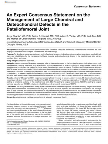
6Chahla et alThe Orthopaedic Journal of Sports MedicineFigure 1. Stacked leaning bar chart representing the breakdown in agreement levels in the third-round Delphi survey. Bars to theleft of the y-axis indicate disagreement, with bars to the right indicating agreement.imaging.8,19,23,47 Location matching has been reported toimprove the congruence between the graft and recipientsurfaces.86Experts agreed that chondrocyte viability is crucial tograft survival. To this point, the preservation technique(fresh grafts have greater chondrocyte viability thancryopreserved grafts), 19,47 timing (implantation in 28 days),2,6,95 and technique of implantation (avoidingimpaction)9,71 can significantly affect chondrocyte viability.In this regard, higher impact loads may be encounteredwhen the graft is thicker than 10 mm and when there is a 2-mm difference between the graft and recipient hole, andthus, thinner plugs are now recommended that match thedepth of the socket.71,91 Laboratory studies demonstratedthat high-impact loads adversely affect cell viability, withless than 50% to 70% of the cells remaining viable in that
The Orthopaedic Journal of Sports MedicineManagement of L
Results: A total of 28 experts (100% response rate) completed 3 rounds of surveys. After 3 rounds, 36 statements achieved a consensus, with over 75% agreement and less than 20% disagreement. A consensus was reached in 100.00% of the statements relating to functional anatomy of the patellofemoral joint, 88.24% relating to surgical indications, 100.00% relating to surgical technical aspects, and .