Transcription
Lin et al. BMC Complementary and Alternative Medicine (2017) 17:88DOI 10.1186/s12906-017-1611-9RESEARCH ARTICLEOpen AccessChlorella sorokiniana inducesmitochondrial-mediated apoptosis inhuman non-small cell lung cancer cells andinhibits xenograft tumor growth in vivoPing-Yi Lin1,2†, Ching-Tsan Tsai3†, Wan-Ling Chuang1, Ya-Hsuan Chao4, I-Horng Pan5, Yu-Kuo Chen6,Chi-Chen Lin2,4,7* and Bing-Yen Wang8,9,10,11*AbstractBackground: Lung cancer is one of the leading causes of cancer related deaths worldwide. Marine microalgae area source of biologically active compounds and are widely consumed as a nutritional supplement in East Asiancountries. Ithas been reported that Chlorella or Chlorella extracts have various beneficial pharmacological compounds thatmodulate immune responses; however, no studies have investigated the anti-cancer effects of Chlorella sorokiniana(CS) on non-small cell lung cancer (NSCLC).Methods: In this study, we evaluated the anti-cancer effects of CS in two human NSCLC cell lines (A549 and CL1-5human lung adenocarcinoma cells), and its effects on tumor growth in a subcutaneous xenograft tumor model. Wealso investigated the possible molecular mechanisms governing the pharmacological function of CS.Results: Our results showed that exposure of the two cell lines to CS resulted in a concentration-dependentreduction in cell viability. In addition, the percentage of apoptotic cells increased in a dose-dependent manner,suggesting that CS might induce apoptosis in human NSCLC cells. Western blot analysis revealed that exposure toCS resulted in increased protein expression of the cleaved/activated forms of caspase-3, caspase-9, and PARP,except caspase-8. ZDEVD (caspase-3 inhibitor) and Z-LEHD (caspase-9 inhibitor) were sufficient at preventingapoptosis in both A549 and CL1-5 cells, proving that CS induced cell death via the mitochondria-mediatedapoptotic pathway. Exposure of A549 and CL1-5 cells to CS for 24 h resulted in decreased expression of Bcl-2protein and increased expression of Bax protein as well as decreased expression of two IAP family proteins, survivinand XIAP.Conclusions: We demonstrated that CS induces mitochondrial-mediated apoptosis in NSCLC cells viadownregulation of Bcl-2, XIAP and survivin. In addition, we also found that the tumors growth of subcutaneousxenograft in vivo was markedly inhibited after oral intake of CS.Keywords: Chlorella sorokiniana, Human non-small cell lung cancer cells, Mitochondrial apoptotic pathway* Correspondence: lincc@dragon.nchu.edu.tw; 156283@cch.org.tw†Equal contributors2Department of Medical Research, China Medical University Hospital,Taichung, Taiwan8Division of Thoracic Surgery, Department of Surgery, Changhua ChristianHospital, Changhua, TaiwanFull list of author information is available at the end of the article The Author(s). 2017 Open Access This article is distributed under the terms of the Creative Commons Attribution 4.0International License (http://creativecommons.org/licenses/by/4.0/), which permits unrestricted use, distribution, andreproduction in any medium, provided you give appropriate credit to the original author(s) and the source, provide a link tothe Creative Commons license, and indicate if changes were made. The Creative Commons Public Domain Dedication o/1.0/) applies to the data made available in this article, unless otherwise stated.
Lin et al. BMC Complementary and Alternative Medicine (2017) 17:88BackgroundLung cancer is one of the leading causes of cancer relateddeaths worldwide. In 2012, an estimated 1.8 million newcases of lung cancer were diagnosed, accounting forapproximately 13% of all cancer diagnoses [1]. Non–smallcell lung cancer (NSCLC) is the most common type oflung cancer and the 5-year survival rate for patientswith this disease is about 22% [2]. Current treatmentsfor NSCLC such as surgery, chemotherapy andradiotherapy are not sufficient and the outcomes oftreatment remain poor because of drug resistance andtoxicity.Marine microalgae contain several groups of biologically active compounds including proteins, minerals,vitamins, fatty acids, and antioxidants [3]. Chlorella is aunicellular green microalgae and is widely consumed asa nutritional supplement in East Asian countries. Chlorella or Chlorella extracts have been demonstrated tomodulate immune response [4, 5], decrease hepatitis Cvirus viral load [6], and to have anti-cancer effects in human prostate cancer cell lines [7], human lung cancercells [8], liver cancer cells [9], and human colon cancercells [10]. However, the underlying molecular mechanisms governing the antitumor activity of Chlorellaremain unclear.Chlorella sorokiniana (CS), a species of green algae ofthe genus chlorella, has been demonstrated to be effectiveat inducing immune responses [4]; however, no studieshave investigated the anti-cancer effects of CS in NSCLCcells. In this study, we evaluated the anti-cancer effects ofCS in two NSCLC cell lines, namely A549 and CL1-5human lung adenocarcinoma cells, and its effects ontumor growth in a subcutaneous xenograft tumor model.We also investigated the possible molecular mechanismsgoverning the pharmacologic function of CS.MethodsPage 2 of 8and vacuum concentrated at 60 C until the solid contentof liquid extract was 5%.Cell viability assayThe cytotoxic effects of CS cellular viability were assessedusing the 3-(4,5-dimethylthiazol-2-y1) -2,5-diphenyltetrathallium bromide (MTT) assay (Sigma-Aldrich, St. Louis,MO). Cells were plated in 24-well plates for 24 h, afterwhich various concentrations of CS (0–1000 ng/ml) wereadded and incubated for 24 h. After removing the mediumand adding 200 μL of 1 MTT solution to each well andallowed to incubate for 4 h. The resulting formazancrystals were then dissolved by adding 600 μL DMSO.The spectrophotometric absorbance was measured at570 nm in a microplate reader (TECAN, Durham, NC).Cell lysis and western blot analysisWhole cell protein was lysed in 2% SDS comprising10 mM EDTA, 50 mM Tris base, 10% SDS pH 8.0 andboiled at 95 C for 10 min. Protein concentrations wereassessed using a BCA protein assay kit (Pierce, Rockford,IL). Equal amounts of protein were loaded on 10–15%SDS PAGE gels, transferred to polyvinylidene difluoride(PVDF, GE Healthcare, Freiburg, Germany) membranesand blocked with 5% nonfat milk in TBST buffer(20 mM Tris-HCl,120mMNaCl, 0.1% Tween 20) for 1 h.Membranes were incubated with various primary antibodies against cleaved caspase-3, cleaved caspase-9,cleaved-8, cleaved-PARP, Bcl-2, Bax, XIAP, survivn, cytochorome c, COX-IV, and β-actin (all from cell signaling,Danvers, MA) at 4 C overnight. After washing, the blotswere incubated with HRP-labeled secondary antibodies(cell signaling, Danvers, MA) for 2 h. The signals on theblots were then developed using the enhanced chemiluminescence system (ECL, Perkin Elmer, Waltham, MA)and analyzed with the Hansor Luminescence Imagesystem (Taichung, Taiwan).Cell culture and reagentsThe human lung adenocarcinoma cell lines A549 andCL1-5 were cultured in Dulbecco’s modified eaglemedium (DMEM, Invitrogen, Rockville, MD) supplemented with 10% fetal bovine serum (FBS, Gibco,Grand Island, NY) and 100 U/ml of penicillin (Gibco,Grand Island, NY) and 100 mg/ml of streptomycin(Gibco, Grand Island, NY). Cultures were maintainedat 37 C in a humidified incubator in an atmosphereof 5% CO2.Chlorella sorokiniana extract (CSE)Chlorella sorokiniana, single-cell thermophilic greenalgae, was provided by International CryptomonadalesBiotechnology (Taiwan). The Chlorella sorokiniana powders were refluxed with distilled water for 1 h. Theextract was filtered by N0.5 filter paper (Tokyo, japan)Mitochondria/Cytosol fractionationThe mitochondrial fraction from the cytosolic fraction ofA549 and CL1-5 cells was isolated using a mitochondria/cytosol fractionation kit (Biovision, Mountain View, CA).Cells were harvested by trypsinization and washed twicewith cold PBS. Then, all cells resuspended in 500 μL ofcytosol extraction buffer and maintained on ice for10 min. Cells were homogenized on ice for 30 to 50 passesusing a Dounce tissue grinder (Biovision, Mountain View,CA) and centrifuged at 3000 rpm for 10 min at 4 C. Thesupernatants were collected and centrifuged again at13,000 rpm for 30 min at 4 C.Cell cycle analysis and sub-G1 measurementCells (3 105 cells/dish) were plated on 6-cm dishesfor 24 h. The cells were then treated with various
Lin et al. BMC Complementary and Alternative Medicine (2017) 17:88concentrations of CS for different of time intervalsand then collected by centrifugation. The pellets werefixed with 75% ethanol at 20 C overnight. Cellswere then centrifuged and resuspended in 500 μL ofPI staining solution comprising 2 mg/mL RNase,1 mg/mL PI and 5% Triton X-100 maintained at 37 C for30 min in the dark. The cells were then analyzed usingAccuri 5 flow cytometry (BD Biosciences, San Jose, CA).The percentages of cells distribution of the cell cycle indifferent phases (sub-G1, G0/G1, S, and G2/M) were calculated using C6 Accuri system software (BD Biosciences,San Jose, CA).Annexin V assayApoptosis assay was measured using a BioVision annexinV-FITC apoptosis detection kit (BioVision, MountainView, CA). A549 and CL1-5 cells were implanted onto 6cm dishes for 24 h and then treated with different doses ofCS for 12 h. Cells were harvested by trypsinization andthen resuspended in 100 μL of binding buffer. Cell suspensions were incubated with 1 μL of annexinV-FITC and1 μL of propidium iodide (PI) for 10 min at roomtemperature in the dark. The cells were assessed immediately by Accuri 5 flow cytometry (BD Biosciences, SanJose, CA). The percentage of apoptotic cells (annexin v )was calculated with C6 Accuri system software (BDBiosciences, San Jose, CA).Mitochondrial membrane potential assayThe mitochondria-specific cationic dye JC-1 (Invitrogen,Carlsbad, CA), which undergoes potential-dependent accumulation in mitochondria, was used to measure membrane potential. When the membrane potential (ΔΨ) isbelow 120 mV, JC-1 is monomeric and emits green light(540 nm) following excitation with blue light (490 nm).At membrane potentials higher than 120 mV, JC-1monomer aggregates and emits red light (590 nm)following excitation with green light (540 nm). For theassay, cells were seeded onto 96-well plates and treatedwith various concentrations of CS for 24 h, followed bystaining with 10 μg/ml JC-1 for 30 min at 37 C. Thesamples were analyzed immediately using Accuri 5 flowcytometry (BD Biosciences, San Jose, CA). The datawere analyzed using C6 Accuri system software (BDBiosciences, San Jose, CA) and displayed in a dot plot ofJC-1 red fluorescence (Y-axis) against JC-1 green fluorescence (X-axis).In vivo antitumor activityIn total, 1 107 CL1-5 cells were subcutaneouslyinjected into the right flank of 16-week-old femaleBALB/c nu/nu mice. Tumor-bearing mice were subdivided into groups of five mice. Therapy was initiated10 days after tumor inoculation when the mean tumorPage 3 of 8volume was 50 mm3. CS was dissolved in ddH2O andmice were fed a daily dose of 50 mg CS/kg body weight.Control mice were fed orally with 100 μL of water.Tumor volumes were calculated as (width)2 x length/2and expressed in mm3.Statistical analysisStatistical analyses were performed by using GraphPadPrism software, version 4.0 (GraphPad Software, Inc., LaJolla, CA, USA). Results are expressed as mean standarddeviation. Statistical analyses were performed by one-wayANOVA followed by Tukey’s post-hoc test. The differences between two groups were compared using theStudent’s t test. A P-value 0.05 was considered to represent statistical significance.ResultsCytotoxic and cell viability effects of CS in A549 andCL1-5 cellsTo determine the cytotoxic effects of CS on cells, A549and CL1-5 cells were treated with 15.625 to 1000 ng/mlCS for 24 h and then cell viability was determined usingthe MTT assay. As shown in Fig. 1, exposure of the twocell lines to CS resulted in a concentration-dependentreduction in cell viability.CS induces apoptosis in A549 and CL1-5 cellsTo examine whether CS causes cell growth inhibition byinducing cell-cycle arrest or apoptosis, A549 and CL1-5cells were assayed using PI staining and subjected toflow cytometric analysis. The results are presented inFig. 2a. No cell cycle arrest was noted after 24 h ofexposure to CS; however, there was a significant dosedependent increase in the number of cells in the sub-G1Fig. 1 Effects of Chlorella sorokiniana (CS) on viability of A549 andCL1-5 cells. Cells were treated with the indicated concentrations ofCS for 24 h following attachment. Cell viability was assessed by theMTT assay. The viability of untreated cells (control) was considered100%. Each point on the graph represents the mean SD of triplicatewells. The data presented are representatives of three independentexperiments with similar results. ***P value 0.001 compared with thecontrol group
Lin et al. BMC Complementary and Alternative Medicine (2017) 17:88Page 4 of 8Fig. 2 Effects of CS on cell-cycle distribution and apoptosis in A549 and CL1-5 cells. a Cell-cycle analysis of CS-treated cells. Cells were treatedwith the indicated concentrations of CS for 24 h and then subjected to cell cycle analysis. b Flow cytometry analysis of CS-induced apoptosis inA549 and CL1-5 cells. The cells were treated with the indicated concentrations of CS for 24 h and then subjected to Annexin V/PI staining. Themeans SD of the experimental triplicates are presented in the bar graph. All data are representative of three independent experiments withsimilar results. *P value 0.05, **P value 0.01, ***P value 0.001 compared with the control groupphase, which is typically considered to indicate apoptosis. To further determine whether CS induced apoptosis, we used flow cytometry after staining with annexinV-FITC and propidium iodide (PI). As shown in Fig. 2b,the percentage of apoptotic cells (annexin-V /PI- andannexin V /PI ) increased in a dose-dependent manner,suggesting that CS might induce apoptotic cell death inhuman NSCLC cells.CS induces caspase-dependent cell death in A549 andCL1-5 cellsChemotherapeutic agents can elicit cell death via oneof two apoptotic signal transduction pathways, namelyan intrinsic (mitochondria-mediated) or extrinsic pathway.These pathways converge at several downstream points,including caspase-3, and/or caspase-7. Activated caspase3 and/or caspase-7 cleave poly (ADP-ribose) polymerase(PARP), which eventually leads to apoptosis [11]. Thus, inorder to clarify the type of a CS-induced apoptotic pathway, the cleaved forms of caspase-8, caspase-9, caspase-3and PARP were measured by Western blotting. As presented in Fig. 3a, the protein expression of the cleaved/activated forms of caspase-9, caspase-3, and PARP, but notcaspase-8, were increased in both cell lines after exposureto CS for 24 h. Activation of caspase-9 and caspase-3proteins suggests that the mitochondrial pathway is involved in apoptosis. Besides, we used various caspase inhibitors to further confirm our finding. As showed inFig. 3b, the specific caspase 8 inhibitor, Z-IETD was insufficient to increase cell viability, thereby excluding the possibility of involvement of the extrinsic pathway in CSinduced apoptosis. However, ZDEVD (caspase-3 inhibitor)and Z-LEHD (caspase-9 inhibitor) were sufficient at maintaining cell viability, implying that the mitochondriamediated apoptotic pathway was the mechanism throughwhich CS induced cell death.CS caused loss of mitochondrial membrane potential andcytochrome C releaseTo confirm whether the mitochondria-mediated apoptotic pathway was involved in the process of apoptosis,the mitochondrial membrane potential (Δψm) in CStreated A549 and CL1-5 cells was measured. Loss ofmitochondrial membrane potential is an indicator ofmitochondrial damage during the process of apoptosis.We used the fluorescent cationic dye JC-1 to evaluatethe effect of CS on membrane potential. A dosedependent decrease was observed in red fluorescence inboth cell lines, suggesting that CS treatment led to areduction in Δψm (Fig. 4a). A loss of Δψm can lead to
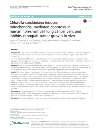
Lin et al. BMC Complementary and Alternative Medicine (2017) 17:88Page 5 of 8Fig. 3 Effects of CS on caspase activation in A549 and CL1-5 cells. a Cells were treated with the indicated concentrations of CS for 24 h. Total cellproteins were extracted and immunoblotted with antibodies to detect the cleaved forms of caspase-8, caspase-9, caspase-3, and PARP. b Cellswere treated with CS (250 ng/ml) and/or the indicated caspase inhibitor for 24 h, and then cell viability was analyzed by the MTT assay. Themeans SD of the experimental triplicates are presented in the bar graph. All data are representative of three independent experiments withsimilar results. **P value 0.01, ***P value 0.001 compared with the control groupcytochrome c release from the mitochondria into thecytosol, an important event for apoptosis induction [12].Thus, the cytosolic fractions of A549 and CL1-5 cellstreated with 125 and 250 ng/ml of CS were preparedand analyzed for cytochrome c release by Westernblotting. As presented in Fig. 5a, the expression of cytochrome c in the cytosolic fractions was observed in bothA549 and CL1-5 cells treated with CS. Taken together,our findings show that CS induces cell death via themitochondrial-mediated pathway.CS reduced Bcl-2, Bcl-xl and IAPs protein expression inA549 and CL1-5 cellsBcl-2 family members are regulators of cytochrome crelease from mitochondria during the process of apoptosis[13]. We assessed the protein expression of Bax, a proapoptotic member of the Bcl-2 family that triggers cytochrome c release, and Bcl-2, an antiapoptotic member ofthe family that inhibits cytochrome c release. As shown inFig. 5b, CS treatment for 24 h resulted in decreased thelevels of Bcl-2 protein and increased the expression of Baxprotein. In addition to the Bcl-2 family, the IAP family ofproteins also regulates caspase activity, thus affectingapoptosis [14]. Therefore, we measured the expression oftwo IAP family proteins, namely survivin and XIAP. Asdemonstrated in Fig. 5b, the levels of XIAP and survivinprotein were markedly reduced after CS treatment, particularly in the A549 and CL1-5 cell lines.CS inhibits CL1-5 tumor growth in a subcutaneousxenograft tumor modelThe prominent inhibitory effect of CS on NSCLC cellproliferation in vitro suggested that CS might suppresstumor growth in vivo. To verify this hypothesis, femaleBALB/c nu/nu mice were subcutaneously inoculatedwith CL1-5 cells and then fed orally with CS starting onday 10 after tumor inoculation. As shown in Fig. 6a, CSat 50 mg/kg resulted in a marked reduction in tumorvolume. Representative images of mice containing CL1-5xenografts on day 21 are presented in Fig. 6b. These results indicate that CS also inhibits the growth of NSCLCcells in vivo.
Lin et al. BMC Complementary and Alternative Medicine (2017) 17:88Page 6 of 8Fig. 4 Effects of CS on mitochondrial membrane potential in A549 and CL1-5 cells. a and b Cells were treated with the indicated concentrationsof CS for 24 h and then subjected to JC-1 fluorescence dye staining. The change in mitochondrial membrane potential (ΔΨm) was examined byflow cytometry. c The means SD of the experimental triplicates are presented in the bar graph. Data are representative of three independent experiments with similar results. *P value 0.05, ***P value 0.001 compared with the control groupDiscussionSeveral species of Chlorella have been reported tohave anti-cancer effects in various cancer cell lines.The most commonly studied Chlorella species areChlorella vulgaris and Chlorella ellipsoidea. C. vulgarishas been shown to have anti-cancer effects by inducing apoptosis signaling cascades in the hepatoma cellline HepG2 [15] and in rat models of liver cancer[16]. A study comparing the anti-proliferative effectsof C. ellipsoidea with those of C. vulgaris in humancolon cancer cells revealed that the apoptosis-inducingeffect of C. ellipsoidea extract was almost 2.5 timesstronger than that of C. vulgaris extract [10]. However, tothe best of our knowledge, no studies have explored theanti-cancer effects of C. sorokiniana (CS). The chemicalconstituents of CS contain three liposoluble fractions andfive compounds namely, (22E, 24R)-5alpha, 3betaepidioxiergosta-6, 22-dien-3beta-ol, (24S)-ergosta-7-en3beta-ol, loliolide, stigmasta-7, 22-dien-3beta, 5alpha,6alpha-triol, and ne [17]. In our study we found that CStreatment resulted in an increase in the Bax/Bcl-2 ratioand a decrease in expression of IAP family proteins XIAPand survivin in human NSCLC cells. We also found thatoral feeding of CS resulted in a marked reduction intumor volume in a xenograft tumor model in mice. Ourresults, therefore, demonstrate that CS induces apoptosisin vitro and in vivo.The anti-cancer effects of some natural compoundshave been shown to be the result of their ability tomodulate apoptosis signaling pathways [18]. In thisstudy, flow cytometric analysis of PI-labeled cells
Lin et al. BMC Complementary and Alternative Medicine (2017) 17:88Page 7 of 8Fig. 5 Effects of CS on the expression of cytosolic cytochrome C,Bcl-2 family and IAP proteins in A549 and CL1-5 cells. Cells weretreated with the indicated concentrations of CS for 24 h. a Cytosolicfractions were extracted and subjected to immunoblotting withanti-cytochrome c antibody. b Total cell proteins were extracted andimmunoblotted with antibodies to detect Bcl-2, Bax, XIAP, andsurvivin. All data are representative of three independent experimentswith similar resultsrevealed that exposure to CS resulted in a significantincrease in percentage of cells showing elevated hypodiploid DNA content (sub-G1) (Fig. 2a), indicating thatCS induces apoptosis in A549 and CL1-5 cells.Cells undergo cell death through two major signalingpathways, namely the mitochondria-dependent pathway(intrinsic pathway) or the cell surface death receptorsignaling pathway (extrinsic pathway) [19]. Caspases playimportant roles in the regulation of cell death andinflammation [20]. In our study, the presence of earlyapoptotic cells (annexin V /PI ) (Fig. 2b), activatedforms of caspase-9 and caspase-3, and PARP cleavageindicated that apoptosis (Fig. 3a), rather than necrosis,was involved in CS-induced A549 and CL1-5 cell death.Bcl-2 family proteins are involved in mitochondrialdysfunction and participate in the mechanism of chemotherapy resistance [21]. The loss of mitochondrialmembrane potential is associated with apoptosis viaregulation of Bcl-2 family members. The Bcl-2 familycomprises anti-apoptotic proteins such as Bcl-2, XIAP,and survivin, and pro-apoptotic proteins such as Bad,Bax, and Bid [13]. Moreover, IAPs (inhibitors of apoptosis) can inhibit apoptosis by directly binding to activated effector caspases, such as caspase-3 and caspase-7,and also inhibit mitochondrial cytochrome c-inducedcaspase-9 activation [14, 22]. Cytochrome c release fromFig. 6 Effects of CS on CL1-5 xenograft tumor growth in vivo.a Mean tumor volume was measured at the indicated days aftertumor implant. b Representative images of mice containing CL1-5xenografts on day 21. Experiments were repeated two times andprovided similar resultsmitochondria into the cytosol acts as a caspase activatorand participates in the mitochondrial apoptotic pathway[23]. In this study we identified apoptotic cells bymeasuring cytochrome c release using a cytometrictechnique with fluorescence microscopy. The releaseof this mitochondrial component was labeled by thestaining pattern (Fig. 5a). Our results proved that CSinduces mitochondrial-mediated apoptosis in humanNSCLC cells.ConclusionsIn conclusion, we demonstrated that caspase-dependentmitochondrial dysfunction is the mechanism throughwhich CS induces apoptosis in NSCLC cells. Downregulation of Bcl-2, XIAP and survivin suggests that CS increases the susceptibility of NSCLC cells to apoptosisinduction. In addition, our data show that the growth ofsubcutaneous xenograft tumors was markedly inhibitedby oral administration of CS.
Lin et al. BMC Complementary and Alternative Medicine (2017) 17:88AbbreviationsCS: Chlorella sorokiniana; CSE: Chlorella sorokiniana extract; NSCLC: Non-smallcell lung cancer; PARP: Poly (ADP-ribose) polymeraseFundingNo financial support for the research.Availability of data and materialsThe materials are available from the authors.Authors’ contributionsCCL, PYL, and BYW designed the study. CTT and YHC performed theexperiments. IHP and YKC contributed data analysis. PYL wrote themanuscript. WLC revised the manuscript. All authors read and approvedthe final manuscript.Page 8 of 89.10.11.12.13.14.Competing interestsThe authors declare that they have no competing interests.15.Consent for publicationThis information is not relevant.16.Ethics approvalAll experimental procedures on the animals were approved by the ResearchCenter for Animal Medicine of National Chung-Hsing University (Taichung,Taiwan).17.Author details1Transplant Medicine & Surgery Research Centre, Changhua ChristianHospital, Changhua, Taiwan. 2Department of Medical Research, ChinaMedical University Hospital, Taichung, Taiwan. 3Department of Public Health,School of Public Health, China Medical University, Taichung, Taiwan.4Institute of Biomedical Science, National Chung-Hsing University, Taichung,Taiwan. 5Biomedical Technology and Device Laboratories, IndustrialTechnology Research Institute, Hsinchu, Taiwan. 6Department of FoodScience, National Pingtung University of Science and Technology, Pingtung,Taiwan. 7Department of Biotechnology, Asia University, Taichung, Taiwan.8Division of Thoracic Surgery, Department of Surgery, Changhua ChristianHospital, Changhua, Taiwan. 9School of Medicine, Chung Shan MedicalUniversity, Taichung, Taiwan. 10School of Medicine, Kaohsiung MedicalUniversity, Kaohsiung, Taiwan. 11Institute of Genomics and Bioinformatics,National Chung Hsing University, Taichung, Taiwan.18.19.20.21.22.23.antioxidant property using supercritical carbon dioxide extraction.Process Biochem. 2010;45(12):1865–72.Mohd Azamai ES, Sulaiman S, Mohd Habib SH, Looi ML, Das S,Abdul Hamid NA, Wan Ngah WZ, Mohd Yusof YA. Chlorella vulgaristriggers apoptosis in hepatocarcinogenesis-induced rats. J Zhejiang UnivSci B. 2009;10(1):14–21.Cha KH, Koo SY, Lee DU. Antiproliferative effects of carotenoids extractedfrom Chlorella ellipsoidea and Chlorella vulgaris on human colon cancercells. J Agric Food Chem. 2008;56(22):10521–6.Wyllie AH, Kerr JF, Currie AR. Cell death: the significance of apoptosis. IntRev Cytol. 1980;68:251–306.Li P, Nijhawan D, Budihardjo I, Srinivasula SM, Ahmad M, Alnemri ES, Wang X.Cytochrome c and dATP-dependent formation of Apaf-1/caspase-9 complexinitiates an apoptotic protease cascade. Cell. 1997;91(4):479–89.Brunelle JK, Letai A. Control of mitochondrial apoptosis by the Bcl-2 family.J Cell Sci. 2009;122(Pt 4):437–41.Yang YL, Li XM. The IAP family: endogenous caspase inhibitors withmultiple biological activities. Cell Res. 2000;10(3):169–77.Yusof YA, Saad SM, Makpol S, Shamaan NA, Ngah WZ. Hot water extract ofChlorella vulgaris induced DNA damage and apoptosis. Clinics (Sao Paulo).2010;65(12):1371–7.Sulaiman S, Shamaan NA, Wan Ngah WZ, Mohd Yusof YA. Chemopreventiveeffect of Chlorella vulgaris in choline deficient diet and ethionine inducedliver carcinogenesis in rats. International Journal of Cancer Research.Int J Cancer Res. 2006;2(3):234–41.Zhang L, Liu PH, Wu JN, Yang GF, Suo YY, Luo N, Chen C. Studies onchemical compounds of Chlorella sorokiniana. Zhongguo Zhong Yao ZaZhi. 2015;40(7):1325–9.Fulda S. Modulation of apoptosis by natural products for cancer therapy.Planta Med. 2010;76(11):1075–9.Jin Z, El-Deiry WS. Overview of cell death signaling pathways. Cancer BiolTher. 2005;4(2):139–63.McIlwain DR, Berger T, Mak TW. Caspase functions in cell death and disease.Cold Spring Harb Perspect Biol. 2013;5(4):a008656.Kirkin V, Joos S, Zornig M. The role of Bcl-2 family members in tumorigenesis.Biochim Biophys Acta. 2004;1644(2–3):229–49.Parrish AB, Freel CD, Kornbluth S. Cellular mechanisms controlling caspaseactivation and function. Cold Spring Harb Perspect Biol. 2013;5:6.Saelens X, Festjens N, Vande Walle L, van Gurp M, van Loo G, Vandenabeele P.Toxic proteins released from mitochondria in cell death. Oncogene.2004;23(16):2861–74.Received: 3 December 2016 Accepted: 27 January 2017References1. Torre LA, Bray F, Siegel RL, Ferlay J, Lortet-Tieulent J, Jemal A. Global cancerstatistics, 2012. CA Cancer J Clin. 2015;65(2):87–108.2. Howlader N, Noone AM, Krapcho M, Miller D, Bishop K, Altekruse SF,Kosary CL, Yu M, Ruhl J, Tatalovich Z, et al. SEER Cancer StatisticsReview, 1975–2013. April 15, 2016th ed. Bethesda: National CancerInstitute; 2016.3. de Jesus Raposo MF, de Morais RM, de Morais AM. Health applications ofbioactive compounds from marine microalgae. Life Sci. 2013;93(15):479–86.4. Chou NT, Cheng CF, Wu HC, Lai CP, Lin LT, Pan IH, Ko CH. Chlorellasorokiniana-induced activation and maturation of human monocyte-deriveddendritic cells through NF-kappaB and PI3K/MAPK pathways. Evid BasedComplement Alternat Med. 2012;2012:735396.5. Bae MJ, Shin HS, Chai OH, Han JG, Shon DH. Inhibitory effect of unicellulargreen algae (Chlorella vulgaris) water extract on allergic immune response.J Sci Food Agric. 2013;93(12):3133–6.6. Azocar J, Diaz A. Efficacy and safety of Chlorella supplementation inadults with chronic hepatitis C virus infection. World J Gastroenterol.2013;19(7):1085–90.7. Renju GL, Muraleedhara Kurup G, Bandugula VR. Effect of lycopene isolatedfrom Chlorella marina on proliferation and apoptosis in human prostatecancer cell line PC-3. Tumour Biol. 2014;35(11):10747–58.8. Wang HM, Pan JL, Chen CY, Chiu CC, Yang MH, Chang HW, Chang JS.Identification of anti-
ately by Accuri 5 flow cytometry (BD Biosciences, San Jose, CA). The percentage of apoptotic cells (annexin v ) was calculated with C6 Accuri system software (BD Biosciences, San Jose, CA). Mitochondrial membrane potential assay The mitochondria-specific cationic dye JC-1 (Invitrogen, Carlsbad, CA), which undergoes potential-dependent ac-