Transcription
REVIEW ARTICLEThoracolumbar Injuries in the AthleteNajeeb Khan, MD, Sohail Husain, MD, and Michael Haak, MDAbstract: Acute thoracolumbar injury in the athlete can be adisabling condition that requires thorough evaluation andtreatment. Although most thoracolumbar spine injuries arebenign myofascial strains that respond well to nonsurgicalmanagement, the spectrum of injuries is broad and includesfractures and bony instability, ligamentous instability, andneurologic compromise. Evaluation of thoracolumbar injuriesrequires a rapid and focused evaluation at the time of injury torule out catastrophic and neurologically threatening injuries; adetailed history and physical examination carried out at a laterpoint in time should be paired with appropriate imaging studies.Initial radiographs may be combined with dynamic radiography, bone scanning, computed tomography, or magneticresonance imaging to delineate the structural extent of injury.Acute treatment may be required and initiated at the time ofinjury; further treatment should be carried out once the natureand extent of the injury is fully understood. Nonoperativetreatment is successful in most of the injuries. Operativetreatment is applied in selected cases of structural instabilityor neurologic compromise.Key Words: athlete, thoracolumbar injury, spondylolysis, parsinterarticularis, herniated disc, myofascial strain(Sports Med Arthrosc Rev 2008;16:16–25)Spine injuries account for 9% to 15% of all athleticinjuries.1–3 Low back pain is a limiting and disablingcondition affecting the athlete’s ability to train andcompete. Fortunately, most thoracolumbar injuries arebenign myofascial strains that are not severe or sportscareer threatening. On occasion, more severe injuriesinvolving fractures and bony instability, ligamentousinstability, or neurologic compromise may occur.A thorough history, physical examination, andappropriate imaging studies are needed to fully evaluateand treat an athlete with a thoracolumbar injury. Eachsport can lead to unique stresses and specific injuries; it isimportant to understand the demands of the sport,mechanisms of injury, and predisposing factors for injuryin each patient.From the Department of Orthopaedic Surgery, Northwestern FeinbergSchool of Medicine, Chicago, IL.No sources of support.Reprints: Michael Haak, MD, Department of Orthopaedic Surgery,Northwestern Feinberg School of Medicine, 675 N. St. Clair, Galter17-100, Chicago, IL 60611 (e-mail: mhaak2@nmff.org).Copyright r 2008 by Lippincott Williams & Wilkins16The purpose of this article is to provide a framework with which physicians, therapists, and trainers mayassess thoracolumbar injuries and develop a plan forevaluation and treatment of the injured athlete. Althoughthe principles used in evaluating and treating thoracolumbar injuries in the general population can be appliedto the athlete, the clinician must also consider the timingand safety of return to sports training and competition.ANATOMY AND BIOMECHANICSKnowledge of the pertinent anatomy is critical inthe evaluation of thoracolumbar injuries. The concept ofthe functional spinal unit (FSU) is helpful in delineatingthe anatomic spinal structures that are at risk to injury,and understanding the biomechanics of injury and loss ofstability. The FSU consists of 2 adjacent vertebral bodies,the interconnecting intervertebral disc ligament, the 2articulating facet joints, and the segmental components ofthe ligaments and muscles.The biomechanical function of the thoracolumbarspine is to allow flexibility and motion while protectingthe spinal cord and nerve roots from compression,tension, shear, and rotational forces. Injury occurs whenextrinsic forces exceed the intrinsic strength of the spineand surrounding elements. The spine requires coactivation of adjacent trunk muscle units to help direct forcesalong the most efficient pathway; this pathway follows thenormal curvature of the spine. Injury occurs when theseforces are misdirected and overwhelm posterior bony andsoft tissue structures. Catastrophic injury occurs whenboth anterior and posterior elements are disrupted, leadingto instability and possible neurologic compromise.Rotational or extension forces may fracture the parsinterarticularis (the connection between the superior andinferior facet joints) and damage the facet joints. Bonyinjury may be associated with extensive forces thatfrequently damage adjacent soft tissues and facet jointstructures. Articular fracture can occur with rapid flexionor extension forces in the thoracic spine. Excessiverotational forces in the lumbar spine can disrupt thefacet ligamentous capsule, with catastrophic failureleading to facet dislocation. Soft tissue injury to muscles,ligaments, and the intervertebral disc may vary dependingon the degree of force transfer, and can be isolated inassociation with bony or joint fracture or disruption.There are several unique anatomic aspects of thethoracic and lumbar anatomy to consider. The thoracicspine, enclosed and protected by the rib cage, hasvertically oriented facet joints to facilitate rotation.Sports Med Arthrosc Rev Volume 16, Number 1, March 2008
Sports Med Arthrosc Rev Volume 16, Number 1, March 2008Strong flexion-extension forces, combined with axial load,are not tolerated as well biomechanically. The lumbarfacet joints are oriented in a coronal fashion to allow flexionand extension, and are prone to failure when stressed withrotational forces. Transitional anatomic segments, such asthe cervico-thoracic junction just above the rib cage and thethoracolumbar junction at T12-L1, are particularly susceptible to shear and flexion-extension forces.Most of the load carrying capacity in the thoracolumbar spine resides in the vertebral body and intervertebral disc. Disruption of the disc and fracture of thebone can lead to macroinstability or microinstability,with pathologically greater motion at the damagedsegment. The thoracic spine demonstrates little flexionextension motion and primarily rotation owing toconnections at the costovertebral joint and linkage tothe sternum; loss of this protective ‘‘cage’’ may expose thethoracic spine to excessive motion and injury. The facetjoints and posterior interspinous ligaments in the lumbarspine carry approximately 10% to 20% of axial loadsthrough the posterior column.The degree of instability caused by the injurydepends on the number of disrupted structures. Acompression fracture involving only the anterior vertebralbody will have a small effect on the stability of the spine.Progressively greater instability will occur with damage tothe entire vertebral body, such as in a burst fracture. Afracture dislocation with damage to the vertebral bodyplus disruption of the posterior complex of facet jointsand interspinous ligaments may lead to catastrophicinstability and likely neurologic involvement.Although weight lifting may cause overload injuries,including compression fractures, contact sports such asfootball or hockey with instantaneous transmission ofhigh force loads may lead to disruption of all themechanical supports. Gymnastics and ballet, with repetitiveflexion-extension forces transmitted through the posteriorelements of the spine, may lead to stress fractures in the parsinterarticularis. Sports such as luge, skiing, and automobileand motorcycle racing place the athlete at risk forcatastrophic injury to the spine because of the high speedsinvolved. The sports-specific mechanism of injury must beconsidered as part of the athlete evaluation.PREDISPOSING CONDITIONSThe greatest predictor of low back pain in athletes isprior low back pain.4,5 Certain athletes have an increasedrisk of chronic back problems. Football players, forexample, have an increased risk of degenerative discdisease, facet degeneration, and chronic low back painthat is proportional to their years of involvement in theirsport.6 Weight lifting, particularly squatting, is associatedwith degenerative disc disease as well. In one study, 80%of male weight lifters showed signs of degenerative discdisease at the age of 40 years.4 Lumbar spondylolysis is asignificant problem for inner linemen and gymnasts whoundergo frequent axial loading and hyperextension of thespine. The incidence of injury to the pars interarticularis isr2008 Lippincott Williams & WilkinsAthletic Thoracolumbar Injuries11% in young gymnasts, as compared with 3% in theage-matched general population.5 The impact of lumbarflexibility on back pain is controversial.7,8 No correlationhas been found between lower-extremity flexibility andback pain.9–11Contact sports may create axial, flexion-extension,or rotational forces that are significant enough tooverload the biomechanical capabilities of the spine.Sports that take place at high speeds are likely to createmore severe injuries than low-speed sports. Gymnastsoccasionally perform on apparatuses where a miss maylead to a fall from a significant height, leading to axialforce overload. It is critical that the training of bothdeveloping and high-level athletes includes learning andreinforcing safe forms of sports-specific contact.Anterior abdominal and paraspinal musculatureplay an important role in stabilizing the spine. Sportsspecific training should be augmented by a progressiveoverall strength and fitness program. In the thoracolumbar area, the program should specifically develop spinalextensors and flexors, along with core abdominal musclesthat help to stabilize the lumbar spine. Rotational andparaspinal extensor musculature should be specificallydeveloped in the thoracic region.Young athletes are at particular risk for injury,especially during the adolescent growth spurt. Athletes incontact sports benefit from separation into various sizeand weight categories to prevent significant mismatches.Trainers should be aware of changes in coordination andagility skills. Athletes in the midst of a growth spurt mayhave muscle and ligament tightness and also diminishedlimb and joint proprioception after a significant heightchange. Appropriate attention should be directed torecovery after performance in all athletes as they maybe at higher risk for thoracolumbar injuries withinadequate rest between sports activities.CLINICAL EVALUATIONThere are 2 different clinical evaluations forthoracolumbar injuries in athletes. There is an on-siteevaluation, at the time of injury, whose sole objective is todetermine whether a serious injury has occurred. Thesecond clinical evaluation is carried out in the officeclinical setting, and is designed to be comprehensive withregard to history of injury and physical examination. Thisincludes appropriate imaging studies and is designed todefine the etiology of the symptoms.On-siteCorrect on-site management is important in preventing further injury. Helmets and shoulder pads aregenerally not removed on the field to avoid injury to thecervical spine. The facemask is removed to allow access tothe airway.The patient should be rapidly surveyed for airway,breathing, and circulatory condition, and resuscitationshould be initiated as indicated. Neck or back pain,weakness, numbness, or paralysis at the time of the injuryshould be assessed, and the neck should be immobilized17
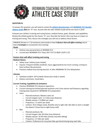
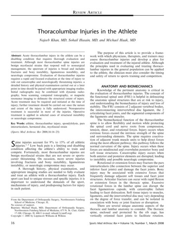
Sports Med Arthrosc RevKhan et alwith a collar and the remainder of the spine with abackboard as indicated. A brief history of the injury, withpossible mechanisms should be elicited from the athlete orother witnesses. The patient should be evaluated for lossof consciousness, altered mental status, and cooperation.Neurologic function should be determined before examining the spine by checking for voluntary motion of thehands and feet, and assuring that the patient has intactsensation on both sides of the body from head to toe.Clearly, any patient with indications for resuscitation has a true medical emergency, and should be urgentlytransported according to the emergency medical plan ofthe sports facility where the injury occurred. Spine painshould be viewed as a significant injury until provenotherwise; any evidence of neurologic deficit such asnumbness, weakness, or paralysis should only furtherindicate the urgency of transport for further evaluation.Athletes with lesser injuries determined by thescreening evaluation on-site should be transported orremoved from the field to a sideline or a locker roomlocation where a more careful examination can be carriedout. Only those patients with a truly minor injury, noneurologic deficit, and the demonstrated ability toperform at optimal level should be allowed to returnto competition. All other patients should be held out ofcompetition until a more comprehensive evaluation hasbeen accomplished, the cause of the symptoms has beendetermined, and they have responded to the treatmentprogram.OFFICE/HOSPITAL SETTINGThe physician’s examination after transfer to aclinical setting should be more comprehensive. Thehistory should include a qualitative description of Volume 16, Number 1, March 2008symptoms, when it started, the mechanism of the injuryfrom the athlete’s standpoint, and the evolution of thesymptoms since the time of injury. Predisposing factorssuch as previous injury and treatment, congenital andchildhood conditions affecting the thoracolumbar spine,and the patient’s practice and performance status beforethe injury may highlight susceptibility because of priorinjury and incomplete rehabilitation, susceptibility because of congenital structural changes, or lack of eitheroverall spinal or core spinal strength.A description of the mechanism of injury may behelpful in determining the injured structures (Table 1).Extension type mechanism places increased forces on theposterior elements; flexion type injuries tend to stress orinjure anterior elements, such as the vertebral bodies orintervertebral discs. Rotational injuries in the lumbarspine may strain or tear the facet capsules. Midline painlocation may indicate the involvement of central bony orligamentous structures; lateral pain or pain over thesacroiliac (SI) joint may be suggestive of ligamentousinjury in the facet or SI joints.A pain drawing allows patients to accurately showthe physician which areas are affected (Fig. 1) and canalso help the clinician to distinguish between radicularand nonradicular pain (Table 2).A careful physical examination should be carried outof the thoracolumbar spine, beginning with inspection ofthe spine for contour asymmetry, evidence of scoliosis orkyphosis, and swelling or ecchymosis over the injured area.A gentle lumbar lordosis is normal, but is often absent withparavertebral muscle spasm. Coronal plane deformity maybe because of congenital or idiopathic scoliosis, but it canalso be acquired from pain and spasm associated withherniated discs and other low back pathology.TABLE 1. Thoracolumbar Injuries in Specific SportsFootballGymnasticsWeight LiftingGolfingTrack and FieldWinter SportsEndurance sports18Football players undergo unique stresses and strains that can lead to thoracolumbar injuries. Blocking entails repeatedaxial loads, hyperextension, and torsional strain on the spine, which can lead to spondylolysis. Interior linemen are atincreased risk for acute spine injury and chronic back pain21,22Gymnasts often undergo axial loading and hyperextension of the spine resulting in lumbar spondylolysis. The incidenceof injury to the pars interarticularis is 11% in young gymnasts, as compared with 3% in the general age-matchedpopulation5Back pain in the weight lifter is usually because of strains and sprains of the spinal musculature and ligaments,spondylolysis, spondylolisthesis, and herniated nucleus pulposus. Rarely, catastrophic injuries can occur when theathlete uses poor technique and tries to lift too much weight. Incorrectly performing squats on Smith machine has beenreported to cause flexion-distraction injury to the thoracolumbar spine, causing cauda equina syndrome in 1 subject23Low back pain in the golfer correlates with decreased lumbar spine extension, lead hip internal rotation, and decreasedFABER distance (measured from the knee to the examination table with the hip in FABER such that the ipsilateralankle rests on the contralateral knee).24 It is suggested, though not proven, that increasing hip range of motion usingphysical therapy modalities can help to prevent low back pain in the golferAlthough the clinical significance is unknown, throwing athletes and high jumpers show more lumbar vertebralosteophytes than other types of athletes25Skiers and ski jumpers tend to have a higher rate of anterior endplate lesions and Schmorl nodes than the generalpopulation. The clinical significance is unknown, but when accompanied by low back pain, the condition is classified as‘‘atypical’’ Scheuermann disease26Butterfly swimmers and rowers are at risk for rib subluxation from the transverse processes of the thoracic spine, usuallyat the sixth and seventh level.27 The defining feature in this rare injury is pain in the posterior rib cage that radiates tothe thoracic spine that is exacerbated with deep breathing, coughing, sneezing, and trunk movement. The proposedetiology of this injury is subluxation of the costo-vertebral joint owing to forces of the serratus anterior muscle on thethoracic ribs. Once cardiac and pulmonary etiologies are ruled out and a diagnosis of rib subluxation is made, thetreatment is manipulation, which involves direct downward pressure on the affected area with the athlete lying proner2008 Lippincott Williams & Wilkins
Sports Med Arthrosc Rev Volume 16, Number 1, March 2008Athletic Thoracolumbar InjuriesPatient:Age:Date:Mark the areas on your body where you feel the described sensations. Use the appropriatesymbol. Mark the areas of radiation and include all affected areas.Ache Numbness OOOOOOOOOPins and NeedlesBurning XXX Radiating pain ///XXX///XXX///Neck Pain: %Arm Pain: %Back Pain: %Leg Pain: %Total 100%Please mark on the line: How bad is your pain now?01No Pain23456Intermediate pain78910Worst painFIGURE 1. A pain drawing that allows patients to accurately show the physician which areas are affected.Palpation examination should cover all superficialstructures, checking carefully for muscle spasm, triggering, soft tissue gaps in the muscle or interspinous processspaces, and bony shift. Range of motion should be testedin all planes.TABLE 2. Red FlagsCauda equina syndromeHerniated discInfectionNeoplasmFractureAbdominal aortic aneurysmr2008 Lippincott Williams & WilkinsA complete lower extremity neurologic examinationincludes evaluating motor strength, sensation, andreflexes. Patients with neurologic deficits should also beevaluated for spinal cord reflexes of myelopathy, such asclonus and the Babinski and Hoffman tests. Pulses andcapillary refill blood flow should be checked andcompared symmetrically. Skin changes over the lowerspine, including hair patches, skin tags, hyperpigmentedareas, and lipomas, can signify underlying congenitalspine pathology that was present before the injury.The straight leg raise test and contralateral straightleg test provoke radicular pain in disc herniations withnerve root impingement. The straight leg raise test isperformed with the patient supine while the physician19
Sports Med Arthrosc RevKhan et alelevates the affected lower extremity. Radicular painwhen raising the unaffected leg provides further evidenceof a herniated lumbar disc.A tobacco history should be elicited and smokersshould be encouraged to quit, as smoking has repeatedlybeen linked to back pain.12 The female athlete triad(disordered eating, amenorrhea, and osteoporosis) shouldbe considered in females, particularly those who participatein endurance sports or activities where appearance is judgedsuch as figure skating, gymnastics, and ballet.13,14 Moststress fractures associated with the female athlete triad areappendicular, although back pain owing to sacral stressfracture or a vertebral compression fracture is possible.Certain symptoms are red flags (Table 1) and canindicate a serious cause of back pain such as caudaequina, herniated disc, infection, neoplasm, fracture, orabdominal aortic aneurysm. The patient should bequestioned regarding saddle anesthesia, progressive neurologic deficit, and any history of bowel or bladderdysfunction, as the development of cauda equinasyndrome is a surgical emergency. Unremitting nightpain or unplanned weight loss and fatigue may besymptoms of neoplastic disease. Infection should beconsidered in any patient with systemic complaints(fevers, chills, fatigue, unplanned weight loss), recentinfections, intravenous drug abuse, night pain, orimmunosuppression. With a history of significant trauma,the clinician should consider the possibility of a fracture.Abdominal aortic aneurysm, although rare in a youngerindividual, can initially present as low back pain. Anabdominal examination would reveal a pulsatile mass.Blood work, including complete blood count, chemistry,sedimentation rate, C-reactive protein, and proteinelectrophoresis, may aid in evaluating infections orpossible neoplastic causes of back pain.IMAGINGIn athletes with back pain that does not respond torest and nonsteroidal anti-inflammatory drugs (NSAIDs),plain radiographs including 2 orthogonal views should beobtained. The anteroposterior (AP) view can be used toassess bony alignment and consistency of interpediculardistance (gradual widening proximal to distal). The lateralview shows vertebral height, sagittal alignment, facet joints,and evidence of spondylolisthesis. AP and lateral radiographs will demonstrate up to 90% of bony fractures andalignment issues. Additionally, oblique and dynamicflexion/extension views are useful to evaluate spondylolisthesis. Oblique views may show a pars defect in spondylolysis,often described as the collar on the Scotty dog.Dynamic radiographs should be taken underpatient-controlled active motion; forceful motion by theexaminer should never be performed. It is important toradiographically evaluate the entire spine in any patientwith a spine fracture, as up to 5% of these individualshave noncontiguous fractures.15Additional imaging studies, such as magneticresonance imaging (MRI) and computerized tomography20 Volume 16, Number 1, March 2008(CT) add another level of detail. CT is best for definingbony anatomy whereas MRI is preferred for delineatingsoft tissue injury, including herniated discs or ligamentousinjury. CT has been shown to be an excellent imagingmodality for identifying and characterizing pars defects,as spondylolysis is often subtle or not visible on radiographs.16 Three-dimensional images can be created withspecial software, and can be useful in the preoperativeplanning for treatment of complex fractures and fracturedislocations. Drawbacks include poorer delineation ofsoft tissues, ligaments, and the nerve root system andrelatively high radiation exposure.After plain radiographs, MRI has become popularas it can potentially diagnose pars abnormalities, discherniation and degeneration, malignancy, infection, andfacet arthropathy.17 However, MRI is limited by its highfalse positive rate. Physicians must interpret MRI resultswith the physical examination in mind. Up to 64% ofasymptomatic adults may have lumbar spine abnormalities on MRI.18Although MRI may not detect a chronic parsdefect, it can detect bone marrow edema and stressreaction that can precede its development. High fieldstrength (1.5 T or above) and fat saturation techniquescan help detect the full range of pars abnormalities fromearly marrow edema to chronic spondylolysis.17 Anemergent MRI should be ordered in patients withprogressive neurologic deficit or suspected cauda equinasyndrome. CT myelogram is the study of choice if MRIis contraindicated and the clinician needs to rule outstenosis of the spinal canal or foramina.Technetium bone scanning is a sensitive, butnonspecific study that is most useful in patients with ahigh clinical suspicion of spondylolysis with negative orequivocal plain radiographs. Single photon emission CThas been shown to be more sensitive than technetiumbone scan.19 Both scans are positive soon after the onsetof back pain as this is when the lesion is mostmetabolically active. A single photon emission CT scancan seem to be normal in chronic pars defects.20With the large number of possibilities for imagingthe thoracolumbar spine, it is critical to develop a rationalplan for which patients to image. Those athletes sustaining severe and catastrophic injuries, including fractures,fracture dislocations, or injuries with neurologic involvement, should obtain radiographs to identify the area ofinjury, and a CT and MRI to define the bony andligamentous damage to help plan possible surgicaldecompression and stabilization. All other patientsshould have a more selective imaging plan, with radiographs being obtained in those patients who fail toimprove or demonstrate progressive neurologic features.DIAGNOSTIC ENTITIESLumbar StrainThe most common cause of low back pain in theathlete is muscular strain. The degree of injury may varyfrom irritation of muscle bundles without disruption all ther2008 Lippincott Williams & Wilkins
Sports Med Arthrosc Rev Volume 16, Number 1, March 2008way to intrasubstance tearing of the muscle fibers or fascia.The athlete typically recalls an event that preceded theonset of symptoms and often has increased pain andstiffness the day after injury. In contrast to a herniated disc,lumbar strain does not cause pain radiating below the knee.Examination reveals midline and paraspinal tenderness on palpation. Pain is exacerbated with flexion andextension. Point tenderness over the gluteal muscles ortensor fascia lata without concomitant radicular symptoms is indicative of a myofascial strain, which is treatedsimilar to lumbar strain. The neurologic examination isnormal and the straight leg raise test is negative. Imagingis not necessary for a diagnosis, although plain radiographs and specialized imaging may be required ifsymptoms do not improve within 1 month.A soft tissue contusion over the thoracolumbarregion can occur because of a direct blow. The muscletissue may swell and present similar findings to the musclestrain. Mild contusions resolve relatively quickly; moresevere injury may require longer time to resolve.Lumbar SprainActing as an elastic stabilizer and extensor assist,the supraspinous and interspinous ligaments in thethoracolumbar spine can be injured by abrupt flexioninjuries. Flexion rotation injuries may damage the facetjoint capsule or costovertebral joints in the thoracic spine.Complete tears may allow hypermobility of the joint anddislocation is the worst-case scenario.Physical examination demonstrates midline tenderness and increased pain with direct pressure over theligamentous interval between spinous processes ordirectly over the process. Lateral radiographs showsplaying of the spinous processes. A ligamentous injurymay require 6 to 8 weeks to heal and requires guided andprogressive therapy and reconditioning to regain segmental stability.PIRIFORMIS SYNDROMEPosttraumatic piriformis syndrome is neuritis of theproximal sciatic nerve that can occur after a blow tothe buttock area. Patients have sitting intolerance andtenderness to palpation at the greater sciatic notch.Although some symptoms are similar to radicular pain,there are key differences in the diagnosis.Pain may be elicited by deep palpation over thepiriformis muscle. The Pace test (resisted abduction andexternal rotation of the lower extremity), the Freiberg test(forced internal rotation of the extended thigh), and theBeatty maneuver (abduction of the affected extremityagainst gravity) all reproduce pain that point towardsciatic nerve irritation by the piriformis.Radiographic and MRI imaging of the spine do notshow other causes for radicular pain, such as a herniateddisc or spinal stenosis. Piriformis syndrome is a diagnosisof exclusion that is best treated conservatively withNSAIDs, physical therapy, and stretching exercises.Surgical release of the sciatic nerve from the piriformisis rarely necessary.28r2008 Lippincott Williams & WilkinsAthletic Thoracolumbar InjuriesINTERVERTEBRAL DISC INJURYInjury to the intervertebral disc encompasses arange of injuries that affect the ligamentous function ofthe disc with regard to rotation (annulus fibrosis)and axial load transmission (nucleus pulposis). Althoughin some cases causing only back pain complaints, theherniation of nucleus pulposis material out of the annulusmay be associated with radicular neurologic complaintsbecause of nerve compression.A traumatic injury or series of repetitive traumas createsmall rents and tears between annular fibers. A final event orseries of events place asymmetric axial loads on the nucleuspulposis, and a portion of the nucleus pulposis migratesalong the zone of annular weakness into the canal. Ninetypercent of herniated discs are posterior or posterolateral;more rarely, the disc material compresses the nerve root in afar lateral location that is outside the nerve root foramen.Disc herniation initially presents as low back pain thatprogresses to radicular pain. Onset can be insidious or relatedto acute trauma. The most common levels are L4-5 and L5S1, accounting for 90% of all symptomatic disc herniation. Itis controversial whether the athletic population is predisposedto acute disc herniation.29 Low back pain in the adult is morelikely to be because of a herniated disc, whereas discogenicback pain is rare in the young athlete.30A very large herniation may compress a significantportion of the cauda equina, and patients with severepain, perineal numbness, or bowel and bladder complaints should be aggressively evaluated with MRI andundergo urgent operative treatment.Examination reveals low back and varying levels ofleg pain that is exacerbated by lumbar flexion and theValsalva maneuver. The most specific tests for discherniation are the straight leg raise and contralateralstraight leg raise tests. Physical e
(Sports Med Arthrosc Rev 2008;16:16-25) Spine injuries account for 9% to 15% of all athletic injuries.1-3 Low back pain is a limiting and disabling condition affecting the athlete's ability to train and compete. Fortunately, most thoracolumbar injuries are benign myofascial strains that are not severe or sports-career threatening.