Transcription
Gao et al. Cell Communication and 5(2022) 20:97Open AccessREVIEWTumor endothelial cell‑derived extracellularvesicles contribute to tumor microenvironmentremodelingJian Gao1,2, Xiaodong Zhang3,4, Lei Jiang5, Yan Li6* and Qianqian Zheng1*AbstractCancer progression involves several biological steps where angiogenesis is a key tumorigenic phenomenon. Extracellular vesicles (EVs) derived from tumor cells and other cells in the tumor microenvironment (TME) help modulateand maintain favorable microenvironments for tumors. Endothelial cells (ECs) activated by cancer-derived EVs haveimportant roles in tumor angiogenesis. Interestingly, EVs from ECs activate tumor cells, i.e. extracellular matrix (ECM)remodeling and provide more supplements for tumor cells. Thus, EV communications between cancer cells and ECsmay be effective therapeutic targets for controlling cancer progression. In this review, we describe the current knowledge on EVs derived from ECs and we examine how these EVs affect TME remodeling.Keywords: Extracellular vesicles, Tumor microenvironment, Angiogenesis, Remodeling, Targeted therapyBackgroundCancer is a complex multifactorial disease where normal cells acquire multiple traits to facilitate prolongedcell survival [1]. Tumor occurrence and development isinseparable from the tumor microenvironment (TME)which is a highly heterogeneous ecosystem incorporating: 1) immunocytes (T/B lymphocytes, tumor-associated macrophages, dendritic cells (DCs), natural killercells (NKs), myeloid-derived suppressor cells, and neutrophils); 2) stromal cells (cancer-associated fibroblasts(CAF), pericytes, and mesenchymal stromal cells); 3)extracellular matrix (ECM) and other secreted molecules (extracellular vesicles (EVs), growth factors,cytokines, and chemokines); and 4) blood and lymph vessel networks [2]. Such cell-to-cell interactions can lead toTME remodeling and may exert a significant impact on*Correspondence: ly135722507hld@126.com; qqzheng@cmu.edu.cn1Department of Pathophysiology, College of Basic Medical Science, ChinaMedical University, Shenyang 110122, China6Department of Radiotherapy, The First Affiliated Hospital of JinzhouMedical University, Jinzhou 121000, ChinaFull list of author information is available at the end of the articlecancer progression and metastasis, drug resistance, andimmunosuppression.Recently EVs, also known as exosomes (Exs), have beenrecognized as crucial signaling mediators in TME regulation. EVs are double-membrane vesicle-like bodies whichare shed from cell membranes or secreted by cells, withdiameters ranging from 40 to 1000 nm. EVs are mainlycomposed of microvesicles (MVs) and Exs [3]. To explorethis topic more comprehensively, the terms “EVs” and“Exs” are used interchangeably. EV-mediated communication networks between tumor and non-tumor cellsappears to be involved from tumor growth to metastasis. For example, a gastric cancer study reported that theEV cargoes inhibin subunit βA and thrombospondin 2were secreted by CAF-derived EVs to the TME to promote cancer [4]. Glioblastoma-derived EVs dramaticallypromoted neural progenitor cell proliferation and migration via the PI3K/Akt pathway [5]. A recent study alsoreported that EV transmitted long-non coding (lnc)ARSRwhich promoted sunitinib resistance in renal cancer[6]. EVs released by tumor cells could also remodel themicroenvironment by activating CAF [7] and promoting The Author(s) 2022. Open Access This article is licensed under a Creative Commons Attribution 4.0 International License, whichpermits use, sharing, adaptation, distribution and reproduction in any medium or format, as long as you give appropriate credit to theoriginal author(s) and the source, provide a link to the Creative Commons licence, and indicate if changes were made. The images orother third party material in this article are included in the article’s Creative Commons licence, unless indicated otherwise in a credit lineto the material. If material is not included in the article’s Creative Commons licence and your intended use is not permitted by statutoryregulation or exceeds the permitted use, you will need to obtain permission directly from the copyright holder. To view a copy of thislicence, visit http:// creat iveco mmons. org/ licen ses/ by/4. 0/. The Creative Commons Public Domain Dedication waiver (http:// creat iveco mmons. org/ publi cdoma in/ zero/1. 0/) applies to the data made available in this article, unless otherwise stated in a credit line to the data.
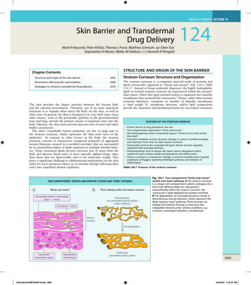
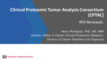
Gao et al. Cell Communication and Signaling(2022) 20:97immune escape [8] and angiogenesis [9]. Among themultiple stromal cell types in the TME, endothelial cells(ECs) are an enriched source of circulating EVs as theytransfer information to adjacent cell types [10].Several studies have now demonstrated that stromalcell-derived EVs promote tumorigenic phenotypes andthat tumor-derived EVs modify the host stroma. However, the effects of EVs released from ECs on TEM arerarely studied. In this review, the role of EC-derived EVsis highlighted to examine their contribution to tumorcells and immune cells.Loading and releasing EVsPrevious studies have confirmed that endosome sortingcomplexes required for transport (ESCRT) are widelyaccepted regulatory mechanisms for EV processing, formation, and release. ESCRTs are composed of ESCRT0, ESCRT-I, ESCRT-II, ESCRT-III, and vacuolar proteinsorting-associated protein 4 (VPS4) [11]. ESCRT-0 initiates pre-ubiquitinated protein sorting and forms intraluminal vesicles (ILVs). Under the action of ESCRT-I/II,ILVs undergo membrane fusion to form multi-vesicularbodies (MVBs). Inside the cell, MVB parts are degradedby lysosomes, while the remaining components movetoward cell membranes and fuse with them. At this time,MVBs are still connected to the membrane surface andESCRT-III forms a spiral structure during this process.This structure shrink the MVB neck and the cell membrane. At the same time, the VPS4 ATPase directly orindirectly hydrolyzes the MVB through hydrolysis, afterwhich MVBs are released extracellularly. EVs are alsoformed via an ESCRT-independent mechanism; Pmel17regulates ILV production and affects EV formation viaits luminal domain [12]. Also, the four-transmembraneCD63 protein mediates melanosome invagination in anESCRT-independent manner [13]. The PLP protein istransferred from lipid-rich endosomal membranes toILVs via an ESCRT-independent manner [14]. In addition to ESCRT-mediated pathways, Wei et al. identifiedphosphorylates RAB31 drives EGFR entry into multivesicular endosomes (MVE) to form ILVs and Exs, which isdependent of flotillin proteins in lipid raft microdomainsinstead of ESCRT [15].EV-release from inside the cell to the outside is synergistically coordinated via several steps which mainly acton MVB and cell membrane separation processes. Studies have confirmed the most important factors mediating EV release are GTPases, including Rab and RALGTPases. Up to now, nine GTPases have been implicated in EV release, including Rab2B, Rab5, Rab7, Rab9A,Rab11, Rab27A, Rab27B, Rab35, and RAL [16]. Rabs havevital regulatory roles transporting MVBs to subcellularlocations and fusing with cell membranes [17]. MVBsPage 2 of 10can be directly or indirectly bound to actin and microtubule scaffolds to facilitate intracellular targeted transport.Interestingly, Rab11 and its family assist MVB transportvia actin and dynein [18]. The process from MVBs to EVsrequires not only motor protein dynamics, but also MVBseparation from cell membranes which is particularlyimportant. Although the specific mechanisms wherebyRab participates in MVB dissociation at the cell membrane have not been confirmed, it is hypothesized Rabinitiates the direct or indirect assembly of soluble N-ethylmaleimide-sensitive factor attachment protein receptor(SNARE) complexes (Fig. 1).The SNARE complex has vital roles in MVB fusion withcell membranes, especially when promoting MVB release.The SNARE complex consists of two components, v- andt-SNAREs. The v-SNARE is located on the vesicle whilethe t-SNARE is located on the presynaptic membrane.Both v- and t-SNAREs pair up and form a complex [19];during this formation, the released energy draws MVBscloser to presynaptic membranes and promotes MVBmembrane fusion. A recent study in human leukemicK562 cells confirmed that VAMP7 (a v-SNARE protein)was involved in MVB plasma membrane fusion and exosomal release [20]. In mammals, SYX-5 (a t-SNARE proteins) was also involved in MVB fusion and promotedEV release [21]. In tumor cells, the key glycolysis enzymePKM2 stabilized SNAREs via SNAP-23 (a t-SNARE protein) phosphorylation to promote external EV release[22]. These studies confirmed that mediating EV releasewas a complex process involving multiple steps and factors. While the regulatory mechanism is not the same indifferent cells, the main processes often involve GTPasesand SNARE complexes. These observations raise anFig. 1 Schematic overview of extracellular vesicle uptake and release
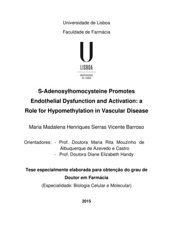
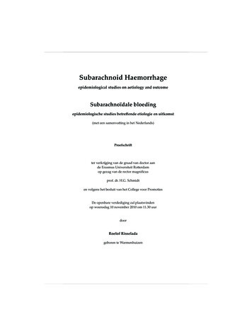
Gao et al. Cell Communication and Signaling(2022) 20:97important question in EV research: do different sortingmechanisms determine the loading of specific moleculesinto EVs? By interfering with this sorting mechanism, it ispossible to influence content loading in EVs to alter intercellular substance exchange in the TME.EV uptakeEVs released into the extracellular space are not only reabsorbed and used by EV-derived origin cells, but are alsotaken up by other cells in the microenvironment. Cellstake up EVs mainly via cell membrane fusion, endocytosis, and binding with specific surface receptors. Additionally, when carrying functional molecules, EVs transferinformation between cells and mediate many physiological and pathological processes. Tian et al. reported thatrat pheochromocytoma PC12 cell-derived Exs enteredand delivered microRNAs into bone marrow-derivedmesenchymal stromal cells and down-regulated theexpression of transforming growth factor β receptor IIand tropomyosin-1 [23]. In non-neoplastic diseases, ECstransduced with HDAdXMoAntimiR33a5p released Exsthat transferred anti-miR-33a-5p to other intimal celltypes, thereby upregulating cholesterol efflux from thesecells. [24]. At present, the decisive factors determiningEV uptake remain unclear. Edgar et al. reported that theprotein tether in mediating the attachment to the cellsurface and signal transmission of EVs [25]. Christianson et al. confirmed that on the surface of receptor cells,the heparin sulfate proteoglycan (HSPG) acted as an EVreceptor rather than just an attachment site on the EVsurface [26]. In mammals, differences in integrin familyexpression often affect EV uptake by receptor cells [27].Importantly, some mechanisms can inhibit uptake; CD47on EV surfaces protects EVs from engulfment by recipient cells and improves EV stability in the microenvironment [28].Tumor-derived EVs taken up by ECs often facilitateangiogenesis signaling and stimulate blood vessel formation [29]. In ECs, EV take-up is interceded via the collaboration of EV surface proteins, e.g., tetraspanins withthe membrane receptors of beneficiary cells [30]. In theTME, cell surface Tspan8-CD49d complexes from EVsderived from tumors are incorporated by vascular rodentECs to improve EC proliferation, migration, and activation [31]. EVs bearing Tspan8-4 complexes bind withintercellular adhesion molecule-1 (ICAM-1) and thenwere compassed by rat ECs [32]. In the absence of content delivery, EVs send messages to beneficial cells viasurface contacts, e.g., EVs containing major histocompatibility complex (MHC)-peptide complexes can activate Tcells via surface receptors [33]. Within 24 h, ECs internalize EVs produced by cancer cells using an internalization pathway. This was previously confirmed in studiesPage 3 of 10where ECs easily captured PKH26-dyed EVs during thefirst 4 h [34]. After internalization, EVs were immediately directed to the perinuclear zone; they moved to thecell periphery and entered advanced pseudopods whentubules were formed in vitro. After complete remodeling,adjacent ECs transported EVs to other ECs and cells inthe TME via nanoparticle structures [35].EC‑derived EV characterization, separation,and roles in tumor progressionEVs derived from ECs are implicated in several physiological and pathological conditions [36–38]. The particulate secretome of endothelia mirrors their moleculardiversity and possibly supports EC plasticity and adaptation within or between different vascular beds whilealso significantly impacting circulating cells, includingimmune cells in blood or lymph [39]. The markers CD63and CD81 are specifically expressed in EVs. In addition,several other EV-specific markers, i.e. CD31, CD54,CD62E, CD105, CD144, CD146, and von Willebrand factor also occur on EVs [32, 33]. Moreover, EC-derived EVsalso deliver proteins such as ICAMs, vascular endothelial (VE)-cadherin, E-selectin, platelet EC adhesion molecule-1, endoglin, and endothelial nitric oxide synthase[40–42]. While the exosomal endothelial markers CD54and CD62E are upregulated by various stimuli, CD31and CD105 are specifically increased by apoptosis [43].CD62E and CD144 are exclusively expressed by ECs,while the aforementioned molecules are not (Fig. 2).To improve specificity and sensitivity, combined multicolor antibodies (CD31 /CD41 , CD31 /CD42b , and CD105 /CD45 ) and monochrome composite markers (CD144 CD105 and C D146 CD105 ) are used toisolate EC-derived EVs. Because ECs express CD62E andCD144 [43], EC-delivered EVs can be specifically separated, identified, and quantified from plasma and varioustissues by immunoaffinity (CD144/CD62E antibodies) ornanoscale fluorescence-activated cell sorting, which is ahighly promising and rapidly advanced method for EVseparation and characterization [44].EV cargoes are closely related to donor cells, and thesubstances in EVs released by different cells often havesignificant differences [45]. Current research has confirmed that EVs released by ECs contain high levels oflncRNAs and protein components. These contents exertdifferent functions during cancer; some inhibit cancer progression, including miR-503 [46] and miR-126[47], while others promote cancer progression, including MALAT1[48], S100A16 [49], delta-like protein 4[50], angiopoietin-2 (Ang2) [51], and carcinoembryonicantigen-related cell adhesion molecule-1 (CEACAM1)[52]. For non-neoplastic diseases, EV contents also exertimportant regulatory roles as they mediate EC function,
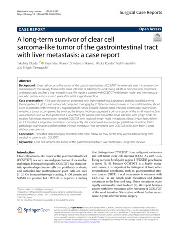
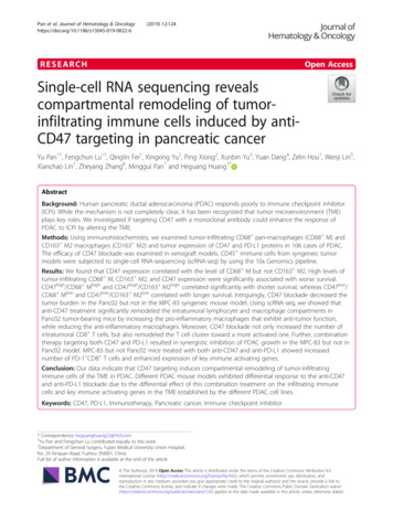
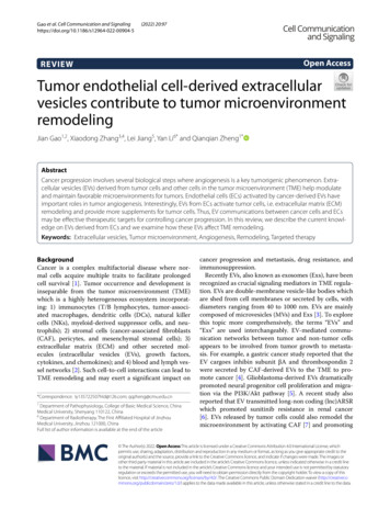
Gao et al. Cell Communication and Signaling(2022) 20:97Page 4 of 10Fig. 2 Specific markers of endothelial cells-derived EVswith miR-125a [53], miR-210 [54], miR-375 [55], miR-214[56], lysyl oxidase like-2 (LOXL2) [57], and HSP70 [58]having significant roles in this process. In heart disease,miR-10b-5p [59], miR-146a [60], and miR-19a [61] areimportant molecules, while for vascular smooth musclecell regulation, miR-143 [62] and versican [63] are highlysignificant. In atherosclerosis, miR-505 [64] and miR-155[65] also have similarly important functions. In chronicobstructive pulmonary disease, R-191 predicts the potential function of Exs as paracrine effectors [53]. TGF-β1could ameliorate renal structure and function [66]. Cytomegalovirus could stimulate allogeneic CD4 memoryT cells [67]. The effects of EV release on tumor cells ismainly facilitated via these cargoes. e.g., miR-503 in EVsregulate the proliferation and invasion of triple-negativebreast cancer cells [46]. Also, stressed human umbilical vein endothelial cells (HUVECs) release EVs containing miR-126, which significantly inhibits tumor cellgrowth [47]. In addition to releasing “tumor-suppressing”EVs, ECs also release “tumor-promoting” EVs. The EVsreleased from human brain microvascular ECs promoteS100A16 expression in lung cancer cells, increase lungcancer cell and anti-apoptotic activity, thereby promoting lung cancer development. ECs also release EVs carrying Ang2 which promotes tumor progression [51].Under stress conditions, CEACAM1 in EVs inhibits Tcell activation, which may be an important factor promoting tumor progression. Our previous studies showedthat YAP1 inhibition in HUVECs was associated withEV release and increased hepatocyte carcinoma (HCC)invasion and metastasis (Fig. 3). Our results suggestedthat during tumor treatment, EVs were a possible reasonfor treatment failure [48] (Table 1).The role of EVs toward immune cellsEVs trigger and regulate immune responses, especiallyin cancer [68]. Several studies have reported that various molecules (adhesions, heat shock proteins, molecules involved in membrane trafficking and ESCRTs),immune response molecules (e.g., MHC class I and IIproteins), immune receptors, ligands (FasL, TRAIL,PD-L1, NKG2D ligands), and co-stimulatory moleculesare distinct cargoes in some specialized EVs. Manyimmunocytes, such as DCs [69], NKs, and T cells [70]are activated by ECs harboring non-classical or HLA-E,MICA, and other NKG2D ligands. In cardiovascular disease, including vascular inflammation and atherosclerotic plaque formation, EVs mediate cross-talk betweenrecipient cells and ECs, and reprogram them toward apro- or anti-inflammatory stance. The pro-inflammatory molecules with chemotactic mediators, including ICAM-1, CCL-2, IL-6, IL-8, CXCL-10, CCL-5, andTNF-α were released by EVs and taken up by monocytes and HUVECs [71]. In anti-inflammatory aspectdisplays, ECs released EVs containing miRNA-222 toreduce endothelial ICAM-1 expression both in vitroand in vivo [72]. In addition, miR-10a was transferredto monocytes from EC-EVs and could repress inflammatory signaling through the targeting of several components of the NF-κB pathway, including IRAK4 [73].
Gao et al. Cell Communication and Signaling(2022) 20:97Page 5 of 10Fig. 3 Schematic molecular components of endothelial cells-derived EVsLipopolysaccharide induces the neutrophil secretion ofEVs containing miR-122-5p which functions in oxidative stress, apoptosis, and increased brain microvascularEC permeability [74]. EVs released by apoptotic ECs arereported as having unique transcriptomic characteristicsand contain non-coding RNA (ncRNA) sequences (mitochondrial transfer RNA, U1 small nuclear RNA, andpathogen-like endogenous retro-elements) with immunostimulatory potential. These EVs from apoptotic ECsmay be recognized by RIGI-like receptors and toll likereceptors (TLRs) (TLR3, TLR7, and TLR8) and possiblyinitiate innate immune responses [75].The circuitous connection between certain EVs andtumors warrants further investigation. Specific ncRNAs have been identified as cargoes in specialized EVsreleased by ECs; interestingly, the same ncRNAs regulatetumor progression in other studies. We have no proof ofa relationship between EC-EV-ncRNA-tumor, yet thisgives us a sensible estimate concerning the connectionamong these elements. Zhao et al. found that miR-503downregulated immune function in esophagus carcinoma [76], while Bovy et al. confirmed miR-503 wascontained in EC EVs during breast cancer neoadjuvantchemotherapy [46]. LncRNAs also have important regulatory roles in tumor immunity. We previously demonstrated that ECs release EVs which carried MALAT1[48], while Hou et al. reported that MALAT1 promotedimmunosuppressive properties in HCC cells [77]. Primary mouse lung ECs may release EVs containing Ang2,while coincidentally, Schmittnaegel et al. verified thatANG-2 inhibition elicited antitumor immunity whichwas enhanced by PD-1 checkpoint blockade [78]. In addition to ncRNAs, proteins from EVs released by ECs alsohave important functions in tumor immunity. S100A6overexpression possibly impaired the infiltration andcytolytic activity of CD8 T cells via the focal adhesionRas-stimulating signaling pathway in pancreatic cancer[79]. Brain microvascular EC-EVs contained S100A16
Gao et al. Cell Communication and Signaling(2022) 20:97Page 6 of 10Table 1 The role of EC-derived EVs in tumor and non-tumor diseasesParent cellBiological functionReferenceEVs on tumor cellsNcRNA miR-503HUVECsInhibits cancer proliferation and invasion[46]miR-126HUVECsInhibits the growth of tumor cells[47]MALAT1Protein S100A16HUVECPromotes invasion in HCC[48]HBMECsPromotes SCLC survival in brain[49]Dll4HMVECsPromotes angiogenesis[50]Ang2Primary mouse lung endothelial cellsPromotes tumor growth[51]CEACAM1human endothelial cell line AS-M.5Modulate immune response, tumor progression, metastasis andangiogenesis[52]HUVECReduced inflammation in cardiovascular disease[59]Human lung microvascularModulate the phenotype of distant endothelial cells of large systemic [53]EVs on non-neoplastic diseasesNcRNA miR-10b-5pmiR-125amiR-143HUVECControl smooth muscle cell phenotype[62]miR-146aHUVECDecrease in metabolic activity in peripartum cardiomyopathy[60]miR-191Human lung microvascularPredicting potential function of exosomes as paracrine effectors inCOPD[53]miR-210Endothelial progenitor cellsProtective effects on ECs against H/R injury[54]miR-375Endothelial progenitor cellRescued the cell protection activity[55]miR-214HMEC-1Blood vessel formation[56]miR-505HUVECInducing Oxidative stress and inflammation in atherosclerosis[64]miR-155HUVECModulate the macrophage phenotype in atherosclerosis[65]miR-19aHUVECImproved vascularization and cardiac function, decreased myocardialfibrosis[61]Protein TGF-β1Glomerular endothelial cellsAmeliorate renal structure and function[66]VersicanHUVECRegulate vascular smooth muscle cells calcification/senescence inhigh glucose condition[63]LOXL2Human microvascular endothelial cells Altered abundances after exposure of their producing cells to cellularstressCytomegalovirus HUVECHSP70Rat aortic endothelial cellsStimulate allogeneic CD4 Memory T CellsInduction of monocyte activation and endothelial cell adhesionwhich is an immune-related prognostic biomarker andtherapeutic target for low-grade glioma [80]. Theseresults suggest EVs released from ECs may be importantfactors regulating tumor immunity. Anti-tumor immunotherapy targeting EC-EVs may an important anti-tumortherapy mechanism in the future.EC derived EV based therapeutic strategiesEVs containing various surface adhesion proteins haveseveral advantages as delivery vectors for cancer genetherapy. As cancers are often associated with aberrantEV formation and release, the inhibition of this pathological process could be an emerging approach for cancertreatment.EVs as novel biocarriers for drug deliveryEC-derived EVs are advantageous for drug targeteddelivery as they carry biological substances to recipientcells. Using in vitro and ex vivo techniques, including[57][67][58]freeze-thaw cycles, incubation, saponin infiltration,sonication, and extrusion procedures, several therapeutic drugs have been loaded into EVs [81]. Thus,EVs are stable, and drug structure and activity remainsunchanged after EV loading, even when stored at freezing temperatures. Recent studies suggested that EVdelivery and effective cellular uptake could be improvedby altering the EV surface with an Arg-Gly-Asp-D-TyrLys peptide [82] or promoting efficient cytosolic releaseusing enhancing cationic lipids [83]. Unfortunately,few studies have reported EC-derived EVs for delivering drugs in experimental or clinical studies, whileother cell studies reported that EVs carrying anticancer drugs are promising new therapeutic strategies inanimal experiments as a promising approach for cancers [84]. In addition to undergoing a phase I clinicaltrial (NCT01294072), these therapeutic strategies arebeing used for EC/EV drug delivery for cardiovasculardiseases.
Gao et al. Cell Communication and Signaling(2022) 20:97EVs carry small interfering RNAs (siRNAs)EVs carry RNAs, which facilitate communicationsbetween cells. Critically, siRNA delivery to ECs andother cells could be an important gene therapy strategy. Because protein expression is suppressed by siRNAs via specific posttranscriptional sequences, siRNAsoffer a unique opportunity to treat multiple ECs relatedto cancer. Recently, across different fields, several siRNAvectors have been used, including viruses, synthetic polymers, and lipid-based carriers. When compared withother RNA vectors, EVs when used as nanoparticles totransfer siRNAs, are more advantageous in retaining bioavailability, delivery efficiency, and compatibility [85].Moreover, siRNA-containing EVs cross the endothelialbarrier and guide siRNAs to target specific tissues andcells [85]. Also, as these molecules easily pass the blood–brain barrier, EVs are extensively used as small RNA carriers to treat brain tumors [86]. A recent study showedthat EVs containing siRNA knocked down target geneexpression by 50%–90% in different cancer cells [87].However, few investigations have provided evidence ofspecific ECs releasing EVs to deliver siRNA. Yang et al.demonstrated that siRNA-VEGF delivery in EC-EVs suppressed VEGF transcription and translation in co-cultured GBM-astrocytoma cells, and showed that siRNAdelivery silenced target genes and could be used as apotential therapeutic strategy for cancer [88].Reducing the abnormal release of EVsIn terms of abnormal EV-release related to cancer, severalstudies emphasized the biological ways of EVs to verifythe key way to suppress the exocrine of EVs. nSMase tookpart in lipid raft and worked as a ubiquitous enzyme toimprove EV formation [89]. These results suggested EVformation and release were blocked by siRNAs againstnSMase2. Menck et al. reported that nSMase-2 inhibitionby GW4869 or RNA interference decreased Exs secretionbut increased MV secretion from plasma membranesboth in vitro and in vivo [90]. Furthermore, GW4869prevented abnormal EV release and improved abnormalvascular remodeling [91]. Rab proteins are key regulatorsof EV secretion, e.g., Rab27 participates in EV exocytosis.Also, EV release is reduced by RAB27 inhibition [92]. Incolorectal cancer cells, RAB27 knockdown inhibited EVrelease and the exosomal-related proliferation and migration of ECs [93].Conclusions and perspectivesAngiogenesis is an essential step in tumorigenesis anddevelopment. Tumor cell metastasis to distant organsand the rapid proliferation of focal tumor cells areinseparable from new blood vessel formation. EVs arePage 7 of 10important mediators during material exchange processes between tumor cells and ECs. EVs released bytumor cells carry proteins and ncRNAs to promote newtumor blood vessel formation. ECs in the TME alsorelease EVs to promote tumor progression. Currently,the function and molecular mechanisms underpinningthe EC release of EVs are unclear and require furtherstudy. However, EV sources, their loading, and relatedprocesses are affected by their cell origin, changes inthe TME, and target cells. Therefore, targeting theseEV characteristics could provide an intervention forregulating tumor angiogenesis. In particular, EVs areproduced in the body and effectively avoid immunesurveillance, therefore, they have a great potential as atargeted therapy for drug delivery in clinical settings.Based on these observations, cross-talk between ECsand other cells can be mediated by EVs and contributeto TME remodeling. EC-derived EVs are expected tobecome important targets for tumor treatments in thefuture.AbbreviationsEVs: Extracellular vesicles; TME: Tumor environment; TAM: Tumor-associatedmacrophages; DC: Dendritic cells; NK: Natural killer cells; MDSC: Myeloidderived suppressor cells; CAFs: Cancer-associated fibroblasts; ECM: Extracellular matrix; MVs: Microvesicles; Exs: Exosomes; MVE: Multivesicular endosomes;ECs: Endothelial cells; HCC: Hepatocyte carcinoma; ESCRT : Endosome sortingcomplexes required for transport; MVB: Multi-vesicular body; VPS4: Vacuolarprotein sorting-associated protein 4; ILV: Intraluminal vesicles; MVB: Multivesicular bodies; SNARE: Soluble N-ethylmaleimide-sensitive factor attachmentprotein receptor; ANGPT1: Angiopoietin-1; BMSCs: Bone marrow-derivedmesenchymal stromal cells; TGFβRII: Transforming growth factor β receptorII; TPM1: Tropomyosin-1; HSPG: Heparin sulfate proteoglycan; CA9: Carbonicanhydrase 9; miRNA: MicroRNA; lncRNA: Long-noncoding RNA; VWF: VonWillebrand factor; ICAM: Intercellular cell adhesion molecule; MHC: Majorhistocompatibility complex; PECAM-1: Platelet EC adhesion molecule-1; eNOS:Endothelial nitric oxide synthase; nanoFACS: Nanoscale fluorescence activatedcell sorting; Dll4: Delta-like protein 4; LOXL2: Lysyl Oxidase Like- 2; Ang2:Angiopoietin-2; CEACAM1: Carcinoembryonic antigen-related cell adhesionmolecule-1.AcknowledgementsWe thank International Science Editing (www. inter natio nalsc ience editi ng. com) for editing this manuscript.Author contributionsYL was a major contributor in writing the manuscript. QZ was a major contributor in revising the manuscript. All authors read and approved the finalmanuscript.FundingThis review was supported by the National Natural Science Foundation ofChina (No. 82103650), the Natural Science Foundation of Beijing Municipality(No. 7214219), the Capital Medical University Natural Science Foundation (No.PYZ20143), and the Natural Science Foundation of The First Affiliated Hospitalof Jinzhou Medical University (No. KYTD-2022012).DeclarationsCompeting interestsThe authors declare that they have no competing interests.
Gao et al. Cell Communication and Signaling(2022) 20:97Author details1Department of Pathophysiology, College of Basic Medical Science, ChinaMedical University, Shenyang 110122, China. 2 Science Experiment Centerof China Medical University, Shenyang 110122, China. 3 Departmentof General Surgery, Beijing Friendship Hospital, Capital Medical University,Beijing 100000, China. 4 National Clinical Research Center for DigestiveDiseases, Beijing 100000, China. 5 Department of General Surgery, AffiliatedHospital of Jinzhou Medical University, Jinzhou 121000, China. 6 Departmentof Radiotherapy, The First Affiliated Hospital of Jinzhou Medical Universit
(SNARE) complexes (Fig. 1). e SNARE complex has vital roles in MVB fusion with cell membranes, especially when promoting MVB release. e SNARE complex consists of two components, v- and t-SNAREs. e v-SNARE is located on the vesicle while the t-SNARE is located on the presynaptic membrane. Both v- and t-SNAREs pair up and form a complex [19];