Transcription
Table of ContentsProtocolPublication NumberWestern BlotNear-Infrared (NIR) Western Blot Detection.988-13627Good Westerns Gone Bad: Tips to Make Your NIR Western Blot Great.988-14772Western Blot Normalization: Challenges and Considerations for Quantitative Analysis.979-15896 Protein Electrotransfer Methods and the Odyssey Imaging Systems.988-16191One Blot Western Optimization Using the MPX Blotting System .988-16189Odyssey Western Blot Blocker Optimization.988-14403ICWCellTag 700 Stain In-Cell Western Assay Kits I and II .988-13623EMSAElectrophoretic Mobility Shift Assay (EMSA) Using IRDye Oligonucleotides.988-12448DNA StainSyto 60 Staining of Nucleic Acids in Gels .988-12449Small Animal ImagingScanning a Mouse on the Odyssey System: Hints and Tips .988-12589Additional ProtocolsImage Studio Software Work Area Folder Best Practices .988-15346Protocols at http://licor.com/bio/supportIn-Gel Western Detection.988-12954In-Cell Western Assay: For Assessing Response of A431 Cells to Stimulation withEpidermal Growth Factor .988-16059 In-Cell Western Assay: IRDye 800CW EGF Competition and Binding Assay Using A431 Cells .988-16066In-Cell Western Assay: Complete Sample Protocol for Measuring IC 50 of Inhibitor PD168393in A431 Cells Responding to Epidermal Growth Factor .988-16060In-Cell Western Assay: Complete Sample Protocol for Measuring IC 50 of Inhibitor U0126in NIH3T3 Responding to Acidic Fibroblast Growth Factor (aFGF-1) .988-16058In-Cell Western Assay: Complete Sample Protocol Detailing the Seeding, Stimulation,and Detection of the HeLa Cellular Response to Epidermal Growth Factor .988-16061In-Cell Western Assay: Complete Sample Protocol Detailing the Seeding, Stimulation,and Detection of the NIH3T3 Cellular Response to Platelet Derived Growth Factor BB (PDGF-BB) .988-16062In-Cell Western Assay: Complete Apoptosis Assay Example Detailing the Seeding, Induction,and Detection of the HeLa Cellular Response to Anisomycin Treatment .988-16057In-Cell Western Assay: Phospho-p53 Detection in COS Cells in Response to Hydroxyurea .988-16063In-Cell Western Assay: Phospho-p38 Detection in HeLa Cells in Response to Anisomycin .988-16193In-Cell Western Assay: Complete Sample Protocol for PMA-induced ERK Activationin Suspension Cell Lines .988-16065Technical Note: FAQs for Suspension Cells for ICW Protocols .988-11461On-Cell Western Plate-Based Assay for Targeted Near-Infrared-Labeled Optical Imaging AgentDevelopment: Receptor-Based Binding and Competition Assays .988-14668 Cell Viability Assay with Sapphire700 Stain and the Odyssey CLx and Sa Imaging Systems .979-14115LI-COR BIOSCIENCES MAKES NO WARRANTY OF ANY KIND WITH REGARD TO THIS MATERIAL, INCLUDING, BUT NOT LIMITEDTO, THE IMPLIED WARRANTIES OF MERCHANTABILITY AND FITNESS FOR A PARTICULAR PURPOSE. LI-COR Biosciences shallnot be liable for errors contained herein or for incidental or consequential damages in connection with the furnishing,performance, or use of this material. Publication Number 984-12556. March, 2012. 2012 LI-COR, Inc. This document containsproprietary information which is protected by copyright. All rights are reserved. No part of this document may be photocopied,reproduced, or translated to another language without prior written consent of LI-COR, Inc. The information contained in thisdocument is subject to change without notice.LI-COR Biosciences is an ISO 9001 registered company. LI-COR, In-Cell Western, MPX, NewBlot, Odyssey, and IRDye areregistered trademarks of LI-COR, Inc. in the United States and other countries. All other trademarks belong to their respectiveowners. The Odyssey Infrared Imaging systems and IRDye reagents are covered by U.S. patents, foreign equivalents, and patentspending.988-162300616
Near-Infrared (NIR)Western Blot DetectionDeveloped for:Odyssey Classic,Odyssey CLx,Odyssey Fc, andOdyssey SaInfrared Imaging SystemsPublished 2003, revised May 2014. Themost recent version of this document isposted at: http://biosupport.licor.com
Page 2 – Near-Infrared (NIR) Western Blot DetectionTable of ContentsPageI.Required Reagents . . . . . . . . . . . . . . . . . . . . . . . . . . . . . . . . . . . .2II.Quick Start Hints & Tips . . . . . . . . . . . . . . . . . . . . . . . . . . . . . . . .2III.Western Blot Detection Methods . . . . . . . . . . . . . . . . . . . . . . . . .3IV.Guidelines for Two-Color Detection . . . . . . . . . . . . . . . . . . . . . . .4V.Adapting Western Blotting Protocols for Odyssey Detection . . .5I. Required Reagents Blotted nitrocellulose (LI-COR, P/N 926-31090/926-31092) or Immobilon -FL PVDF membrane (LI-COR, P/N 926-31099 or 926-31100) Odyssey Blocking Buffer (PBS) (LI-COR, P/N 927-40000, 927-40100) or Odyssey BlockingBuffer (TBS) (LI-COR, P/N 927-50000, 927-50100)** Primary antibodies IRDye 800CW, 680RD, or 680LT secondary antibodies (LI-COR) Tween 20 PBS/TBS** Ultrapure water Methanol for wetting of PVDF SDS** IMPORTANT! PBS-based and TBS-based blocking buffers may be used with this protocol.Odyssey Blocking Buffer is available in both formulations.Be sure to keep your buffer system consistent throughout the protocol for blocking,antibody dilutions, and washes. For example, if you use a TBS-based buffer system, chooseOdyssey Blocking Buffer (TBS). If you use a PBS-based buffer system, choose Odyssey BlockingBuffer (PBS).II. Quick Start Hints and TipsInfrared fluorescence detection with Odyssey Family Imaging Systems provides a quantitativetwo-color detection method for Western blots. Following, you will find some basic Hints and Tipsto help you get started. For a more comprehensive guide, see Good Westerns Gone Bad: Tips toMake Your NIR Western Blot Great at www.licor.com/GWGBIR1. Store the IRDye secondary antibody vial in darkness at 4 C. Minimize exposure to light andtake care not to introduce contamination into the vial. Dilute immediately prior to use. Ifparticulates are seen in the antibody solution, centrifuge before use.
Near-Infrared (NIR) Western Blot Detection – Page 32. The best transfer conditions, membrane, and blocking agent for experiments will vary, depending on the antigen and antibody.3. Do not write on blot with a pen or Sharpie marker. Ink from most pens and markers willfluoresce in the 700 nm channel of all Odyssey Family Imaging Systems. The ink maywash off and re-deposit elsewhere on the membrane, causing increased background. Usea pencil to mark membranes. Ink from the Odyssey Pen does not fluoresce and can beused to mark nitrocellulose membranes. If the Odyssey Pen is used for PVDF membranes,the ink will dissolve and wash off when the blot is wetted in methanol.4. Let the membrane dry after transfer for 1 hour or overnight, to maximize protein retentionon the membrane.5. Handle blot with clean forceps only.6. Before using forceps, incubation trays, and the Odyssey scanning surface or sample tray(if applicable), clean with 100% methanol to remove any residual dye signal from previoususe. Rinse with a small volume of distilled water, followed by isopropanol. Dry with a lintfree wipe.7. When processing Western blots, do not use dishes/boxes that have ever been used forCoomassie staining. The Odyssey imagers are very sensitive to Coomassie (which is astrongly-fluorescent dye), and use of dishes with small traces of Coomassie will add atremendous amount of background in the 700 nm channel.Maintain the same buffer system throughout the Western blot process. For example, if youblock your blot in Odyssey Blocking Buffer (PBS), use PBS-based buffers throughout theprotocol.8. Do not include detergents during the blocking step.9. For 1-color blots, use IRDye 800CW secondary antibody for detection of the protein forbest sensitivity.10. For 2-color blots: Make sure primary antibodies are from different species (for example, Rabbit andMouse) Use the IRDye 800CW secondary antibody to detect the protein target with lowestabundance, and IRDye 680RD secondary antibody to detect the more abundant protein.11. If you are using PVDF, add 0.01% SDS to the diluted secondary antibody. Do not add SDS ifusing nitrocellulose membrane.12. Incubate with secondary antibodies in the dark for one hour with gentle shaking. The incubation box can be covered with aluminum foil.
Page 4 – Near-Infrared (NIR) Western Blot DetectionIII. Western Blot Detection MethodsThis protocol is designed to help you achieve success with NIR Western blot detection methods.Read the entire protocol carefully before beginning your optimization experiments.Membrane GuidelinesA low-background membrane is essential for NIR Western blot success. Background can resultfrom membrane autofluorescence or from non-specific binding of antibodies. Polyvinylidene fluoride (PVDF) and nitrocellulose membranes are typically used for Western blotting applications.There are many brands and vendors for both types of membrane.Before using your blotting membrane for the full Odyssey Western blot protocol, cut a smallsample of membrane for testing. Image this sample (both wet and dry) to evaluate the level ofmembrane autofluorescence. If possible, include a sample of membrane that is known to workwell with the Odyssey system, so you can compare background levels.To learn more about optimizing your Western blots, see Good Westerns Gone Bad: Tips to MakeYour NIR Western Blot Great (www.licor.com/GWGBIR)For detailed gel transfer information, see Protein Electrotransfer Methods and the Odyssey InfraredImaging Systems (www.licor.com/proteintransfer)Western Blot Detection ProtocolAfter membrane transfer, perform the following steps:1. For PVDF membrane:a. Pre-wet 1 minute in 100% methanol.b. Rinse with ultra pure water.c. Wet in 1X PBS or TBS for 2 minutes (using the appropriate buffer system).For nitrocellulose membrane:a. Wet in 1X PBS or TBS for 2 minutes, or until fully hydrated (using the appropriatebuffer system).2. Place membrane in incubation box and block the membrane in Odyssey Blocking Buffer(PBS or TBS) for 1 hour with gentle shaking. Be sure to use sufficient blocking buffer tocover the membrane (a minimum of 0.4 mL/cm2 is suggested). For a detailed Westernblot blocker optimization protocol, see Odyssey Western Blot Blocker Optimization(www.licor.com/optimize)3. Prepare primary antibody:a. Primary antibody diluent: Odyssey Blocking Buffer (PBS or TBS) 0.2% Tween 20 (finalconcentration).b. Dilute primary antibody in Odyssey Blocking Buffer 0.2% Tween 20, using the vendor’s recommended dilution for Western blot applications. Dilutions typically rangefrom 1:200 - 1:5,000, depending on the primary antibody.c. Use enough antibody solution to completely cover the membrane.
Near-Infrared (NIR) Western Blot Detection – Page 5For further details about primary antibody optimization, see One Blot Western OptimizationUsing the MPX Blotting System (www.licor.com/oneblot)4. Incubate blot in diluted primary antibody for 1 to 4 hours* at room temperature with gentleshaking, or overnight at 4 C.*Optimal incubation times vary for different primary antibodies.If the procedure cannot be completed in full, this is a good place to stop until the followingday. Incubate the blot in primary antibody overnight at 4 C with gentle shaking, and resume the protocol the next day.5. Wash membranes: Pour off primary antibody solution. Rinse membrane with appropriate buffer – 1X TBS-T (0.1% Tween 20) or 1X PBS-T(0.1% Tween 20). Cover blot with 1X TBS-T (0.1% Tween 20) or 1X PBS-T (0.1% Tween 20). Shake vigorously on platform shaker at room temperature for 5 minutes. Pour off wash solution. Repeat 3 additional times.6. Dilute IRDye secondary antibody in the appropriate diluent listed below:a. Secondary antibody diluent: PVDF membrane To blocking buffer, add Tween 20 to a final concentration of 0.2% and SDS to a finalconcentration of 0.01%.b. Secondary antibody diluent: nitrocellulose membrane To blocking buffer, add Tween 20 to a final concentration of 0.2%. Do not add SDS.NOTE: A Suggested dilution range for secondary antibodies is typically 1:5,000 to1:25,000. Recommended dilutions can be found on the secondary antibody pack insert.Use 1:20,000 as a suggested starting point if using LI-COR secondary antibodies.7. Protect membrane from light during incubation. Incubate blot in diluted secondaryantibody for one hour at room temperature with gentle shaking.8. Protect membrane from light during washes.Wash membranes: Pour off secondary antibody solution. Rinse membrane with the appropriate buffer – 1X TBS-T (0.1% Tween 20) or1X PBS-T (0.1% Tween 20). Cover blot with 1X TBS-T (0.1% Tween 20) or 1X PBS-T (0.1% Tween 20). Shake vigorously on platform shaker at room temperature for 5 minutes. Pour off wash solution. Repeat 3 additional times.9. Rinse membrane with 1X TBS/PBS (as appropriate) to remove residual Tween 20.
Page 6 – Near-Infrared (NIR) Western Blot Detection10. The Western blot is now ready to image. The membrane can be stored in 1X TBS or 1X PBS for up to 48 hours in the dark at 4 C.If the blot is prepared more than 48 hours in advance, air-dry the blot and store in thedark at room temperature until ready to image.11. Image with an Odyssey Family Imaging System. If signal on membrane is too strong or too weak, adjust the imaging parameters to optimize the image.– Odyssey Classic: Re-image the membrane at a lower or higher scan intensity setting,respectively.– Odyssey Fc: Adjust image acquisition time.– Odyssey CLx: Use the AutoScan function to improve the dynamic range of theimage.IV. Guidelines for Two-Color DetectionTwo different antigens can be detected simultaneously on the same blot using IRDye secondaryantibodies. When performing a two-color blot, use the standard Western blot protocol with thefollowing modifications: Combine the two primary antibodies in the diluted antibody solution (Step 3, Section III).Incubate simultaneously with membrane (Step 4, Section III). The primary antibodies mustbe from two different host species. Combine the two IRDye secondary antibodies in the diluted antibody solution (Step 6,Section III). Incubate simultaneously with membrane (Step 7, Section III).Two-color detection requires careful selection of primary and secondary antibodies. The followingguidelines will help you successfully design two-color experiments: The two primary antibodies must be derived from different host species so that they can bediscriminated by secondary antibodies of different specificities (for example, primary antibodies from rabbit and mouse will be discriminated by anti-rabbit IgG and anti-mouse IgGsecondary antibodies, respectively). If the two primary antibodies are mouse monoclonals from different IgG subclasses (IgG1,IgG2a, or IgG2b), IRDye Subclass-Specific secondary antibodies can be used for multiplexdetection. The same subclasses cannot be combined in a two-color Western blot (for example, two IgG1 primary antibodies). For details, refer to Western Blot and In-Cell Western Assay Detection Using IRDye Subclass-Specific Antibodies (www.licor.com/subclass) Anti-Goat secondary antibodies cannot be multiplexed with Goat-derived secondary antibodies (Example: Donkey anti-Goat and Goat anti-Rabbit). The secondary antibodies willcross-react. One secondary antibody must be labeled with a 700 nm channel dye and the other with800 nm channel dye. In general, it is recommended that the IRDye 800CW secondary antibody (800 nmchannel) be used to detect the lower-abundance protein target and IRDye 680RD secondary antibody (700 nm) to detect the more abundant protein.
Near-Infrared (NIR) Western Blot Detection – Page 7 Always use highly cross-adsorbed secondary antibodies for two-color detection. Failureto use cross-adsorbed antibodies may result in increased cross-reactivity of the secondary antibodies. For best results, avoid using primary antibodies from mouse and rat together in a twocolor experiment. The two species are so closely related that it is not possible to completely adsorb away all cross-reactivity. If there is no other option, it is crucial to run single-color blots first with each individual antibody to be certain of expected band sizes.V. Adapting Western Blot Protocols for Odyssey Imaging SystemsWhen adapting Western blotting protocols for Odyssey detection or using a new primary antibody, it is important to determine the optimal antibody concentrations. Optimization will helpachieve maximum sensitivity and consistency, while minimizing background. Three parametersshould be optimized: primary antibody concentration, IRDye secondary antibody concentration,and detergent concentration in the diluted antibodies.Primary Antibody ConcentrationPrimary antibodies vary widely in quality, affinity, and concentration. The correct working rangefor antibody dilution depends on the characteristics of the primary antibody and the amount oftarget antigen to detect. Start with the vendor’s dilution recommendation for Western blotting orwith the dilution normally used for chemiluminescent detection.Secondary Antibody ConcentrationOptimal dilutions of IRDye secondary antibodies should also be determined. Refer to the appropriate pack insert for recommendations at http://www.licor.com/packinsert. The amount of secondary antibody required varies depending on how much antigen is being detected. Abundantproteins with strong signals may require less secondary antibody. Using too much secondaryantibody may increase membrane background and/or non-specific banding.Detergent ConcentrationAddition of detergents to diluted antibodies can significantly reduce background on the blot.Optimal detergent concentration will vary, depending on the antibodies, membrane type, andblocker used. Keep in mind that some primary antibodies do not bind as tightly as others and maybe washed away by too much detergent. Never expose the membrane to detergent until blockingis complete, as this may cause high membrane background.Tween 20: Blocking buffer – do not add Tween 20 during blocking. Diluted primary and secondary antibodies should contain Tween 20.Use a final concentration of 0.1 - 0.2% Tween 20 for PVDF or nitrocellulose membranes. Wash solutions should contain 0.1% Tween 20.
Page 8 – Near-Infrared (NIR) Western Blot DetectionSDS:When using PVDF membrane, addition of SDS will dramatically reduce overall membrane background in the 700 nm channel. It is critical to use only a very small amount,because SDS is an ionic detergent and can disrupt antibody-antigen interactions if toomuch is present at any time during the protocol.SDS should not be used with nitrocellulose membranes. Blocking buffer - do not add SDS to the blocking reagent during blocking. Diluted primary antibodies should not contain SDS. Diluted secondary antibodies should contain a final concentration of 0.01 – 0.02%SDS and 0.1 – 0.2% Tween 20, when PVDF membrane and IRDye 680LT secondaryantibodies are used. SDS is optional when using IRDye 680RD antibodies with PVDF membrane, but isessential when using IRDye 680LT antibodies with PVDF. Wash solutions should not contain SDS. 2014 LI-COR, Inc. LI-COR, Odyssey, IRDye, MPX, and In-Cell Western are trademarks or registered trademarks of LI-COR, Inc. in the United Statesand other countries. All other trademarks belong to their respective owners.LI-COR is an ISO 9001 registered company.Global Headquarters, Serving the United States 4647 Superior Street Lincoln, NE 68504Toll free: 800-645-4267 Fax: 1-402-467-0819 biosales@licor.comLI-COR GmbH, Germany, Serving Europe, Africa, and the Middle East.LI-COR Biosciences GmbH Siemensstraße 25A 61352 Bad Homburg GermanyPhone: 49 (0) 6172 17 17 771 Fax: 49 (0) 6172 17 17 799 bio-eu@licor.comLI-COR Ltd., United Kingdom, Serving Denmark, Finland, Iceland, Ireland, Norway, Sweden, and UKLI-COR Biosciences UK Ltd. St. John’s Innovation Centre Cowley Road Cambridge CB4 0WSUnited Kingdom Phone: 44 (0) 1223 422104 Fax: 44 (0) 1223 422105 bio-eu@licor.comLI-COR Biotechnology Distributor Network: www.licor.com/bio/distributorswww.licor.com/bioDoc # 988-136270514
Good Westerns Gone Bad:Tips to Make Your NIR Western Blot GreatDeveloped for:Aerius,Odyssey Classic,Odyssey CLx, andOdyssey Sa Infrared Imaging Systems,and the Odyssey Fc Imaging SystemPlease refer to your manual to confirmthat this protocol is appropriate for theapplications compatible with yourOdyssey Imager model.Published December 2008. Updated July 2014.The most recent version of this document isposted at http://biosupport.licor.com
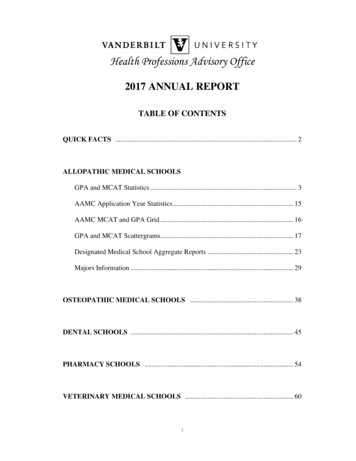
Page 2 – Good Westerns Gone Bad: Tips to Make Your NIR Western Blot GreatContentsI.II.III.IV.V.VI.Introduction to Western Blotting . . . . . . . . . . . . . . . . . . . . . . . . . . . .Important Factors That Affect Western Blot Results . . . . . . . . . . . . .Imaging Factors That Can Affect Western Blot Results . . . . . . . . . . .Software Adjustments for Image Optimization . . . . . . . . . . . . . . . . .Data Analysis with Image Studio Software: Background SubtractionData Analysis with Odyssey Applications Software:Background Subtraction . . . . . . . . . . . . . . . . . . . . . . . . . . . . . . . . . . .VII. Summary . . . . . . . . . . . . . . . . . . . . . . . . . . . . . . . . . . . . . . . . . . . . . .VIII. References . . . . . . . . . . . . . . . . . . . . . . . . . . . . . . . . . . . . . . . . . . . . .Page. .2. .2.11.13.15. .15. .16. .16I. Introduction to Western BlottingWestern blotting is used to positively identify a protein from a complex mixture. It was first introduced by Towbin, et al. in 1979, as a simple method of electrophoretic blotting of proteins tonitrocellulose sheets.1 Since then, Western blotting methods for immobilizing proteins onto amembrane have become a common laboratory technique. Although many alterations to the original protocol have also been made, the general premise still exists. Macromolecules are separated using gel electrophoresis and transferred to a membrane, typically nitrocellulose or polyvinylidene fluoride (PVDF). The membrane is blocked to prevent non-specific binding of antibodies and probed with some form of detection antibody or conjugate.Near-infrared fluorescence detection on the Odyssey Classic, Odyssey CLx, Odyssey Fc, orOdyssey Sa Imaging Systems provides a quantitative two-color detection method for WesternBlots. This document will discuss some factors that may alter the performance of a nearinfrared (NIR) Western blot, resulting in “good Westerns, gone bad.”II. Important Factors That Affect Western Blot ResultsA. MembraneA low-background membrane is essential for NIR Western blot success. Background can resultfrom membrane autofluorescence or non-specific binding of antibodies. Polyvinylidene fluoride(PVDF) and nitrocellulose membranes are typically used for Western blotting applications. Thereare many brands and vendors for both types of membranes.Before using your blotting membrane for the full Odyssey Western blot protocol, cut a small sample of membrane for testing. Image this sample (both wet and dry) to evaluate membrane autofluorescence. If possible, include a sample of membrane that is known to work well with theOdyssey system, so you can compare background levels.LI-COR has evaluated many different transfer membranes for Western blotting; examples of membrane performance are shown in Figure 1. PVDF membranes typically display higher membraneautofluorescence than nitrocellulose, and more variability in background levels. In general, nitrocellulose membranes offer the lowest membrane autofluorescence. However, PVDF has many
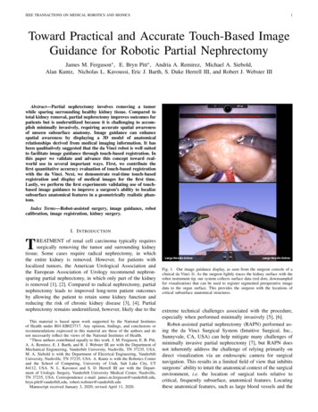
Good Westerns Gone Bad: Tips to Make Your NIR Western Blot Great – Page 3advantages (including higher binding capacity, higher target retention, and better tensile strength)that make it an appropriate choice for many experiments.NOTE: Not all sources of PVDF and nitrocellulose have been evaluated by LI-COR, and lot-to-lotvariation can occur. It is important to evaluate the membrane before use. Samples of membranecan be quickly evaluated by imaging them both wet and dry with any Odyssey System.700 nm ChannelScan Intensity 7800 nm ChannelScan Intensity 8Millipore Immobilon -FLMillipore Immobilon-PBioRad Immun-Blot Pall BioTrace PVDFPerkin Elmer PolyScreen Amersham Hybond -PFigure 1. Membrane autofluorescence from PVDF affects Westernblot performance. Transferrin was detected by Western blotting, usingvarious vendors and brands of PVDF membrane. Blots were imaged withthe Odyssey Classic Infrared Imaging System in both 700 and 800 nmchannels.B. Blocking ReagentMany different sources and types of blocking reagents are used for Western blot applications.Antibody performance can sometimes be compromised by the blocking reagent chosen. Blockingbuffer choice may affect antibody specificity and non-specific binding, and can dramatically increase the number of background bands (Figure 2).2 Excessive blocking (for example, with highconcentrations of nonfat dry milk) may cause loss of blotted proteins or mask the desired antibody-antigen interactions.3
Page 4 – Good Westerns Gone Bad: Tips to Make Your NIR Western Blot GreatDetection reagents may cross-react with certain blocking buffers. Milk-based blockers may contain IgG that can cross-react with anti-goat antibodies. This can significantly increase backgroundand reduce sensitivity. Milk-based blockers may also contain endogenous biotin or phosphoepitopes that can cause higher background.If an antibody fails with one blocking condition, trying another blocker may solve the problem.No single blocking reagent will be suitable for ALL antigen-antibody pairs. Refer to the Odyssey Western Blot Blocker Optimization Protocol (www.licor.com/optimize) to help design an experiment to optimize your blocking conditions. Figure 2 shows the behavior of a PKCα antibody in5% BSA, 5% Milk, and Odyssey Blocking Buffer on nitrocellulose membrane. For this primaryantibody, choice of blocking buffer had a dramatic effect on non-specific banding, particularlywhen 5% BSA was used.PKCaFigure 2. Effects of blocking buffer on detection of PKCα. Western blots were detected with anti-PKCα primary antibody and IRDye 800CW Goat anti-Mouse IgG, usingdifferent blocking buffers. All blots were processed identically, with the exception ofblocking reagent. Images were generated with an Odyssey Classic Infrared Imager(scan intensity 5, sensitivity 5).C. TBS- vs. PBS-Based Blocking BuffersMany blocking reagents are available in Phosphate Buffer or Tris Buffer (PBS/TBS) systems. Ingeneral, TBS blocking reagents are used for detection of phospho-proteins, because the phosphate present in PBS blocking reagents may competitively bind with antibodies to phosphoproteins. Some phospho-proteins can be detected with PBS-based blocking reagents, dependingon the antibody specificity and affinity; however, it is important to optimize the blocking reagentfor the specific antibody that is being used for optimal Western blot performance.The same buffer system should be used throughout the Western blot process; for example, if youare using a TBS-based buffer system, choose Odyssey Blocking Buffer (TBS). If you are using aPBS-based buffer system, use Odyssey Blocking Buffer (PBS).
Good Westerns Gone Bad: Tips to Make Your NIR Western Blot Great – Page 5The experiments below show the importance of optimizing for the proper blocking reagent.Figure 3 shows Jurkat cell lysate stimulated with Calyculin ( ) and a non-stimulated control (-).Phospho-Akt (green bands) was clearly detected with TBS-based Odyssey Blocking Buffer, butcould not be detected with the PBS-based Odyssey Blocking Buffer. This suggests that binding ofthe pAkt primary antibody may be affected by the phosphates in the PBS blocking reagent. Detection of total Akt (red bands) was also less robust with the PBS buffer system.Figure 3. Phospho-Akt detection is affected by TBS and PBS blocking buffers systems.Jurkat cells were treated with Calyculin A ( ) or left untreated (-), and lysates were loaded at 10 µg,5 µg, and 2.5 µg/well. Odyssey Blocking Buffer (TBS) and Odyssey Blocking Buffer (PBS) were usedfor blocking and antibody dilution, as indicated; appropriate TBS and PBS buffers were used forwashes. Blot was probed with anti-phospho-Akt mA
III. Western Blot Detection Methods This protocol is designed to help you achieve success with NIR Western blot detection methods. Read the entire protocol carefully before beginning your optimization experiments. Membrane Guidelines A low-background membrane is essential for NIR Western blot success. Background can result