Transcription
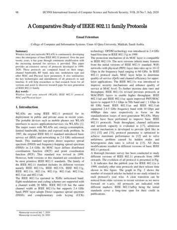
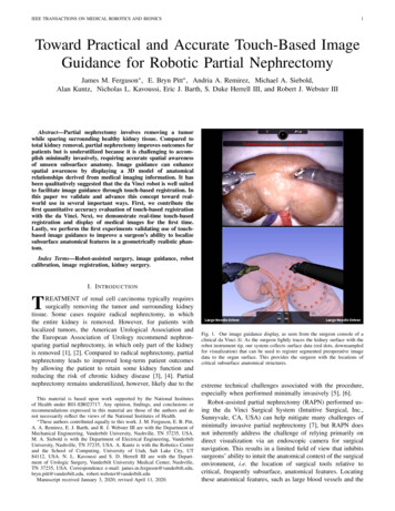
IEEE TRANSACTIONS ON MEDICAL ROBOTICS AND BIONICS1Toward Practical and Accurate Touch-Based ImageGuidance for Robotic Partial NephrectomyJames M. Ferguson , E. Bryn Pitt , Andria A. Remirez, Michael A. Siebold,Alan Kuntz, Nicholas L. Kavoussi, Eric J. Barth, S. Duke Herrell III, and Robert J. Webster IIIAbstract—Partial nephrectomy involves removing a tumorwhile sparing surrounding healthy kidney tissue. Compared tototal kidney removal, partial nephrectomy improves outcomes forpatients but is underutilized because it is challenging to accomplish minimally invasively, requiring accurate spatial awarenessof unseen subsurface anatomy. Image guidance can enhancespatial awareness by displaying a 3D model of anatomicalrelationships derived from medical imaging information. It hasbeen qualitatively suggested that the da Vinci robot is well suitedto facilitate image guidance through touch-based registration. Inthis paper we validate and advance this concept toward realworld use in several important ways. First, we contribute thefirst quantitative accuracy evaluation of touch-based registrationwith the da Vinci. Next, we demonstrate real-time touch-basedregistration and display of medical images for the first time.Lastly, we perform the first experiments validating use of touchbased image guidance to improve a surgeon’s ability to localizesubsurface anatomical features in a geometrically realistic phantom.Index Terms—Robot-assisted surgery, image guidance, robotcalibration, image registration, kidney surgery.I. I NTRODUCTIONTREATMENT of renal cell carcinoma typically requiressurgically removing the tumor and surrounding kidneytissue. Some cases require radical nephrectomy, in whichthe entire kidney is removed. However, for patients withlocalized tumors, the American Urological Association andthe European Association of Urology recommend nephronsparing partial nephrectomy, in which only part of the kidneyis removed [1], [2]. Compared to radical nephrectomy, partialnephrectomy leads to improved long-term patient outcomesby allowing the patient to retain some kidney function andreducing the risk of chronic kidney disease [3], [4]. Partialnephrectomy remains underutilized, however, likely due to theThis material is based upon work supported by the National Institutesof Health under R01-EB023717. Any opinion, findings, and conclusions orrecommendations expressed in this material are those of the authors and donot necessarily reflect the views of the National Institutes of Health. These authors contributed equally to this work. J. M. Ferguson, E. B. Pitt,A. A. Remirez, E. J. Barth, and R. J. Webster III are with the Department ofMechanical Engineering, Vanderbilt University, Nashville, TN 37235, USA.M. A. Siebold is with the Department of Electrical Engineering, VanderbiltUniversity, Nashville, TN 37235, USA. A. Kuntz is with the Robotics Centerand the School of Computing, University of Utah, Salt Lake City, UT84112, USA. N. L. Kavoussi and S. D. Herrell III are with the Department of Urologic Surgery, Vanderbilt University Medical Center, Nashville,TN 37235, USA. Correspondence e-mail: lt.edu, robert.webster@vanderbilt.eduManuscript received January 3, 2020; revised April 11, 2020.Fig. 1. Our image guidance display, as seen from the surgeon console of aclinical da Vinci Si. As the surgeon lightly traces the kidney surface with therobot instrument tip, our system collects surface data (red dots, downsampledfor visualization) that can be used to register segmented preoperative imagedata to the organ surface. This provides the surgeon with the locations ofcritical subsurface anatomical structures.extreme technical challenges associated with the procedure,especially when performed minimally invasively [5], [6].Robot-assisted partial nephrectomy (RAPN) performed using the da Vinci Surgical System (Intuitive Surgical, Inc.,Sunnyvale, CA, USA) can help mitigate many challenges ofminimally invasive partial nephrectomy [7], but RAPN doesnot inherently address the challenge of relying primarily ondirect visualization via an endoscopic camera for surgicalnavigation. This results in a limited field of view that inhibitssurgeons’ ability to intuit the anatomical context of the surgicalenvironment, i.e. the location of surgical tools relative tocritical, frequently subsurface, anatomical features. Locatingthese anatomical features, such as large blood vessels and the
IEEE TRANSACTIONS ON MEDICAL ROBOTICS AND BIONICStumor itself, is critical to safely and successfully performingRAPN. Surgical image guidance can help surgeons locatethese features, providing additional anatomical context byaccurately registering 3D anatomical volumes (typically generated by segmentation of preoperative computed tomography(CT) or magnetic resonance (MR) images) to the surgicalenvironment and displaying this information to the surgeonduring the procedure (see Fig. 1). Accurate image guidancehas the potential to improve patient outcomes by makinglocalization, dissection, and isolation of critical vascular andorgan structures, as well as correct margin selection, easier forsurgeons.It has been suggested that the da Vinci robot’s kinematicscould be used to achieve accurate registration for image guidance [8], [9]. In this work, we create such a system, quantify itsperformance, and demonstrate its ability to improve an experienced surgeon’s performance. Our image guidance system usesthe instruments of the da Vinci robot itself as 3D localizers fordigitizing anatomical surfaces. By lightly tracing an instrumenttip over the surface of the target anatomy while recording therobot’s joint values, our system generates a set of points on thesurface of the anatomy. Our system computes a surface-basedregistration between the preoperative images and the patient’sanatomy during the surgery. Using this registration, we thendisplay a 3D model of the patient’s anatomy segmented fromthe preoperative imaging to the surgeon in the da Vinci’ssurgeon console, enabling the surgeon to visualize the locationof subsurface anatomy that is not visible via the endoscope(see Fig. 1). By using the inherent capabilities of the daVinci for registration, our system provides an image guidanceapproach that is well suited to the clinical workflow.This paper presents our touch-based image guidance systemand analyzes its accuracy. We also describe practical steps fordeploying it in the operating room using a clinical da Vinci Sisystem. We present a series of phantom experiments to providea thorough accuracy analysis of a touch-based registration forimage guidance. Finally, we present a phantom experimentdemonstrating the utility of our image guidance system forimproving an experienced surgeon’s ability to localize subsurface anatomical features important in partial nephrectomy. Byproviding practical and accurate image guidance, our methodhas the potential to improve surgeons’ ability to accuratelyaccomplish partial nephrectomy. Success in achieving this hasthe potential to increase utilization of partial nephrectomy,thereby providing enhanced health outcomes to many morepatients.II. R ELATED W ORKImage guidance has previously been recognized as potentially useful in facilitating partial nephrectomy, and numerousresearch groups have sought to implement such image guidance systems. One approach to image guidance in laparoscopicpartial nephrectomy involved inserting fiducial markers onbarbed needles directly into the kidney [10], [11]. The kidneysand fiducials were then imaged and segmented intraoperativelyto enable registration by direct point-to-point correspondencebetween the fiducials in the segmented images and those same2fiducials in the endoscopic video. While providing highlyaccurate, real-time guidance, these fiducial-based methodsincrease the risk and complexity of surgery by requiringforeign objects to be manually inserted into the kidney by thesurgeon. Furthermore, the need for intraoperative imaging andsegmentation represents a time-intensive interruption of thesurgical workflow. Indeed, the robotic system is not compatiblewith intraoperative CT, and thus one would have to fullyremove the robot to register the image set.A less invasive approach to registration is fiducial-free manual registration. In manual registration, the surgeon is taskedwith visually aligning 3D images or models to the surgicalfield. In [12] and [13], preoperative MR and CT images and3D anatomical models were displayed alongside endoscopicvideo in the da Vinci’s surgeon console, and surgeons couldmanually adjust the orientation of the images to match theendoscopic view. Ukimura et. al. [14] and Nakamura et.al. [15] presented augmented reality systems in which surgeonsmanually aligned translucent 3D anatomical models directlyoverlaying the image feed from a laparoscopic endoscope.These studies found that surgeons benefited from havingpreoperative imaging information more readily available withrespect to the live camera images. However, this approach increases cognitive burden on surgeons and provides no accuracyguarantees. Indeed, relying on human hand-eye coordinationand spatial reasoning to perform registration makes accuracyhighly dependent on an individual user’s skill, resulting inlow registration precision, as evidenced by large variations inregistration accuracy from trial-to-trial in these studies.To enhance precision and facilitate objective accuracy, others have sought to employ stereo endoscopes for instrumenttracking and registration to patient anatomy. Su et. al. [16]proposed a multi-step CT-to-endoscope registration methodwhere the segmented kidney surface was first manually alignedwith the stereoscopic video. Surface-based video trackingtechniques were then used to refine and stabilize the registration during system operation. Pratt et. al. [17] utilized anaugmented endoscope overlay by first identifying a matchingfeature in both of the stereo images and the preoperativescans to align the translational degrees of freedom and thenusing a rolling-ball interface to manually align the rotationaldegrees of freedom. We direct the reader to [18] for athorough overview of research aimed at using computer visionalgorithms to automatically detect and track the da Vinciinstruments in the stereo endoscope video. These vision-basedapproaches are limited by a requirement for persistent, directline of sight between the endoscope camera and either theanatomical surface or the surgical instruments. During surgery,line of sight is often obstructed by blood, smoke, and othersurgical tools. Furthermore, endoscope-based methods typically require accurate tracking of the endoscope position itselfto localize tracked objects in the surgical field. Accurate endoscope tracking usually requires an external tracking systemand a calibration process to determine the rigid transformationfrom the tracked frame to the camera frame, such as themethod presented in [19]. Some researchers have sought toaugment camera-based tracking methods by combining themwith either geometric or kinematic information to improve
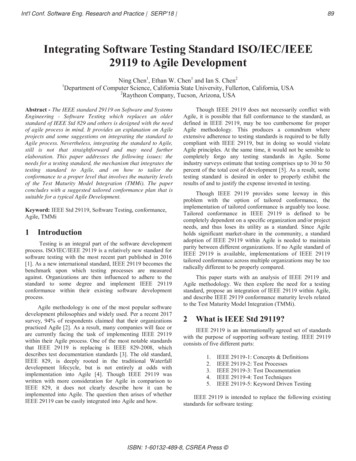
IEEE TRANSACTIONS ON MEDICAL ROBOTICS AND BIONICSaccuracy [20]–[22]. These results show promise and warrantfurther investigation, but have yet to fully address the abovelimitations of endoscope-based methods.The kinematic information inherently available in the daVinci surgical system represents a means of 3D localizationthat relies neither on intraoperative use of external trackersnor on processing endoscopic video. Previous research foundthat the da Vinci’s active joints (which control motion of thelaparoscopic instruments during operation) can be localizedwith sufficient accuracy for image guidance; however, theaccuracy of the passive setup joints (used for gross manipulator positioning) was not suitable [23]–[25]. Kwartowitzet. al. [25] proposed to address this shortcoming by using a“hybrid” tracking scheme that combines two tracking modalities (specifically kinematic tracking and optical tracking) tomore accurately track the multiple manipulators of the daVinci in a common coordinate system. In this hybrid trackingscheme, the base frames of the active kinematic chains areregistered to external, optically tracked frames attached to thebase of the da Vinci. Thus, all base frames can be localizedwithin the coordinate system of the optical tracker and themanipulator tips can then be kinematically tracked relative totheir respective base frames. Fiducial localization experimentsin [26] later validated the accuracy of hybrid tracking withthe da Vinci for image guidance applications. In this paper,we implement this hybrid tracking approach as part of anew calibration method that also simultaneously estimateskinematic parameters of the da Vinci system.Kinematic tracking of the da Vinci instruments has alsoshown particular promise in combination with “drop-in” ultrasound probes. In [27], registering the image frame of theultrasound to the kinematic frames of the robot enabled theultrasound plane to be displayed in the live endoscopic video.Researchers also combined automatic detection of the robotinstruments in ultrasound images [28] with kinematic trackingto produce semi-autonomous ultrasound guidance that trackedinstrument motions [29]. Later work in [30] demonstratedregistration between kinematically tracked ultrasound and preoperative CT images for application to partial nephrectomy.The feasibility of this ultrasound-based registration techniquein the context of the operating room is, however, inherentlycoupled to the accuracy of intraoperative segmentation of theultrasound images, and represents a skill- and time-intensiveaddition to the surgical workflow. As an alternative, the touchbased method examined in this work uses the da Vinci’s kinematically tracked instruments directly to digitize anatomicalsurfaces to enable registration.The idea of a touch-based registration for image guidancewith the da Vinci system was first introduced by Ong et. al. [8].During a partial nephrectomy case, the instrument tool tipwas lightly traced over the kidney surface while recording therobotic joint values; the data was processed postoperativelyto generate a sparse set of surface points that were usedfor a standard surface-based registration. The concept showedqualitative merit; however, the authors noted they were unableto perform quantitative analysis of the touch-based methoddue to the unavailability of a ground truth comparison duringthe human trial. Building upon this concept, we have further3assessed surface-based registration with the da Vinci usingrigid phantoms [31]; however, thorough analysis of registrationerror for this touch-based method using anatomically accuratephantoms has thus far remained unstudied. In this paper wetake essential steps toward practical and accurate deploymentof this touch-based registration concept by presenting a systemthat is suitable for deployment in a real-world operatingroom and accomplishes registration in real time. We alsorigorously evaluate the accuracy of touch-based registrationon anatomically accurate soft-tissue phantom models, anddemonstrate its ability to improve the localization accuracyof an experienced surgeon.III. S YSTEM OVERVIEWA. Preoperative System Setup and CalibrationPreoperative calibration of the da Vinci Si system is necessary to achieve sufficient kinematic tracking accuracy to enableour touch-based registration. Figure 2 shows a clinical da VinciSi deployed for preoperative calibration, which takes placeas the da Vinci system is draped prior to surgery. Additivelymanufactured reference frames designed to interface with theda Vinci system are clamped rigidly to the distal ends of thesetup arms (Fig. 2, upper right). These reference frames areequipped with reflective optical tracking markers. To maintainthe sterile field, the reference frames are first clamped withoutreflective spheres before deploying the sterile drapes. Afterdraping the robot, sterile, disposable, commercially availablespheres are attached to mounting posts through the sterileplastic drapes. This process ensures a sterile barrier betweenthe clamping system and the sterile surgical environment.As shown in Fig. 2 (lower right), the da Vinci instrumentsgrasp sterile, optically tracked calibration tools. Each calibration tool is previously pivot-calibrated so that the positionof the interface with the instrument tip is accurately knownrelative to the optical markers. This enables measurement ofthe instrument position relative to the optically tracked reference frames at the base of each serial chain. Our system usesa Polaris Spectra (Northern Digital Inc., Waterloo, Ontario,Canada) optical tracking system, which is currently availablefor use in many operating rooms, including any operatingroom at our institution. The Polaris system has a reportedtracking accuracy of 0.25 mm, and for this work, we considermeasurements with the optical tracker to be ground truth [32].To collect calibration data prior to surgery, a robot operator simply moves the calibration tools throughout the robotworkspace while recording data. In our current implementation, the operator momentarily pauses at discrete locations toensure synchronization of optical tracking and robot encoderdata streams. This is necessary only because we did not havedirect access into the robot software to enable synchronizationbetween the data streams of the optical tracker and robot.In future clinical implementations, the markers can simplybe waved in front of the optical tracking system to collectcalibration data. This data collection process is repeated foreach da Vinci manipulator and each instrument to be usedduring surgery.Our system employs a hybrid tracking technique [25] forcalibration that enables the da Vinci’s manipulators to be
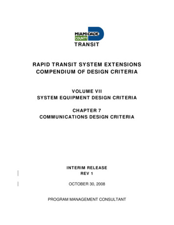
IEEE TRANSACTIONS ON MEDICAL ROBOTICS AND BIONICSkinematically tracked relative to external, optically trackedreference frames (rather than the internal base frames of the daVinci system). Using hybrid tracking bypasses the comparatively inaccurate setup joints of the da Vinci system, shorteningthe effective kinematic chain and improving tracking accuracybeyond the inherent capabilities of the da Vinci system. Ourcalibration process also simultaneously calibrates the parameters of the kinematic model using standard techniques [33]–[35]. The result of calibration is that each robotic instrumentcan be accurately tracked with respect to the location of thereference frames attached at the base of the active serial chain.B. Touch-Based RegistrationOur touch-based registration method aligns two sets of data:a densely sampled point set describing the organ surface inimage space and a sparsely sampled point set of surface datadescribing the organ surface in physical space. The denseimage space point set is obtained preoperatively from volumetric imaging. For the experiments presented in this paper,the kidney surface was manually segmented from CT imagesusing 3D Slicer, an open-source medical image computing andvisualization software platform [36]. In the future, however,when an image guidance system like ours is developed intoa commercial product, it is likely that manual segmentationwould be replaced by an automatic segmentation algorithm.Any existing or future segmentation algorithm would bestraightforward to incorporate into the framework describedin this paper, since our system assumes only the existence ofa segmentation without regard for how the segmentation wasaccomplished.The physical space point set is obtained intraoperativelyby lightly tracing the surface of the patient’s organ with thetip of the da Vinci’s instrument. We track the instrument’stip position in physical space during this process using thepreviously calibrated kinematics. Surface tracing is quick andnon-disruptive to surgical workflow: acquiring a sufficientnumber of surface points for accurate registration requires onlyabout 30 seconds. After tracing, the data are automaticallydownsampled to exclude data points within 2 mm of neighboring points to eliminate variations in point cloud densitycaused by variable tracing speed. This results in a set of pointsin physical space that lie on the surface of the patient’s kidney.Previous work concluded that the physical-space data usedfor surface-based registration should include at least 28% ofthe anterior surface area of the kidney to ensure accurateregistration [37]. Therefore, once surface tracing is complete,our system automatically analyzes the tracing data to verifythat the tracing covers a sufficient area. Our system determinesthe surface area corresponding to a tracing by constructing asurface mesh from the tracing data using the ball-pivoting surface reconstruction algorithm [38] (illustrated in Fig. 3). Thearea of the reconstructed surface is compared to the kidney’ssurface area, which is determined from the preoperative CTimages.Registration between the image space point set with thephysical space point set is computed using the globally optimal4iterative closest point (GoICP) algorithm [39]. GoICP doesnot require user initialization and as such is not subject tosuboptimal initialization concerns associated with standardICP algorithms. The resulting registration between the imagespace and the physical space relates knowledge of the patient’sanatomy present in the preoperative images to the currentposition of the robot with respect to the patient.C. Real-Time Data Streaming and VisualizationUsing the computed registration, we display the position ofanatomical structures segmented from preoperative imagingto the surgeon in real time directly in the da Vinci surgeonconsole (see Fig. 1).We built our image guidance system as a submodule of 3DSlicer, an open-source medical image computing and visualization platform that enables patient image segmentation, preoperative planning, and real-time model rendering for imageguidance [36]. Our system interfaces with the clinical da VinciSi application programming interface (API) [40] through a dataacquisition module built with the open-source Plus Toolkit [41]that streams kinematic data output by the API to 3D Slicerusing the standardized OpenIGTLink messaging protocol [42].This enables the endoscope camera view and graphical modelsof the da Vinci’s manipulators in the image guidance displayto track the movement of the physical instruments in real time.Our image guidance (see Fig. 1) is displayed directly in the daVinci surgeon console through the console’s TilePro interface.IV. S YSTEM VALIDATION E XPERIMENTSWe first evaluate the efficacy of our calibration method toimprove the overall kinematic accuracy of the da Vinci robot.We then evaluate the accuracy of our touch-based registrationmethod.A. Calibration AccuracyWe wish to determine the number of measurements thatmust be collected during preoperative setup to ensure goodcalibration results. In the context of robot calibration, thisnumber is generally difficult to predict, as it varies from systemto system and also depends on the measurement methodused [43]. We performed a series of trials to determine therelationship between tracking accuracy and the number ofmeasurements used in calibration, as described below.For our touch-based application, the da Vinci instrument tipserves as the localizer. Given that surface-based registrationrelies only on discrete points of position data, only thepositional (not rotational) accuracy of the localizer needs tobe considered. Therefore, the accuracy of our system canbe quantified by the fiducial localization error (FLE) of theda Vinci instruments, i.e. the distance between the modelpredicted tip location and the true tip location:robotFLE probot.model ptrue(1)In practice, the FLE cannot be directly measured becauseour model-predicted position is measured in a different coordinate frame from our “true” position (as measured by the
IEEE TRANSACTIONS ON MEDICAL ROBOTICS AND BIONICS5Fig. 2. Hybrid tracking implemented with the da Vinci Si in the operating room. Optically tracked markers (top right) are rigidly clamped to the base ofeach manipulator, and sterile tracking spheres are attached to the markers over the robot drapes ensuring sterility. Calibration is achieved by gripping sterilecalibration objects (bottom right) in the manipulators (or pressing them onto the endoscope) and waving them in front of the tracker preoperatively.Fig. 3. Surface reconstruction from surface tracing data. A. Original pointset from an example robotic instrument tracing. B. Reconstructed surface forsurface area computation to ensure adequate model coverage.optical tracker). However, it is possible to indirectly estimatethe expected value of the FLE from these data, as describedbelow. The transformation between the two coordinate frames(the robot and the optical tracker) can be estimated from astandard, rigid, point-based registration between the modelpredicted positions and the true measured positions [44]. Theerror associated with such a registration can be quantifiedby the fiducial registration error (FRE), which is the rootmean-square error in the alignment of the registered points.Performing numerous registrations using different sets of pointsamples provides a good estimate for the expected value of theFRE for registrations between the two frames. The expectedvalue of the FRE can be used to estimate the expected valueof the FLE, according to the following relationship derived in[44]:FLE2 FRE2,(1 2/N )(2)where N is the number of points used in the registrationand the angle bracket operator denotes the expected valueof a random variable. This formulation relies on standardassumptions that the components of FRE are independent,isotropic, 3D normal random variables.Our evaluation data set comprised 130 calibration measurements, collected at distinct poses representing a sparsesampling of the entire da Vinci Si active workspace. Eachcalibration measurement consists of a set of robot joint valuesand a corresponding Cartesian position, measured in the optical tracker’s workspace. All data was collected using the daVinci’s EndoWrist Large Needle Driver instrument.To determine the relationship between the model-predictedposition accuracy and the number of data points used in modelcalibration, we performed a Monte Carlo cross-validationanalysis of the evaluation data set. Each iteration of the crossvalidation was performed as follows: A number M {10, 15, 20, . . . , 95, 100} of “trainingpoints” were selected uniformly at random from thecomplete set of 130 points.The kinematic model was calibrated using the trainingpoints.K 30 “validation points” were selected uniformly atrandom from the remaining 130 M points.A number N {5, 6, 7, . . . , 19, 20} of “registrationpoints” were selected uniformly at random from thevalidation points. This process was repeated 1000 timesfor each value of N , resulting in a total of 16, 000 distinctsets of registration points per iteration.Using each set of registration points, a rigid point-basedregistration between the (calibrated) model-predicted positions and the measured “true” positions was performed.The mean value of FLE for the calibrated system wascomputed from the average FRE of each registrationaccording to Eq. (2).This process was repeated a total of 1000 times for each
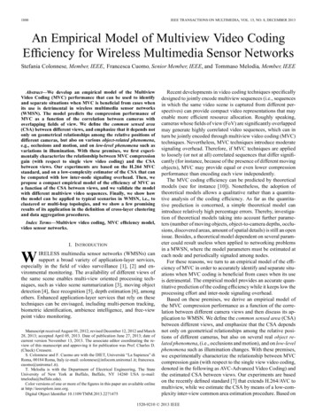
IEEE TRANSACTIONS ON MEDICAL ROBOTICS AND BIONICSFig. 4. Fiducial localization error (FLE) of the da Vinci Si vs. the numberof measurements used for calibration of the hybrid tracking model. The redarea indicates the standard deviation for each respective trial. Only marginalimprovements to accuracy can be seen past M 60.6Fig. 6. Optically tracked phantom platform used for evaluating registrationaccuracy. Surface data for registration is acquired by tracing the phantomsurface (illustrated as red dots). The location of the phantom relative to thetracked platform is known, enabling evaluation of our touch-based registrationtechnique.34% ErrorReductionreflect only the deviation between the model-predicted positionof a robot manipulator and the measured position (i.e. wherethe robot “thinks” the manipulator is versus where it trulyis). These accuracy results do not describe the accuracy withwhich a surgeon can direct the da Vinci manipulators duringteleoperation (i.e. where the surgeon wants the manipulatorto be versus where it truly is). While touch-based imageguidance relies on a highly accurate model-predicted position,teleoperation with visual feedback and a human in the loopdoes not.Fig. 5. Distribution of RMS errors between the model-predicted robot tipposition using hybrid tracking and the ground truth, optically tracked tipposition. A significant decrease in error is seen when using our calibrationmethod (red) over using the nominal robot parameters (blue). Results are for1000 calibration trials with M 60 measurements per trial.value of M . Figure 4 shows the results of this analysis, whichindicates that using more than 60 data points to compute thehybrid tracking calibration offers only marginal improvementsto localization accuracy.Figure 5 shows the accuracy improvement of the calibratedda Vinci model compared to the nominal model from [25]. Themean and standard deviation of the calibrated system’s FLEare 0.95 mm and 0.14 mm, respectively, representing a 34%reduction in mean localization error. While no well-definedlocalization accuracy threshold exists for image guidance applications, it is clear that increased accuracy is always desired.Our calibrated system accuracy is comparable to prior methodsused to track the absolute position of the da Vinci’s instrumenttips (1.31 mm in [24] and 1.39/1.95 mm for PSM1/PSM2 in[25]).We wish to emphasize that the accuracy values reported hereB. Registration AccuracyWe evaluated the accuracy of our touch-based registrationmethod in a series of experiments using a commerc
and the School of Computing, University of Utah, Salt Lake City, UT 84112, USA. N. L. Kavoussi and S. D. Herrell III are with the Depart- . Correspondence e-mail: james.m.ferguson@vanderbilt.edu, bryn.pitt@vanderbilt.edu, robert.webster@vanderbilt.edu Manuscript received January 3, 2020; revised April 11, 2020. Fig. 1. Our image guidance .