Transcription
Fava et al. Arthritis Research & Therapy (2016) 18:99DOI 10.1186/s13075-016-0993-2RESEARCH ARTICLEOpen AccessFrequency of circulating topoisomerase-Ispecific CD4 T cells predicts presence andprogression of interstitial lung disease insclerodermaAndrea Fava1, Raffaello Cimbro1, Fredrick M. Wigley1, Qing-Rong Liu2, Antony Rosen1 and Francesco Boin3*AbstractBackground: Scleroderma is an antigen-driven T cell-mediated autoimmune disease. Presence of anti-topoisomerase-Iantibodies is associated with pulmonary fibrosis and predicts increased mortality. Characterization of autoreactive Tlymphocytes may shed light on disease pathogenesis and serve as a biomarker for disease activity. Here, we aimed toquantify and functionally characterize circulating topoisomerase I (topo-I)-specific CD4 T cells and to define theirassociation with presence and progression of interstitial lung disease (ILD) in patients with scleroderma.Methods: Using flow cytometry, circulating topo-I-reactive CD4 T cells were identified by the expression of specificactivation markers (CD154 and CD69) upon stimulation with purified topo-I and quantified in 27 SSc patients and 4healthy donors (HD). Polarization of autoreactive T cells (Th1, Th2, Th17, Th1–17) was defined using surface expressionof specific chemokine receptors. Presence and progression of ILD were determined using high-resolution chest CTand pulmonary function tests.Results: Topo-I-reactive CD4 T cells were found in all topo-I-positive patients compared to one topo-I-negativesubject and no HD. Topo-I-specific CD4 T cells exhibited a distinct Th17 polarized phenotype. Autoreactive T cellswere significantly increased in subjects with evidence of ILD and were quantitatively associated with the decline oflung volumes.Conclusions: Topo-I-specific T cells can be reliably quantified in the peripheral blood of patients with scleroderma,exhibit a pro-inflammatory Th17 phenotype, and predict progression of ILD.Keywords: Systemic sclerosis, Interstitial lung disease, T lymphocytes, Th17, AutoantigenBackgroundSystemic sclerosis (SSc) is characterized by obliterativemicroangiopathy, progressive fibrosis of affected organs,and autoimmunity [1]. Early in the disease, an inflammatory mononuclear cell infiltrate precedes fibrosis andparallels the development of vasculopathy in affected tissues, suggesting that a disrupted immune response is amajor driver in SSc pathogenesis [2]. A more precise understanding of the immune effector pathways would* Correspondence: francesco.boin@ucsf.edu3Department of Medicine, Division of Rheumatology, University of California,San Francisco, 513 Parnassus Avenue, Med Sci, S-847, San Francisco, CA94143, USAFull list of author information is available at the end of the articleallow identifying reliable markers to monitor disease activity and to define suitable targets for a selective therapeutic intervention.In scleroderma, the presence of specific autoantibodiesis strongly associated with distinct clinical phenotypes[3]. In particular, anti-topoisomerase-I (topo-I) antibodies have high specificity for scleroderma, and arepresent in 20–45 % of patients [4, 5]. They are associated with diffuse skin involvement and pulmonary fibrosis, and independently predict more severe disease andmortality [6, 7]. Despite these robust associations, theserum anti-topo-I antibody levels are not useful tomonitor disease activity and progression in SSc patients[8–10], and there are no convincing data supporting a 2016 Fava et al. Open Access This article is distributed under the terms of the Creative Commons Attribution 4.0International License (http://creativecommons.org/licenses/by/4.0/), which permits unrestricted use, distribution, andreproduction in any medium, provided you give appropriate credit to the original author(s) and the source, provide a link tothe Creative Commons license, and indicate if changes were made. The Creative Commons Public Domain Dedication o/1.0/) applies to the data made available in this article, unless otherwise stated.
Fava et al. Arthritis Research & Therapy (2016) 18:99direct role for these autoantibodies in the pathogenesisof SSc suggesting that the anti-topo-I response is involved but not antibody mediated.Importantly, anti-topo-I antibodies undergo classswitching and exhibit a strong association with humanleukocyte antigen (HLA) alleles across different ethnicgroups, implicating T cells in this response [11, 12]. Thepresence of an antigen-driven, T cell-dependent processin SSc is supported by several studies [12–16]. Definingthe functional phenotype of topo-I-reactive T cells wouldbe relevant to determine whether and how this cellularsubset may contribute to inflammation and tissue damage in SSc. However, no conclusive data have been provided to define whether autoantigen-specific T cellresponses are amplified in individuals with active diseaseand contribute to the generation of distinct phenotypesthrough their effector function. These aspects have beendifficult to investigate due to poor accessibility of targettissues and technical challenges to detect ex vivo lowfrequency antigen-specific T cells [12–16]. In this study,we employed a novel assay to identify, quantify andfunctionally characterize topo-I-specific CD4 T cells.This is based on the detection upon antigen stimulationof reliable markers of early activation (i.e., CD154,CD69) as well as of T cell polarization. We found thattopo-I-responsive T cells are selectively increased inanti-topo-I-positive SSc patients and exhibit a T helper(Th)17 pro-inflammatory functional phenotype, with frequency quantitatively associated with presence and progression of interstitial lung disease (ILD). These findingsimplicate amplified topo-I-specific T cell responses inthe pathogenesis of SSc lung disease, and suggest thatthey may be appropriate targets to monitor disease activity and predict outcome.MethodsPatients evaluated at the Johns Hopkins SclerodermaCenter were included in the study after providing written informed consent if they met the American Collegeof Rheumatology preliminary criteria for the classification of SSc [17]. The Johns Hopkins institutional reviewboard approved the study.Clinical phenotypingDemographic and clinical data including age, sex, ethnicity,smoking status, disease duration, scleroderma subtype,specific organ involvement, use of immunosuppressiveagents, and autoantibody status were obtained at the timeof the visit. Severity of skin involvement was quantifiedusing the modified Rodnan skin score [18]. Pulmonary involvement was determined based on abnormal pulmonaryfunction tests (PFT), including measurements of forcedvital capacity (FVC) and single-breath carbon monoxidediffusing capacity (DLCO), calculated according to thePage 2 of 12American Thoracic Society recommendations [19]. AllPFT data were standardized to same reference values[20, 21]. Presence of ILD was confirmed by the detectionof established fibrotic lung changes on computerized tomography of the chest. ILD progression was defined as %change in FVC in the year preceding the test date or inthe subsequent 10 months in those patients for whomfollow-up data became available. Lung disease severitywas assessed using the Medsger severity scale [22].T cell culture and stimulationPeripheral blood mononuclear cells (PBMC) were isolated from whole blood through density-gradient centrifugation (Ficoll-Paque Plus, GE Healthcare, Chicago, IL,USA), resuspended in RPMI medium supplemented with5 % autologous serum, 2 mM L-glutamine, 100 U/mlpenicillin, and 100 ug/ml streptomycin, transferred ontoa 96-well plate (1.5 106 cells/well) and cultured at 37 C.To prevent CD154 internalization, PBMCs were preincubated with anti-human CD40 blocking antibody (1 ug/ml;G28.5, BioLegend, San Diego, CA, USA) for 15 minutes.Subsequently, cells were stimulated for 18 hours usingalternatively 1 ug/ml staphylococcal enterotoxin B (SEB)/staphylococcal enterotoxin A (SEA), 15 ug/ml tetanustoxoid (TT) (NIBSC), 17 ug/ml human recombinanttopoisomerase-I purified from baculovirus-infected insectcells (GenScript Biotech Corp, Piscataway, NJ, USA),18 ug/ml purified human recombinant (baculovirus)protein arginine methyltransferase 6 (PRMT6) (CaymanChemicals, Ann Arbor, MI, USA) or human peptidyl arginine deiminase type 4 (PAD4) (gift from Dr. Erika Darrah).Flow cytometryAfter stimulation and washings with PBS, cells were thenstained with Live/Dead Fixable Blue Dead Cell Stain Kit(Molecular Probes, Eugene, OR, USA), BV510-conjugatedanti-CD3 (OKT3, BioLegend), Pacific Blue-conjugatedanti-CD4 (RPA-T4, BD Pharmingen Franklin Lakes, NJ,USA), APC-H7-conjugated anti-CD8 (SK1, BD, FranklinLakes, NJ, USA), PE-conjugated anti-CD154 (TRAP1, BDPharmingen), APC-conjugated anti-CD69 (FN50, BDPharmingen), Alexa-fluor 488-conjugated anti-CXCR3(TG1/CXCR3, BioLegend), PerCP-Cy5.5-conjugated antiCCR6 (TG7/CCR6, BioLegend), PE-Cy7-conjugated antiCCR4 (1G1, BD Pharmingen) antibodies, and flowcytometry acquisition was performed on a FACSAria(BD) instrument. Analysis was conducted using FlowJosoftware (Tree Star Inc., Ashland, OR, USA).Cytokine quantitationFreshly isolated PBMCs were sorted accordingsurface expression of the chemokine receptorsCXCR3, and CCR4 (Fig. 3a), resuspended inmedium supplemented with 10 % fetal bovineto theCCR6,RPMIserum
Fava et al. Arthritis Research & Therapy (2016) 18:99(FBS), 2 mM L-glutamine, 100 U/ml penicillin and 100ug/ml streptomycin, and then stimulated with anti-CD3/CD28 beads (Life Technologies, Carlsbad, CA, USA) at abead-to-cell ratio of 2:1 for 48 hours (37 C). Supernatants were collected and stored at -80 C. Quantitationof interferon (IFN)γ, interleukin (IL)-4 and IL-17A wasperformed in duplicate using DuoSet-ELISA kits (R&DSystems, Minneapolis, MN, USA) according to manufacturer’s instructions.Page 3 of 12Calculations of relative expression from Ct data werecarried out according to User Bulletin #2 for ABI Prism7900 Sequence Detection System.Enzyme-linked immunosorbent assay (ELISA)Anti-topoisomerase-I antibody concentration was measuredin the serum of SSc patients using QUANTA Lite Scl70enzyme-linked immunosorbent assay (Inova Diagnostics,San Diego, CA, USA) according to the manufacturer’sinstructions.RNA isolation and cDNA synthesisTopo-I specific CD4 T cells identified after 18 h stimulation as described in the Methods section of the mainmanuscript were sorted according to their chemokinereceptor expression pattern into 50 μl of RNA extractionbuffer (PicoPure RNA isolation kit, Arcturus Bioscience,Inc., Mountain View, CA, USA), incubated at 42 C for30 min and centrifuged (800 g) at RT for 2 min. Thesupernatant was then collected in an RNase-free tube.Subsequent column filtration, washing, and elution ofRNA from the columns were performed according tosection C of the PicoPure RNA isolation protocol(Protocol for Use with CapSure Macro Laser CaptureMicroscope Caps). RNA integrity numbers were measuredusing Agilent RNA 600 Pico kit (Agilent Technologies,Santa Clara, CA, USA). Single-strand cDNAs were synthesized with the Superscript III first-strand cDNA synthesiskit following the manufacturer’s protocol (Invitrogen, LifeTechnologies, Waltham, MA, USA).Pre-amplification and quantitative real-time polymerasechain reaction (RT-qPCR)TaqMan PreAmp Master Mix Kit was used for cDNApreamplification (Applied Biosystems, Life Technologies,Waltham, MA, USA) using pooled primer mixes (200times dilution) of the predesigned TaqMan assays ofIL17A (Hs00174383 m1), IL4 (Hs00174122 m1), IFNG(Hs00989291 m1) and endogenous control beta-actin(4326315E). cDNAs were preamplified in an ABI 9700Thermal Cycler using the program: 95 C hold for10 min, and then 14 cycles of denaturation at 90 C for15 sec and annealing and extension at 60 C for 4 min.The pre-amplification PCR products were immediatelydiluted five times with molecular biology-grade water (5PRIME, Gaithersbrug, MD, USA) and stored at -20 C orimmediately processed for qPCR. Duplex qPCR assayswere performed on technical duplicates using a Famlabeled probe for each target gene and a Vic-labeledprobe for the endogenous control gene, along with TaqMan Advanced Fast PCR Master Mix (Life Technology).RT-qPCR reactions were run in a 7500 Fast TaqMan instrument using the program: 95 C hold for 20 secfollowed by 40 cycles of denaturation at 95 C for 3 secand annealing and extension at 60 C for 30 sec.Statistical analysisStatistical analysis was performed using Prism 6.0 Softwarefor Macintosh (GraphPad Software, San Diego, CA, USA).Differences between variables were determined usingStudent t test and Wilcoxon rank-sum test for continuousvariables, and the χ2 or Fisher’s exact test for categoricalvariables. Multiple comparisons were performed by analysis of variance (ANOVA) with Bonferroni correction fornormally distributed variables or Kruskal-Wallis andDunn’s test otherwise. Linear associations were analyzedusing Pearson correlation coefficient upon assessment forGaussian distribution with Shapiro-Wilk test. Throughout,a two-tailed α of 0.05 was used.ResultsWe studied 27 consecutive SSc patients: 15 anti-topo-Ipositive and 12 anti-topo-I-negative. The two groupswere similar with regards to the main demographic anddisease characteristics (Table 1). Consistent with previous studies, anti-topo-I positive patients exhibited a significant higher prevalence of ILD (p 0.005), worsemodified Rodnan Skin Score, and a trend for lower diffusion capacity of lung for carbon monoxide (DLco).CD154 and CD69 upregulation identifies topoisomerase-Ireactive CD4 T cellsIn order to detect with reliability and specificity antigenspecific T cells, we developed a flow cytometry-basedprotocol measuring the expression of early activationmarkers CD69 and CD154 on CD4 T cells. First, we optimized the assay using PBMCs obtained from a healthydonor recently immunized with combined tetanus, diphtheria, and pertussis (Tdap) vaccine. After stimulation for18 hours with tetanus toxoid (TT) in the presence of antiCD40 blocking antibodies and 5 % autologous serum, a selective upregulation of both CD69 and CD154 was detected on CD4 T cells (0.8 %), while no significantresponse was observed in unstimulated cells (0.04 %) orafter exposure to a negative control protein (0.09 %)(Fig. 1a). The gating strategy for this experiment is shownin Additional file 1: Figure S1. This approach was then applied to SSc patients using the whole topo-I molecule purified from baculovirus-infected insect cells and two
Fava et al. Arthritis Research & Therapy (2016) 18:99Page 4 of 12Table 1 Patient demographics and clinical characteristicsSSc PatientsTopo-I negative (N 12)Topo-I positive (N 15)P value§Age (years)51.3 9.655.1 7.448.2 10.3.093Female21 (78)9 (75)12 (80).557White22 (81)9 (75)13 (87).338Black5 (19)3 (25)2 (13)Variables*RaceSmoking statusNever16 (62)9 (82)7 (47)Past1 (4)0 (0)1 (7)Current9 (34)2 (18)7 (47)15 (56)5 (42)10 (67).1827 85 99 7.037Diffuse SSc skin type*mRSS (range 0–51).174SSc durationRP onset*, years11.1 7.811.2 7.310.9 8.5.5161st non-RP symptom*, years10.2 7.111.0 7.29.6 7.3.792Lung severity score* (range 0–4)1.2 1.30.9 1.51.5 1.1.168FVC* (% predicted)77.9 14.781.4 19.877.07 11.1.106DLCO (% predicted)75.3 16.683.8 15.771.3 17.3.051ILD†11 (46)1 (10)10 (71).00528.8 12.532.5 17.525.9 5.9.522**eRVSPAutoantibody statusACA3 (11)3 (25)0 (0).075Anti-RNA-polymerase III5 (21)3 (25)1 (8).29511 (41)4 (33)7 (47).381Immunosuppression (current)‡All values are given as number (%) unless otherwise specified. Frequencies (%) are calculated based on available data. SSc disease duration was calculated at thetime of serum sampling from the onset of RP or from the first non-RP symptom. Lung severity score is reported as previously defined by Medsger et al. [29]ACA anticentromere antibody, DLCO, diffusion capacity of lung for carbon monoxide, eRVSP right ventricular systolic pressure estimated by echocardiography,FVC forced vital capacity, ILD interstitial restrictive lung disease, mRSS modified Rodnan skin score, RP Raynaud’s phenomenon, SSc systemic sclerosis*Mean SD†The presence of ILD was defined by presence of fibrosis on computed tomography of the chest (CT chest was available in 10 topo-I-negative and 14 topo-I-positive subjects)‡Use of immunosuppressants includes cyclophosphamide, mycophenolate, methotrexate, or prednisone§P values were determined by Fisher’s exact test, Pearson chi-square, or the Wilcoxon rank-sum test, as appropriateunrelated proteins for which no T cell autoreactivitywas expected in SSc (PRMT6 and PAD4) as negativecontrols. A representative experiment is shown inFig. 1b. Topo-I-reactive CD154 CD69 CD4 T cellswere specifically identified in topo-I-positive SSc patients, while no response was triggered by eitherPRMT6 or PAD4.Topoisomerase-I-specific CD4 T cells are increased inanti-topo-I-positive SSc patientsNext, we sought to define the magnitude of autoreactiveCD4 T cell expansion in the peripheral blood of SScpatients. CD4 T cells from anti-topo-I-positive subjectsconsistently exhibited a significant upregulation of CD69and CD154 upon topo-I challenge compared to unstimulated cells and those from anti-topo-I-negative patients (Fig. 1c). Autoreactive CD154 CD69 CD4 topo-I-specific T cells were found in all 15 topo-I-positive patients compared to only one topo-I-negativesubject and no healthy donors (HD), with a frequencyranging between 0.136 % and 0.417 % of total CD4 Tcells (topo-I-positive vs topo-I-negative p 0.001; topoI-positive vs HD p 0.005; topo-I-negative vs HD p ns)(Fig. 2).Recognition of topoisomerase-I by autoreactive CD4 Tcells is HLA-DR restrictedAs CD4 T cells recognize antigens in the context ofmajor histocompatibility complex (MHC) class II molecules, we asked whether T cell response to topo-I wasMHC-restricted and in particular mediated by HLA-DRalleles [23]. PBMCs from topo-I-positive patients werestimulated with topo-I in presence of anti-HLA-DRor -HLA-DP blocking antibodies. Positive responses totopo-I were significantly decreased by HLA-DR blockade(p 0.013), while anti-HLA-DP antibodies did not impact
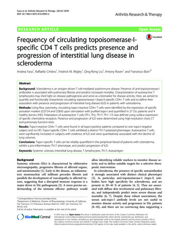
Fava et al. Arthritis Research & Therapy (2016) 18:99Fig. 1 (See legend on next page.)Page 5 of 12
Fava et al. Arthritis Research & Therapy (2016) 18:99Page 6 of 12(See figure on previous page.)Fig. 1 Detection of circulating topoisomerase-I-specific CD4 T cells by CD154 and CD69 expression and upregulation. After incubation (15 min)with anti-CD40 blocking antibodies, freshly isolated peripheral blood mononuclear cells (PBMCs) were stimulated for 18 h in presence of 5 %autologous serum as indicated below. Gating was set on lymphocytes, singlets, live, CD3 and CD4 cells. Numbers in quadrants indicate thepercent (%) cells in the parent CD4 population. a PBMCs from a healthy donor recently immunized with combined tetanus, diphtheria, andpertussis (Tdap) vaccine were stimulated with PRMT6 (negative control), SEA SEB (positive control) and tetanus toxoid (TT). b PBMCs from atopo-I SSc patient were stimulated with PRMT6, PAD4 (negative controls), or topo-I. c Representative experiments on two anti-topo-I-negativeand two anti-topo-I-positive SSc patients. The gating strategy for this experiment is shown in Additional file 1: Figure S1. PRMT6 protein argininemethyltransferase 6, PAD4 human peptidyl arginine deiminase type 4T cell activation (Fig. 3), implicating that specific topo-Iepitopes presented to the T cell receptor (TCR) by HLADR molecules can selectively induce and maintain autoreactive T helper immune responses.Topoisomerase-I-specific CD4 T cells exhibit a Th17polarized phenotypeTo characterize the functional properties of topo-Ispecific CD4 T cells, we validated and employed an approach to identify polarized T helper subsets based onthe surface expression of specific chemokine receptors[24]. Th1, Th2, Th17 and Th1-Th17 CD4 T cells weredefined by different combinations of CXCR3, CCR6 andCCR4 expression (Fig. 4a). After cell sorting and stimulation of each individual subset with anti-CD3/CD28beads, the production of specific cytokines was assessedin the supernatants, confirming their polarized status(Fig. 4b). CCR6 CCR4 CXCR3- T cells (Th17) selectively secreted IL-17A, CCR6 CCR4-CXCR3 T cells(Th1–17) both IFNγ and IL-17A, CCR6-CCR4 CXCR3- T cells (Th2) exclusively IL-4 and CCR6-CCR4CXCR3 T cells (Th1) mainly IFNγ. Based on thesefindings, we found that CD4 T cells responding totopo-I stimulation, exhibited a distinct enrichment inthe Th17 subset (p 0.001) compared to the total CD4 population, while their Th1 polarization was decreased(p 0.002) (Fig. 4c). The frequency of Th2 and Th1–17subsets did not differ between topo-I-specific and totalCD4 T cells. To further test the specificity of these phenotypes, IFNγ, IL-4 and IL-17A mRNA expression wasquantified in topo-I-responsive T cells sorted accordingto their polarized status. The cytokine specificity ofautoreactive polarized subsets was confirmed at thetranscriptional level (Fig. 4d). In particular, topo-Ispecific CCR6 CCR4 CXCR3-CD4 T cells selectivelyexpressed IL-17A mRNA.Topoisomerase-I specific CD4 T cells are quantitativelyassociated with presence and progression of ILDSeveral studies have confirmed that anti-topo-I positivityis significantly more prevalent among SSc patients withpulmonary fibrosis and entails worse clinical outcome.Fig. 2 Increased frequencies of topoisomerase-I-specific CD4 T cellsin the blood of anti-topo-I-positive SSc patients. Topo-I-specific CD4 T cells frequency was measured in the peripheral blood of 15 antitopo-I-positive, 12 anti-topo-I-negative SSc patients, and 4 healthydonors (HD). Values represent the percentage of CD154 CD69 cellswithin the CD4 population. To minimize any noise from the background,the frequency of topo-I-reactive T cells was calculated after subtractingthe small percentage of CD154 CD69 CD4 T cells detected withinunstimulated PBMCs from the same subject. Horizontal lines indicatethe mean frequency for each groupFig. 3 Topoisomerase-I-specific CD4 T cells activation is HLA-DRrestricted. PBMC from topo-I-positive SSc patients were cultured for 18 hwith topo-I and anti-HLA-DR or anti-HLA-DP blocking antibodies orisotype control (1 ug/ml). Values are represented as percent of responserelative to the frequency of CD154 CD69 CD4 cells detected afterstimulation with topo-I alone as assessed by flow cytometry. Data arerepresentative of three separate experiments (mean SD)
Fava et al. Arthritis Research & Therapy (2016) 18:99Page 7 of 12Fig. 4 Topoisomerase-I-specific CD4 T cells show a Th17 polarized functional phenotype. a CD4 T cell subsets were identified by flowcytometry using the combined surface expression of chemokine receptors CCR6, CXCR3, and CCR4. b After sorting, the different T helper subsetswere stimulated with anti-CD3/CD28 beads for 48 h and secretion of IFNγ, IL-4, and IL-17A was measured in their supernatants by enzyme-linkedimmunosorbent assay (bars represent mean SE, n 3). c The distribution of polarized T helper subsets within topo-I-specific CD4 T cells (closedcircles) is shown in comparison to the general CD4 population (open circles). T test with Bonferroni correction was used for multiple comparisons.Lines indicate mean SD. d After sorting topo-I-specific CD4 T cells according to their polarized T helper phenotype (range 16–2064 cells),mRNA expression levels of IFNγ, IL-4, and IL-17A were measured by qPCR using the Arcturus PicoPure RNA Isolation system, which is designedto recover high-quality RNA from a very low number of cells (10–500 cells) and to accomplish cDNA synthesis from minimal amounts of cDNA.IFNγ and IL-17A expression was calculated as fold change in reference to a mix of topo-I-reactive CD4 T cells from three patients; IL-4 expressionis reported as delta Ct from actin expression since it was not detectable in the reference population. Data are representative of four separatepatients (mean SE)
Fava et al. Arthritis Research & Therapy (2016) 18:99Therefore, we next addressed these important clinical associations and sought to define whether the frequency oftopo-I-specific T cell responses might have clinical utility.Topo-I-reactive T cells were significantly increased in theperipheral blood of patients with ILD compared to thosewithout lung involvement (p 0.002) (Fig. 5a). A lowerDLco was associated with higher number of topo-Ispecific T cells (Pearson r -0.6, p 0.018) (Fig. 6) but noassociation was found with the FVC (Fig. 5b). In contrast,the proportion of circulating topo-I-specific CD4 T cellswas quantitatively associated with the progression of lungfibrosis as defined by the loss of lung volumes within theyear preceding the test date (Pearson r 0.721, p 0.002)(Fig. 5c) or during the subsequent one (Pearson r 0.764,p 0.017) (Fig. 5d). None of these clinical associationswere found with anti-topo-I serum titers (Additional file 2:Figure S2). Importantly, the frequency of topo-I-specific Tcells did not show any linear relationship with anti-topo-Ititer or with disease duration (Figs. 7 and 8).These datasuggest that autoreactive T cell activation against topo-I issustained during progression of SSc-ILD and predictsfurther loss of lung volumes.DiscussionThe evidence that a distinct immune-mediated processmay drive target tissue injury in scleroderma has notPage 8 of 12Fig. 6 Frequency of topoisomerase-I-specific CD4 T cells exhibitsnegative association with the diffusion capacity of lung for carbonmonoxide (DLco). Association of topo-I-specific CD4 T cells withDLco (% predicted) in SSc patients. Pearson correlation coefficient rand p values are displayedbeen paralleled by the ability to define with precision itsmagnitude and the functional features of involved cellular and molecular effectors. In this study, we havedeveloped a reliable method to identify and quantifytopoisomerase-I-specific CD4 T cells with high specificity and precision, determining that in SSc patientstopo-I-specific T cells are strongly polarized toward aFig. 5 Frequency of topoisomerase-I-specific CD4 T cells is associated with presence and progression of interstitial lung disease (ILD). a Frequency oftopo-I reactive CD4 T cells in anti-topo-I-positive SSc patients stratified by presence of ILD. Lines indicate means. P value was calculated using Wilcoxonrank-sum test. b Association of topo-I-specific CD4 T cells with FVC (% predicted). c and d Associations between topo-I-specific CD4 T cells frequencyand the degree of ILD progression defined as % change in forced vital capacity (FVC) in the year preceding the test date (c) or in the subsequent10 months (d) in those patients for whom follow-up data became available. Pearson correlation coefficient r and p values are displayed in b, c, and d
Fava et al. Arthritis Research & Therapy (2016) 18:99Fig. 7 Frequency of topoisomerase-I-specific CD4 T cells does notcorrelate with anti-topoisomerase-I antibody serum concentration.Serum anti-topoisomerase-I antibody concentration is measured inunits of reactivity compared to a standard low positive sample asper manufacturer’s instructions. Pearson correlation coefficient r andp value are displayedpro-inflammatory Th17 phenotype and quantitatively associated with the presence and progression of ILD.Lung fibrosis is the most common pulmonary manifestation in SSc and a leading cause of morbidity and mortality [25, 26]. A substantial group of SSc-ILD patients(15–25 %) progress towards end-stage lung disease [27].Regrettably, treatment options for ILD have been limitedto nonselective immunosuppression. Therapeutic efficacyhas been hindered by our poor ability to diagnose lung involvement earlier, to effectively monitor the disease courseand to identify highly selective pathogenetic targets.Currently, the only reliable tool to predict disease activityand progression in SSc-ILD is the prospective measurement of lung volumes, as high-resolution chest computedFig. 8 Frequency of topoisomerase-I-specific CD4 T cells is notassociated with disease duration. Disease duration is calculated inyears from the first non-Raynaud’s phenomenon symptoms to thetime of blood sampling. Pearson correlation coefficient r andp values are displayedPage 9 of 12tomography or bronchoalveolar lavage fluid analysis haveshown lack of sensitivity and/or specificity both before orafter therapeutic intervention [28, 29]. Importantly, thefact that ILD progression is often identified by spirometrycriteria only after irreversible lung damage has already occurred remains one of the major obstacles to the promptinitiation of therapy and ultimately to its efficacy. Thefindings of this study indicate that quantification andfunctional profiling of topo-I-specific CD4 T cells mayrepresent a novel, useful, and noninvasive tool to assessand predict disease activity in SSc-ILD. Moreover, thequantitative association of these autoreactive T cells withthe degree of progressive FVC decline suggests that expansion of the topo-I autoreactive T cells and ongoinglung injury may be mechanistically connected.The identification of circulating autoreactive CD4 Tcells is challenging due to their very low frequency. Previous studies pursuing the detection of topo-I-specific Tcells in SSc patients have yielded limited results due torelatively weak cellular responses to topo-I as well as poorspecificity, as autoreactive T cells have been found also inanti-topo-I-negative patients as well as healthy controls[12–14, 16]. Most of these investigations used bulk assaysmeasuring proliferation of mononuclear cells by 3H-thymidine incorporation after several days of in vitro stimulation and could not reliably determine the frequency ofcirculating topo-
RT-qPCR reactions were run in a 7500 Fast TaqMan in-strument using the program: 95 C hold for 20 sec followed by 40 cycles of denaturation at 95 C for 3 sec and annealing and extension at 60 C for 30 sec. Calculations of relative expression from Ct data were carried out according to User Bulletin #2 for ABI Prism 7900 Sequence Detection .