Transcription
(2022) 21:30Zuo et al. Molecular en AccessREVIEWThe crosstalk between reactive oxygenspecies and noncoding RNAs: from cancer codeto drug roleJing Zuo1†, Zhe Zhang1†, Maomao Li2†, Yun Yang2†, Bohao Zheng2, Ping Wang2*, Canhua Huang1* andShengtao Zhou2*AbstractOxidative stress (OS), characterized by the excessive accumulation of reactive oxygen species (ROS), is an emerging hallmark of cancer. Tumorigenesis and development driven by ROS require an aberrant redox homeostasis, thatactivates onco-signaling and avoids ROS-induced programmed death by orchestrating antioxidant systems. Theseprocesses are revealed to closely associate with noncoding RNAs (ncRNAs). On the basis of the available evidence,ncRNAs have been widely identified as multifarious modulators with the involvement of several key redox sensingpathways, such as NF-κB and Nrf2 signaling, therefore potentially becoming effective targets for cancer therapy.Furthermore, the vast majority of ncRNAs with property of easy detected in fluid samples (e.g., blood and urine)facilitate clinicians to monitor redox homeostasis, indicating a novel method for cancer diagnosis. Herein, focusingon carcinoma initiation, metastasis and chemoradiotherapy resistance, we aimed to discuss the ncRNAs-ROS networkinvolved in cancer progression, and the potential clinical application as biomarkers and therapeutic targets.Keywords: Reactive oxygen species, Oxidative stress, Noncoding RNAs, Cancer diagnosis, Cancer therapyIntroductionIntracellular oxidative reactions play a vital role in metabolic processes, by which plenty of biomolecular compounds like reactive species are produced. Under normalphysiological conditions, these products maintain properconcentrations, with a close relation to enzyme activation[1–3], protein synthesis [4, 5], signal transduction [6, 7]*Correspondence: wangping 886@126.com; hcanhua@scu.edu.cn;taotaovip2005@163.com†Jing Zuo, Zhe Zhang, Maomao Li and Yun Yang contributed equally tothis work.1State Key Laboratory of Biotherapy and Cancer Center, West ChinaHospital, and West China School of Basic Medical Sciences & ForensicMedicine, Sichuan University, and Collaborative Innovation Centerfor Biotherapy, Chengdu 610041, People’s Republic of China2Department of Obstetrics and Gynecology, Key Laboratory of BirthDefects and Related Diseases of Women and Children of MOE and StateKey Laboratory of Biotherapy, West China Second University Hospital,Sichuan University and Collaborative Innovation Center, Chengdu,People’s Republic of Chinaand gene expression [8, 9]. However, excessive productshave some detrimental impacts, probably leading to thedysregulation of the above biological events and causingall different kinds of diseases, including inflammation[10], diabetes [11], cardiovascular disease [12] and evenmalignancies [13]. Therefore, the maintenance of redoxhomeostasis is essential to normal cellular activities andhuman health. There are four main reactive species:reactive oxygen species (ROS), reactive nitrogen species(RNS), reactive sulfur species (RSS) and reactive chlorinespecies (RCS). ROS is the most abundantly produced andfrequently explored, involving superoxide anion (O2 ),hydrogen peroxide ( H2O2), hydroxyl radical (OH ), singlet oxygen (1O2) and ozone (O3) [14]. ROS originatesfrom internal oxygen metabolism and external environmental changes. Internal sources stem from oxygen,which is catalyzed by different enzymes, and primarilyoccurs in mitochondria, peroxisomes and endoplasmic The Author(s) 2022. Open Access This article is licensed under a Creative Commons Attribution 4.0 International License, whichpermits use, sharing, adaptation, distribution and reproduction in any medium or format, as long as you give appropriate credit to theoriginal author(s) and the source, provide a link to the Creative Commons licence, and indicate if changes were made. The images orother third party material in this article are included in the article’s Creative Commons licence, unless indicated otherwise in a credit lineto the material. If material is not included in the article’s Creative Commons licence and your intended use is not permitted by statutoryregulation or exceeds the permitted use, you will need to obtain permission directly from the copyright holder. To view a copy of thislicence, visit http:// creat iveco mmons. org/ licen ses/ by/4. 0/. The Creative Commons Public Domain Dedication waiver (http:// creat iveco mmons. org/ publi cdoma in/ zero/1. 0/) applies to the data made available in this article, unless otherwise stated in a credit line to the data.
Zuo et al. Molecular Cancer(2022) 21:30reticulum (ER) [15] . Meanwhile, external inducers arecomposed of ultraviolet radiation, ionizing radiation andtoxic compounds, which generate accumulated ROS andprobably cause cancerous transformation [16–18].OS refers to the equilibrium disturbance of oxidativeand anti-oxidative systems in favor of oxidant burden,which is generally mediated by ROS. Normally, intracellular antioxidants are enough to neutralize extra oxidesto maintain homeostasis and ensure a balanced status.However, when redox balance is broken, extra ROS willinduce OS and damage biomacromolecules, includingDNA [19–21], RNA [22, 23], proteins [24] and lipids[25–27], engendering cell death or provoking the malignant transformation of normal cells. Intriguingly, excessive ROS seems to preferentially accumulate in cancercells. Thus, there are two interesting issues worthy of discussion: how cancer cells protect themselves from ROStoxicity and whether ROS-based anticancer strategiesbenefit cancer therapy.NcRNAs are defined as transcripts that cannot betranslated into proteins or functional proteins at least.In recent years, ncRNAs have attracted much interest and been proved to be direct or indirect elements ofgene transcriptional and post-transcriptional regulation[28–31]. A large number of ncRNAs have been identified, which are classified into two major groups in length:short ncRNAs ( 200 bp) and long ncRNAs (lncRNAs)( 200 bp). Short ncRNAs mainly comprise microRNAs(miRNAs), circular RNAs (circRNAs), small interferingRNAs (siRNAs), small nuclear RNAs (snRNAs), smallnucleolar RNAs (snoRNAs), PIWI-interacting RNAs(piRNAs), tRNA-derived small RNAs (tsRNAs) andenhancer noncoding RNAs (eRNAs) [32, 33]. A growing number of ncRNAs are regarded as possible diagnostic and prognostic biomarkers of cancer dynamicsand treatment monitoring, for instance, circulating U2small nuclear RNA functioned as a biomarker in epithelial ovarian cancer and lymphoma [34, 35]. Furthermore,considering the widespread functions of ncRNAs in cancer, the enormous therapeutic potential of ncRNAs isnonnegligible. siRNAs possess the advantages of smallersize, targetable features and simplex working principle,making it possible to treat cancer by silencing key oncogenes with the extensive application of nanotechnology[36, 37]. Additionally, the interplay of ncRNAs with ROShas also been proved extensively, thereby a considerabletreatment method referring to ncRNAs could be built onthe basis of ROS. CircRNA-101,036, identified as a tumorsuppressor, represses cancer development by inducing ER stress and substantial ROS accumulation in oralsquamous cell carcinoma [38]. These results reveal thatncRNAs can become promising therapeutic targets andagents based on ROS.Page 2 of 17For many years, most studies have been addicted to thedrivers of cancer initiation and progression, such as aberrant expression of oncogenes and tumor suppressors ordysregulation of pivotal signaling pathways. They are everconsidered as promising targets for cancer therapy, andsome pharmaceutical companies have developed drugsbased on these targets. Nevertheless, severe side-effects,low sensitivity and therapeutic resistance greatly limittheir further application. Cancer cells have many uniquecharacteristics compared with normal cells, includingimmortalization, metastasis, aerobic glycolysis, genemutation and immune evasion. Targeting these characteristics may provide more optional treatment methodsto kill cancer cells while sparing normal cells. OS is oneof the most significant hallmarks within cancer cells, andhas been reported to influence ncRNAs. NcRNAs canalso modulate ROS in many ways, so profoundly understanding the crosstalk between ROS and ncRNAs willfacilitate the development of new ROS-based ncRNAtargeted or ncRNA medicine. Herein, we systematicallyreview the progress of ncRNAs and ROS interacting witheach other in cancer mainly from a therapeutic perspective, emphasizing the potential of ncRNA-based agents inclinical application.The production and therapeutic potential of ROSin cancerROS is primarily derived from mitochondria, whose oxidative metabolism efficiently produces ATPs to meet theenergy demand of normal cells in the presence of oxygen.As a result of oxidative metabolism, the reactive speciesbyproducts are generated, such as ROS. However, whenmitochondrial dysregulation occurs, high ROS levels willrelease a ROS burst, causing mitochondrial destructionand even damaging the whole cell [39]. Generally, theantioxidant system can neutralize excessive ROS to prevent oxidative damage. While in cancer cells, elevatedlevels of ROS are commonly found, hence, it is confusingwhy cancer cells produce extra ROS and how they easily escape ROS damage and keep their malignant featuresof rapid proliferation, migration, invasion and apoptoticinhibition.Metabolic stress, persistently occurs in tumor microenvironments, which implies a lack of nutrients, oxygenand growth factors because of excessive consumption viacontinuous proliferation and relatively insufficient angiogenesis of cancer cells [40], is a cause of ROS overload.Glucose deprivation of cancer cells directly leads to glycolysis blockade and ATP synthesis reduction, resultingin glycolysis-relevant antioxidant deficiency and provoking oxidative metabolism reoccurrence. Under suchconditions, ROS production accelerates and eliminationdecreases, directly contributing to ROS accumulation.
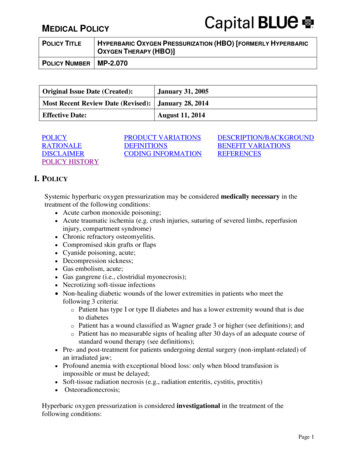
Zuo et al. Molecular Cancer(2022) 21:30Nevertheless, ROS levels within cancer cells can bemodulated to a proper degree through their robustantioxidant systems to utilize oncogenic roles. Cancercells exhibit a powerful antioxidant system consisting ofreductants such as the antioxidant enzymes-superoxidedismutase (SOD), catalase (CAT), glutathione peroxidase (GPX) and the antioxidant agents-nicotinamide adenine dinucleotide phosphate (NADPH) and glutathione(GSH). Antioxidants sustain a moderate ROS level toprotect cells from ROS attack and facilitate cancer progression. Antioxidant production is regulated by multiple signaling pathways and genes, such as nuclear factorerythroid 2-related factor 2 (Nrf2) [41]. Moderate ROSlevels can decrease the toxicity of natural killer cells (NKcells) [42] and increase the self-renewal capacity of cancer cells. In addition, it can also activate multiple protumorigenic signaling pathways such as NF-κB, TGF-β,JAK2-STAT1 and PI3K/Akt/ERK or activate oncogenesand inhibit tumor suppressors to enhance cancer progression [43–47]. Accordingly, through above intracellular self-adaptation alterations, ROS will be partiallycounteracted and cancer cells survive even can be furtherpromoted.There should be a threshold value for ROS levels inregulating cell fate – malignant transformation anddeath, which may suggest underlying treatment regimensPage 3 of 17(Fig. 1). Normal cells, cancer cells and cancer stem cells(CSCs) have different intracellular ROS loads that may bethe basis of ROS-elevated cancer therapy [48]. A higherlevel of ROS prompts a lower dose demand of ROS-elevated drugs/radiation in killing cancer cells thus, this is acomplicated and significant course to explore appropriatetherapeutic doses and ensure treatment benefits. Whilein most cases, patients who accept traditional chemo/radiotherapy show different side effects, including infections, anemia, fever, gastrointestinal discomfort and hairloss [49]. These impacts are probably derived from excessive treatments that cause ROS levels in normal cellsto reach a toxic threshold and damage normal tissues.Moreover, decreasing treatment doses has difficulty killing most cancer cells and leads to cancer relapse. Additionally, CSCs have been universally acknowledged to bea pivotal element of cancer relapse. Stemness is a toughchallenge for cancer therapy due to a lower ROS level inCSCs than that in normal cells.In conclusion, although it shows great potential incancer therapy, severe side effects are equally harmful. Hence, other replaceable therapeutic strategies onthe foundation of ROS are urgently developed. Notably,ROS-based ncRNA-targeted or ncRNA medicine haveexhibited some impressive achievements in cancer therapy, thus deeply understanding the crosstalk betweenFig. 1 The production and therapeutic potential of ROS in cancer. In normal cells, the endogenous antioxidant system is enough to counteractexcessive ROS to maintain redox homeostasis which benefits enzyme activation, protein synthesis, signal transduction and gene expression.Nevertheless, when the redox balance is broken, accumulated ROS will damage biological molecules and render malignant transformation. Notably,further elevated ROS levels can prevent cancer development even cause cancer cell death. The dual roles of ROS in cancer development remind usof two opposite treatments - antioxidant and pro-oxidant therapy
Zuo et al. Molecular Cancer(2022) 21:30ROS and ncRNAs will greatly promote the developmentof ROS-based agents and benefit cancer patients.The mechanisms of ROS‑ncRNAs axis in cancerprogressionOxidative stress, that induces oxidative damage of biomacromolecules and aberrant modulation of redoxsignaling, is a key biological event throughout cancer initiation and development [50–52]. ROS as a second messenger during the moderate OS supports dysregulationof various pro-tumorigenic signaling pathways for malignant disease, in contrast, induction of excessive OS leadsto cancer cell death by triggering ROS-driven apoptosisand ferroptosis [53, 54], thus indicating a potential development of OS-dependent strategy in clinical treatment.Accordingly, understanding the exquisite mechanism ofredox modulation patterns facilitates a better therapeutic purpose by breaking redox homeostasis in cancercells. NcRNAs occupy a majority of all transcripts, andare defined as dark matters in gene transcription at first.With in-depth investigation on the role of ncRNAs, theyare commonly identified as being significantly involved inmodulating carcinoma development [55–58], thereforedisplaying a promising potential as biomarkers or targets in cancer diagnosis and therapy [34, 35, 59, 60]. Inthis section, focusing on the detailed mechanism of thecrosstalk between ncRNAs and ROS, we summarize theprogress in recent years of ncRNAs involved in cancerinitiation, metastasis and chemoradiotherapy resistancebased on redox signaling regulation.Cancer initiationCancer initiation, featuring uncontrollable cell proliferation and growth, is the best stage for cancer prevention.Carcinogenic environmental factors with ROS induction properties have been proved to be one of the maincauses in many cancer patients [61, 62]. A growing bodyof studies has revealed the underlying mechanisms associated with ncRNAs-ROS axis-driven cancer initiation.For example, hexavalent chromium and arsenic werereported to increase ROS-dependent miR-21 transcription and compromise the expression of programmedcell death 4 (PDCD4), thus inducing the upregulation ofdownstream genes like E-cad, c-myc and uPAR and subsequently malignant transformation of bronchial epithelial cells [16, 63]. Furthermore, a previous study indicatedthat ionizing radiation (IR)-induced malignant transformation resulted in excessive OS by miR-21-mediateddownregulation of the antioxidant enzyme superoxidedismutase 2 (SOD2) [17]. In line with this, UVB-triggered cancer initiation was revealed to hold a close relationship with hexavalent chromium and arsenic-derivedOS based on modulation of miR-21 [18]. In addition,Page 4 of 17bronchial epithelial cells under chronic arsenic exposurewere found to have accelerated cancerization, which wasattributed to high ROS levels promoted the expression ofhypoxia-inducible factor-1α (HIF-1α) and cyclooxygenases-2 (COX-2) by mitigating miR-199a-5p transcription [64].In addition to miRNAs, lncRNAs involved in redoxregulation also function in cancer initiation. The c-mycdownregulated lncRNA transcript IDH1-AS1 renderedthe homodimerization of IDH1 and enhanced its enzymatic activity, which directly caused α-KG accumulationand OS and glycolysis blockage, preventing cancer proliferation at an early stage [65]. ROS-dependent lncRNAAX800134 was upregulated by TNF-α in hepatocellularcarcinoma (HCC), which was elucidated to be responsible for the growth of cancer cells [66]. MACC1-AS1, anantisense of MACC1, was found to be increased in gastric cancer (GC). MACC1-AS1 maintained redox balanceby stabilizing MACC1 mRNA and enhancing metabolicplasticity to promote the proliferation of cancer cells,which was indicated to be involved in AMPK/Lin28 signaling [67]. In melanoma, Wei et al found that lncRNAgrowth arrest-specific transcript 5 (GAS5) was significantly downregulated. The reduced transcription ofGAS5 greatly alleviated oxidative stress and acceleratedcell cycle progression through EZH2/CDKN1C axis [68].Although miRNAs and lncRNAs dominate in OSmediated cancer initiation based upon available reports(Fig. 2), other ncRNAs still remain a large potentialin the process. Although few studies have reportedtheir interaction with ROS in cancer initiation, circRNAs have also been largely found to interact with ROSin other diseases and exhibit a promising role in cancerinitiation. For instance, circRNA-101,036, identified asa tumor suppressor, induced ER stress and redox imbalance in oral squamous cell carcinoma [38]. As a spongeof miR-330-5p, tumor-suppressive human circular RNA(CircITCH) significantly reduced OS and alleviateddoxorubicin-induced cardiotoxicity via elevating theexpression levels of SIRT6, Survivin and SERCA2a [69].Likewise, in intestinal ischemia/reperfusion, circ-PRKCBwas reported to sponge endogenous miR-339-5p, modulating p66Shc expression and redox signaling [70].Cancer metastasisMetastasis is one of the biggest obstacles in the treatmentof terminal cancer patients. Understanding the detailedmechanisms in cancer metastasis may benefit the discovery of valuable therapeutic targets and the developmentof efficient interventions. Similarly, along with ROS, multiple types of ncRNAs have been confirmed to participate in cancer metastasis judging from current evidence(Fig. 3).
Zuo et al. Molecular Cancer(2022) 21:30Page 5 of 17Fig. 2 The crosstalk of ROS with ncRNAs in cancer initiation. The external factors, such as ultraviolet ray, ionizing radiation and toxic compounds,and internal factors caused by the aberrant gene regulation can both lead to cancer initiation through the complex interplay of ROS with ncRNAsEpithelial-mesenchymal transition (EMT), characterized by the transition from epithelial cells to cells witha mesenchymal (M) phenotype, is a prerequisite of themetastasis for most solid cancers. EMT is triggered bysome EMT-activating transcription factors (EMT-TFs),mainly of the SNAIL, TWIST and ZEB families and theirtargets like metalloproteinase-9 (MMP-9) [71]. During breast cancer progression, miR-373 quenched ROSlevels by downregulating the ROS inducer thioredoxininteracting protein (TXNIP), stimulating HIF-1α andTWIST expression [72]. Notably, TWIST bound to thepromoter of the miR-371/373 cluster, which remarkablyenhanced miR-373 transcription. Hence, the miR-373TXNIP-ROS-HIF1α-TWIST positive feedback signalingaxis showed a powerful EMT-activating capacity. MiR125b was found to be downregulated in HCC tissues,which prompted mitochondrial protein 18 kDa (MTP18)expression, leading to mitochondrial fission. Mitochondrial damage caused much ROS accumulation andincreased MMP-9 expression, directly promoting EMTand cancer metastasis [73]. MiR-206 was reported to bemodulated by nuclear factor NRF2 to drive tumorigenesis [74]. Simultaneously, miR-206 blocked TGF-β signaling and downstream neuropilin-1 (NRP1) and smad2expression, inhibiting EMT, migration and invasion ofbreast cancer cells [75]. Oncogenic miR-21 level wasreduced via kallistatin protein-mediated ROS downregulation, which further suppressed Akt signaling and EMTin cancer cells [76].In addition to miRNAs, mounting evidence indicatesthat lncRNAs are also engaged in cancer metastasis.LncRNA UCA1 worked as a ceRNA, which preventedthe effects of miR-1 and miR-203a on Slug expression,promoting EMT and invasion in breast cancer [77]. Similarly, lncRNA-MUF (mesenchymal stem cell-upregulatedfactor) can also act as a ceRNA of miR-34a, activatingSnail1 expression and Wnt/β-catenin signaling to facilitate HCC metastasis [78]. Lnc-SNHG1 sponged miR302/372/373/520 to enhance the expression of targetgenes-TGF-β receptor 2 (TGFBR2) and RAB11A, thuscontributing to EMT in pituitary cancer [79]. Althoughthe underlying mechanism remains unclear, we speculate
Zuo et al. Molecular Cancer(2022) 21:30Page 6 of 17Fig. 3 The crosstalk of ROS with ncRNAs in cancer metastasis. Cancer metastasis is tightly regulated by the intricate interaction of ROS with ncRNAswhich involved multiple pivotal molecules and signaling pathwaysthat it is related to miR-1/34a/372-triggered ROS regulation from available reports [74, 80, 81]. LncRNAMALAT1 was found to upregulate ROS levels via keap1/Nrf1/2 signaling [82], which induced EMT and metastasis in head and neck squamous cell carcinoma (HNSCC)through STAT3 activation [83]. PM2.5 exposure of lungcancer cells significantly increased ROS levels and upregulated subsequent lncRNA loc146880 expression, causing malignant metastasis of lung cancer cells [84].Besides miRNAs and LncRNAs, current studies haveimplied circRNAs are also capable of participating inOS-mediated cancer metastasis. CircSCAF11 overexpression inhibited ROS production by acting as a spongeof miR-145-5p in glioma, which is a cause of glioma initiation and metastasis [85]. MiR-30c-5p was reportedto be an important OS regulator by multiple studies [86–88]. In colorectal cancer, circ3823 functionedas a ceRNA of miR-30c-5p, alleviating the repressiveeffect of miR-30c-5p on TCF7 which upregulated MYCand CCND1 and eventually led to cancer initiation andmetastasis [89]. Hu et al identified an oncogenic circASAP1 through circRNA sequencing in patients withHCC, which was proved to be associated with cancer initiation and metastasis via miR-326/miR-532-5p-MAPK1/CSF-1 signaling, respectively [90]. The result suggestedthat the underlying mechanism was also modulated byOS [91, 92].Cancer chemoradiotherapy resistanceChemoradiotherapy resistance is the leading threat forcancer patients. Reversing chemoradiotherapy resistance is a pivotal bottleneck problem to be resolved.OS-induced chemoradiotherapy resistance has beencommonly identified, targeting which will be conduciveto overcoming multidrug resistance (MDR) and radiotherapy failure [93].
Zuo et al. Molecular Cancer(2022) 21:30Increasing evidence indicates that CSCs are at topand responsible for cancer chemoradiotherapy resistance, relapse and treatment failure [94]. It was reportedthat CSCs possessed a lower ROS level, which may beexploited to develop ROS-elevated treatments to reserveCSC-mediated chemoradiotherapy resistance. As a target of miR-223, the inhibition of HAX-1 led to mitochondrial damage and accelerated ROS generation intriple-negative breast cancer stem cells (TNBCSCs),which ultimately enhanced TRAIL-induced apoptosis[95]. MiR-153 downregulated Nrf2 and GPX1 expression, by which the elevated ROS levels decreased thestemness of glioma stem cells (GSCs) and relievedradiation resistance [96]. However, Yang et al reportedthat the decreased ROS levels resulting from miR-210were able to weaken the stemness of hypoxic GSCs andreverse radiation resistance [97], which implied thatCSC-induced chemoradiotherapy resistance should befurther considered before applying redox agents. LncRNAs were also reported to participate in the process. Forexample, lncRNA H19 was upregulated in HCC tissues,knockdown of which caused OS and reversed the chemotherapy resistance of CD133 CSCs via MAPK/ERKsignaling [98].Independent of CSCs, we find that the ncRNA-ROSaxis can also influence chemoradiotherapy resistancePage 7 of 17through other biological processes on the basis of available evidence (Fig. 4). In breast cancer and melanoma,let-7a increased mitochondrial ROS production andimproved the toxic effects of doxorubicin on cancercells, although the underlying mechanism remainedunclear [99]. MiR-17-3p elevated ROS levels by inhibiting three primary mitochondrial antioxidants, MnSOD,glutathione peroxidase 2 (Gpx2) and thioredoxin reductase 2 (TrxR2), remarkably strengthening the sensitivityof prostate cancer cells to IR [100]. MiR-34b/c preventedubiquitin-specific protease 2a (USP2a)-c-myc-GSH signaling in prostate cancer, reversing cisplatin and doxorubicin resistance through ROS-induced apoptosis [101].The promoters of miR-200c-3p and miR-34a-3p wereoccupied by zinc finger E-box binding homeobox 1(ZEB1), which inactivated their transcription. Intriguingly, ROS-induced miR-200c/34a transcription targetedZEB1 in turn, which greatly upregulated miR-200c/34alevels, inhibiting P-glycoprotein (P-gp) expression andreversing MDR [102]. Simultaneously, ROS-inducedmiR-34a transcription prevented c-Met expression andenhanced the cisplatin sensitivity of HCC cells [103].LncRNAs were also found to play a pivotal part inenhancing the sensitivity of chemoradiotherapy inmultiple studies. LncRNA NLUCAT1, identified fromthe hypoxic status of lung adenocarcinoma cell lines,Fig. 4 The crosstalk of ROS with ncRNAs in cancer chemoradiotherapy resistance. ROS interacts with ncRNAs, influencing the effects ofchemoradiotherapy. The elaborate regulatory network provides us some possible therapeutic intervention targets in chemoradiotherapy resistance
Zuo et al. Molecular Cancer(2022) 21:30facilitated proliferation and invasion via decreasingROS levels with lower sensitivity of lung cancer cellsto cisplatin-induced apoptosis [104]. LINC00963 wasfound to be elevated in several cancer types and associated with breast cancer invasion. Through spongingmiR-324-3p, LINC00963 greatly relieved DNA damageand OS, rendering cancer progression and radiotherapyresistance [105]. LncRNA SLC7A11-AS1 was identifiedin gemcitabine-resistant pancreatic ductal adenocarcinoma (PDAC) cells, which bound with β-TRCP1 andprevented the ubiquitination and degradation of Nrf2in the nucleus. A lower level of ROS mediated by Nrf2increased the stemness of cancer cells, which benefiteddrug resistance [106].Several reports have exhibited other ncRNAs were alsoinvolved in OS-modulated chemoradiotherapy resistance. CircCCND1 was remarkably upregulated in lungcancer cells and patients, which endowed cisplatin resistance of lung cancer cells by sponging miR-187-30 tomodulate OS [107]. MiR-23b-3p has been proved to beclosely related to chemotherapy resistance in multiplestudies [108–111]. It suggested that circZNF292, acting as a ceRNA of miR-23b-3p to regulate anti-oxidativegenes, may be a potential target in reversing chemotherapy resistance [112].Therapeutic implications based on the ROS‑ncRNAsaxis in cancerConsidering the great power of ROS and ncRNAs inregulating tumorigenesis and cancer progression, multiple preclinical studies targeting the ncRNAs-ROS axishave been conducted and achieved impressing treatmentoutcomes. ROS inducers and ncRNA-targeted or ncRNAagents are two primary druggable patterns with ncRNAsserving as both redox sensors and regulators, implyingthe large potential of modulating the ncRNAs-OS signaling axis in cancer therapy.ROS inducers in cancer therapyNatural/synthetic compoundsButyrate was found to be a ROS inducer through upregulating miR-22 expression and provoking mitochondrialdysfunction in HCC, directly causing cytochrome crelease and caspase-3-dependent apoptosis [113]. Sulindac sulfide-triggered ROS decreased miR-27a expression,which prevented colon cancer cell growth via miR-27a/ZBTB10/specificity protein (Sp) axis. A similar process also occurred in Ethyl 2-((2,3-bis (nitrooxy) propyl)disulfanyl) benzoate (GT-094)-treated colorectal cancer[114, 115]. Phenethylisothiocyanate (PEITC) stimulatedROS generation, downregulating miR-27a/miR-20a/miR-17 expression. The process induced miRNA-modulated ZBTB10/ZBTB4/ZBTB34 expression, inhibitingPage 8 of 17Sp transcription factors-Sp1, Sp3, and Sp4 in pancreaticcancer cells. ROS-mediated Sp inhibition facilitated theapoptotic process, which may be a general mechanism ofROS-elevated cancer therapy [116]. However, quinacrineacted as a ROS inducer to upregulate FOXP3 by activating p38 MAPK and inactivating ERK. FOXP3-mediatedmiR-183 decreased β-TrCP mRNA stability, protectingSp1 from degradation which enhanced pro-apoptoticprotein Bax expression and the apoptosis of leukemiacells [117]. Besides, curcumin, betulinic acid (BA) andpolygonatum odoratum lectin (POL) can also exert theirantitumor effects via miRNA-mediated ROS accumulation [118–120].NanomaterialsWith the development of nanotechnology, nano-deliverysystems are promising in cancer diagnosis and therapywith lower toxicity and fewer side effects to normal cells.Among which the OS-mediated nanodrugs occupy amajority in cancer treatment. Mounting evidence hasconfirmed their effectiveness in preclinical studies. Forexample, in tumor acidy microenvironment, FeS@BSAnanocluster was degraded and produced much H2S andFe2 , which synergis
Jing Zuo1†, Zhe Zhang1†, Maomao Li2†, Yun Yang 2†, Bohao Zheng2, Ping Wang 2*, Canhua Huang1* and Shengtao Zhou2* Abstract Oxidative stress (OS), characterized by the excessive accumulation of reactive oxygen species (ROS), is an emerg-ing hallmark of cancer. Tumorigenesis and development driven by ROS require an aberrant redox .