Transcription
www.jcimcr.orgJournal ofClinical Images and Medical Case ReportsISSN 2766-7820Case ReportOpen Access, Volume 3A case of Raynaud’s phenomenon in Arizona, USAMerhavy ZI1, 2*; LaSon DS1,2; Varkey TC2,3¹Ross University School of Medicine; Bridgetown, Barbados, USA.²Dell Medical School at the University of Texas at Austin; Austin, TX, USA.³Colangelo College of Business at Grand Canyon University; Phoenix, AZ, USA.*Corresponding Author: Merhavy ZIRoss University School of Medicine; Bridgetown,Barbados, USA.Email: ZackMerhavy@gmail.comReceived: Jun 20, 2022Accepted: Jul 15, 2022Published: Jul 22, 2022Archived: www.jcimcr.orgCopyright: Merhavy ZI (2022).DOI: www.doi.org/10.52768/2766-7820/1965AbstractThis case presentation assesses one example of a patient sufferingfrom Raynaud’s phenomenon without having previously sought outmedical intervention. This study focuses on the patient’s perspectivein hopes of providing insight to future providers in handling patientinteractions for those suffering with the same disease. As little aboutthe disease is widely known, this study will address the etiology, pathophysiology, diagnostic tests, current and experimental treatment options, and known genetic components of Raynaud’s disease. The goalof the authorial team is to bring attention to the patient’s perspectiveto increase rapport, treatment compliance, and increase provider understanding of the language they use to bring up symptomology. Thiscase also looks to help primary care physicians better characterize thepatient’s disease state. It is the hope of this research team that available treatments and therapies will become more widely known andunderstood for the purpose of healthcare professionals to incorporatethem into their potential appointments with patients suffering fromthese conditions.Keywords: Raynaud; Raynaud’s phenomenon; Raynaud’s disease.Case presentationThe patient is a 24-year-old male, living in south Phoenix,Arizona, USA. He has been suffering with Raynaud’s for roughly8 years at the time this case study was written. The patient initially presented with his chief complaint of hand color changesand painful stimuli when exposed to the cold. He states thattwo to three of the digits of the hands bilaterally will becomepale to white with any minor stimuli of cold exposure and thatthis leads to increased pain sensation when his hands rewarm.He states the symptoms developed roughly around sophomoreyear of high school and that they were initially only right sided,but have now become bilateral. He endorses that stress andnegative emotional mindsets may cause the phenomena to occur at higher temperatures or become worse, sometimes withhim experiencing symptoms up to the mid 70-degree Fahrenheit ( 24-degree Celsius) range.The patient stated that his mother suffered with similarsymptoms, but could not remember the name of the disorder,only stating that, “her hands would start to get ‘frostbitten’ veryeasily, and the first symptom was her hands turning white”. Thepatient stated that when his mother sought medical attentionfor her condition, the physician did not know anything aboutthe condition, telling her to “dress intelligently for cold temperatures”. Remembering this experience, the patient had notpreviously sought out medical attention for himself for the assumption that he would simply be told the same thing.The patient noted that in addition to the spread and decreasing temperatures of the symptoms in his hands, the onset
Citation: Merhavy ZI, LaSon DS, Varkey TC. A case of Raynaud’s phenomenon in Arizona, USA. J Clin Images Med Case Rep.2022; 3(7): 1965.of symptoms is beginning to occur at faster rates. The patientstated “ because of this, I have to take frequent trips inside torun my hands under warm water so I can avoid any possibility ofnerve damage”. He continues to state “I like to go snowboarding,which unfortunately means expensive heated gloves and handwarmers are a must. One time I forgot to bring hand warmerson the mountain and the battery in one of my gloves stoppedworking after I’d gotten a bunch of snow inside it. After an hour,one finger was turning from white to purple and I had to returnto the car for fear of suffering permanent nerve damage. Mysymptomatic episodes are always confined to my hands and willtypically last indefinitely, until I make a real effort to warm upmy hands. Just going inside, unless it’s really hot, won’t helpmost of the time. The fastest method is to run my fingers underwarm water which can stop symptoms within a minute or two.The next best option is to hold my fingers in front of the hot airvents in my car, and this can take about 3-5 minutes. If that’s notpossible, then I can hold my hands between my armpits, or inside my sleeves, and this can take anywhere from 5-20 minutes.One caveat to all of this is that once symptoms have started thefirst time, it’s almost like my hands are primed and the issue willreturn quickly if I don’t stay inside.”Physical examination of the patient demonstrated hands thatwould quickly change to white, then with some minor time, theskin would become bluer in coloration. With some rewarming,the patient’s hands would become ruddy in coloration and thenreturn to the patient’s baseline. Upon further investigation, thepatient’s pain scale is stated to be at a 2/10 most of the time,stating “this is what makes it scary since sometimes I don’t evennotice my fingers have been without blood for a long time”.Differential diagnosisBased on the patient’s constellation of symptoms, his familyhistory, and his physical examination the team found that thiswas most likely a classic case of Raynaud’s syndrome. However, because of the age of the patient, his male biological sex,and the severity of the disease, he was recommended to seean outpatient rheumatologist for further diagnostic work upand treatment. Raynaud’s phenomena can be secondary to anumber of disorders and at the time of writing, the patient wasworking on getting in to see outpatient physicians for treatmentof his condition.The differential for the underlying cause of this particularcase includes autoimmune diseases such as Lupus or Scleroderma, medication side effects, especially from drugs that affectsmooth muscle contractions (i.e., beta blockers or stimulants),smoking or other allergen side effects, and mechanical injuriesto the affected extremities [3,7,8]. Lesser mimics of the diseaseinclude Carpal Tunnel Syndrome, minor mechanical injuriesfrom repetitive motions, and increased plaque burden from peripheral arterial disease [3,7,8]. Raynaud’s phenomenon can besecondary to several disorders, and these should be addressedbefore starting treatment.End diagnosis and treatment planBased on the constellation of symptoms, the patient was diagnosed with Raynaud’s phenomenon and was given instruction and referral paperwork to meet with an outpatient Rheumatologist. As part of the American healthcare system at thewww.jcimcr.orgtime of writing, the patient was working on making an appointment to see an outpatient Rheumatologist for treatment of hiscondition. He was advised to avoid situations which would impact his digits that may lead to severe nerve damage and wasencouraged to dress warmly until the baseline problem was discovered and addressed. As the work-up was still in process, nomedications were started at that time.Summary and etiologyRaynaud’s phenomenon is now a well-established diagnosis,first described in 1862, in a doctoral thesis by Dr. Auguste Gabriel Maurice Raynaud, which consists of a contraction of thesmooth muscles of the arteries leading to a decreased perfusion of the capillary beds of downstream tissues. This particulardisease affects around five percent (5%) of the global population and is often associated with autoimmune disorders including hypo- or hyperthyroidism, hypo- or hyperparathyroidism,Multiple Sclerosis, and other such disorders [1-5]. The mostcommon location for Raynaud’s phenomenon symptomatology is that of the extremities, particularly the fingers and toes.While more rare, patients with severe disease that need moreurgent work describe experiencing symptoms on the tip of thenose, and the ear lobes [1,2,4,5]. Nevertheless, while not oftendiscussed in medical schools, the actual action of this on thesmooth muscles of the arteries can be seen all throughout thebody and can play a pivotal role in acute care medicine, leadingto physicians placing arterial lines in large bore vessels in thesepatients for fear of auto-amputation [6-8].The vasoconstriction of the cutaneous arterioles in the extremities and skin is a normal and physiological process whichleads to the redirection of the circulation from the surface levelcapillaries to that of deeper tissues and internal organs. This asa mechanism, teleologically, promotes the conservation of energy by preventing the loss of heat energy from the body and ismediated by the release of norepinephrine, an adrenergic hormone, by the sympathetic nervous system [4-8]. Nevertheless,Raynaud’s phenomenon, a pathological condition, whether primary or secondary, occurs when there is an exaggeration of thisnormal response in the extremity’s vascular system [1,2,9,10].Primary Raynaud’s is the disorder itself without a secondarycause [1-5]. Like that of essential or primary hypertension, thediagnosis of Primary Raynaud’s cannot be given until secondarycauses, such as autoimmune disorders are ruled out [1-5]. Secondary Raynaud’s occurs when the symptoms of the disorderare caused by another underlying disorder [1-5]. Unlike PrimaryRaynaud’s, because of the underlying disease state, SecondaryRaynaud’s disease often has more severe features and can morequickly progress, leading to soft tissue and/or nerve damage inelevated temperatures as was described in the patient’s casereport [1-5].PathophysiologyThe major biopatho physiological mechanism of action forthis disease is directed through the Alpha 2C adrenergic receptorthat is found on the vascular smooth muscle cells [3,11]. Whenthese cells are stimulated by the sympathetic nervous system’srelease of catecholamines, they increase the amount of calciumions that enter the smooth muscle cells leading to contractionof the muscle cell itself [12,13]. Normally, on the outside of thePage 2
cells, there are Alpha 2A and Alpha 2B receptors which inducevasoconstriction and Beta 2A receptors which induce vasodilation [14,16]. Nevertheless, when induced by stress conditions,including cold or other environmental factors, the trans-Golgireceptor, Alpha 2C, is translocated to the cell membrane. Whenthis occurs, the ability of the smooth muscle cells to respond tocatecholamines increases exponentially [14,16].Mechanism of cold-induced mobilization of Alpha 2C adrenergic receptorsDuring a sudden decrease in temperature, the cutaneousarteriole smooth muscle cells sense this decrease due to changes in the energetics of the breakdown of food molecules [17].This then causes the mitochondria to release Reactive OxygenSpecies (ROS), which then proceeds to activate the Rho/ROCKpathway leading to the rearrangement and reconfiguration ofthe cytoskeleton [17]. This rearrangement of F-actin and filamin-2 proteins of the cytoskeleton allow for the cell to mobilize the α2C-AR from the endoplasmic reticulum/Golgi to thecell surface [17]. This whole process leads to an increase in thesensitivity of that particular smooth muscle cell, and as a sumtotal, the entire cutaneous arteriole [17,18]. Other mechanismsby which this process is mediated include the inhibition of genetranscription for the α2C-AR by cyclic AMP’s effect on protein kinase A and the increase in production of α2C-AR by cyclic AMPacting on the exchange protein activated by cyclic AMP (EPAC)and the GTP-binding protein Rap1 [17-18]. The gross exaggeration of these normal biochemical processes and the resultingpathological pain, autoamputation, and/or nerve damage iswhat is known as Raynaud’s phenomenon or Raynaud’s disease[19].Tests and diagnosisIn the primary work-up of Raynaud’s disease, similar to othermajor concerns of medicine is making sure that all the potentialcauses of the disease are considered. The authors include themajor screening tests for the work-up of a patient with Raynaud’s to rule in or out the major baseline etiologies such as thedifferent autoimmune diseases listed above in the Differentialsection. The main goal of any clinician is to consider the likelihood of the different disease states based on the patient’s history and physical examination, including iatrogenic causes, andthen only order those tests which will change the managementof the patient [41,42].Antinuclear Antibody (ANA) testAntinuclear antibodies are specified antibodies that arefound in the blood, programmed to attack the nucleus of thecells within the body’s tissues [20,21]. By performing an ANAtest, a positive result can assist healthcare professionals inidentifying an autoimmune disorder (i.e., Lupus, Sjogren’s syndrome, Raynaud’s, Scleroderma, etc.) [21]. Although a positiveresult from an ANA test does not specifically signify Raynaud’s,it may light the way for healthcare professionals to explorefurther tests to identify the individual’s specific disease. Mostautoimmune diseases that occur with secondary Raynaud’s areANA positive, but many patients with a positive ANA are typically healthy and will remain so [21].Erythrocyte Sedimentation Rate (ESR)An Erythrocyte Sedimentation Rate, or ESR, is a type of bloodtest that measures how quickly red blood cells, or erythrocytes,settle at the bottom of a test tube that contains a blood samplewww.jcimcr.org[22]. Normally, red blood cells settle relatively slowly, and thus,a faster-than-normal rate may be caused by inflammation in thebody, potentially indicating a sign of a chronic disease, an immune disorder, or other medical condition [22]. An ESR test canhelp determine if a patient has a condition that causes chronicinflammation such as forms of arthritis, vasculitis, or inflammatory bowel disease [22]. Although an abnormal range from anESR test does not specifically signify Raynaud’s, it can be used tohelp a healthcare professional order further tests to identify theindividual’s specific disease.C - Reactive Protein (CRP) TestA C-Reactive Protein test, or CRP, is often used in conjunctionwith ESR to indicate a probable chronic inflammatory condition[22,23]. Similarly to ESR, a high CSR value may be a sign of a serious infection or inflammation [22,23]. Both ESR and CRP lackspecificity, and thus, should still be used in combination withmedical history and a physical exam for diagnosis and monitoring [22]. As many physiological factors such as non-infectiousconditions or a resolve of inflammation can contribute to a highESR and a low CRP or vice versa, it can be incredibly beneficialto a healthcare provider to order both tests [22].UrinalysisA urinalysis, or urine dipstick test, can be an invaluable toolfor a healthcare provider as it is a cheap and quick method ofassisting in a diagnosis [24,25]. A urine dipstick test is a wayin which many genitourinary and systemic conditions can potentially be diagnosed [25]. Similar to a CRP test, a urinalysiscan detect proteins produced by the liver in response to inflammation and may ultimately help providers determine similarabnormalities seen in autoimmune disorders [24,25]. As theurine dipstick test cannot directly signify whether a patient hasRaynaud’s or not, this tool provides means for ruling out otherpossible chronic autoimmune disorders such as Lupus or glomerulonephritis [24,25].Complement levelsTesting for complement levels in a patient’s blood can signifywhether the pathologic effect is due to an increased or persistent activation, caused by immune complexes as seen in Lupus[28]. Complement levels also help to identify a decreased function or presence of complement inhibitors as seen in cases ofischemia [28]. Based on the specific rise or fall in complementlevels that are seen, healthcare providers can determine if a patient likely has a recurrent infection or an autoimmune disease[26]. Again, as much is still unknown about Raynaud’s this testwill only further rule out other possible autoimmune disordersas their specific complement indicators have been identified,whereas Raynaud’s has not [26].Other testsThere are various other lesser-used tests that healthcareprofessionals can still utilize in order to diagnose Raynaud’s.Many of these highly effective tests, some newer and someolder, include Computed Tomography (CT)-guided percutaneous thoracic sympathetic chain radiofrequency thermocoagulation, testing for Th1- and Th17-related cytokines in venous andarterial blood, infrared thermography, photoacoustic and highfrequency ultrasound imaging, and more [12,29-31].Page 3
Table 1: This table contains the different tests utilized in the work up of Raynaud’s disease, what they look for, the benefits of ordering thetest, and the draw backs to ordering them.Test NameWhat does it look for?BenefitsAntinuclear Antibody (ANA))Specific antibodies found in theblood, programmed to attack thenucleus of the cells within the body’stissues [20-21].Can assist in identifying an autoimmune disorder (i.e., Lupus, Sjogren’ssyndrome, Raynaud’s, Scleroderma).Is a screening test [20-21].Not specific for any particulardisease [20-21]A faster-than-normal rate may becaused by inflammation in the body,potentially indicating a sign of achronic disease, an immune disorder,or other medical condition. Is ascreening test [22]Not specific for any particulardisease [22].Measures how quickly red bloodcells, or erythrocytes, settle at theErythrocyte Sedimentation Rate (ESR)bottom of a test tube that contains ablood sample [22]DrawbacksC-Reactive Protein (CRP)Levels of C-reactive Protein, an acutephase reactant released by the liversecondary to inflammation [22-23].A high CSR value may be a sign of aNot specific for any particularserious infection or inflammation. Is adisease [22-23].screening test [22-23].UrinalysisAbnormal levels of Glucose, Proteins,Ketones, Red Blood Cells, WhiteBlood Cells, or Bacteria within theUrine [24-25].A urinalysis can detect proteinsproduced by the liver in responseto inflammation and may ultimatelyhelp providers determine similarabnormalities seen in autoimmunedisorders [24-25]Levels of C3 and C4, the major components of the compliment system,activated by antibodies, lectin, orautoactivation [26-28].Can signify whether the pathologiceffect is due to an increased or persistent activation of the complimentsystem, caused by immune complexes as seen in Lupus. Can also helpto identify a decreased function orpresence of complement inhibitors asseen in cases of ischemia [26-28].Complement LevelsNot specific for any particulardisease.This test will only further ruleout other possible autoimmune disorders as their specificcomplement indicators havebeen identified [26-28].Table 2: This table includes the major treatment options, their mechanism of action, benefits, and contraindications to use.Treatment or therapyMechanism of actionAlpha Blockers (i.e. Doxazosin,Prazosin, terazosin,tamsulosin,alfuzosin, silodosin, phenoxybenzamine)Keeping the norepinephrine from tightening the muscles in the walls of smallerarteries and veins, and thus, the vesselsremain open and relaxed, ultimatelyimproving blood flow and lowering bloodpressure [32].Calcium Channel Blockers(i.e.Amlodipine, Diltiazem,Felodipine, Isradipine, Nicardipine, Nifedipine, Nisoldipine, andVerapamil)Prevent calcium from entering the cellsof the heart and arteries [33]. As calciumcauses the heart and arteries to contractstronger, by blocking the calcium, bloodvessels are allowed to relax and open[1,33]increases peripheral blood flowand reduces levels of pain experienced during episodes [33]Sick sinus syndrome (except inthose with an artificial pacemaker),pulmonary congestion, acutemyocardial infarction,and severehypotension [1,33].Increases bioavailability of Nitric oxide[34].NO-mediated vasodilation, cutaneous vascular conductance andnormalized skin temperature following local cooling, and increasesystemic anti-inflammatory status[34].Congenital heart disease characterized by ductal dependent systemic blood flow,hypersensitivity,and severe left ventricular dysfunction [34].Nitrate Rich Beetroot JuiceSingle-Port Thoracoscopic SympathectomySulodexide ( 80% heparan sulfateand 20% dermatan sulfate)www.jcimcr.orgRemoval of the sympathetic nervous connection to that of the digital extremities[35].Components of the arterial wall, or basalmembrane, and extracellular matrix, whichultimately influences the function and activity of heparin-binding proteins decreasing the likelihood of clot formation withinconstricted blood vessels and capillaries ofthe extremities [36].BenefitImproved the recovery time offinger temperature after beingexposed to cold temperatures[32].Decrease in the number and duration of Raynaud’s attacks [35].Useful for those with treatmentresistant Raynaud’s phenomenon[35].Significant increase in bloodperfusion in the capillary vessels,a reduction in the frequency ofpain episodes, and a decreaseof pain intensity during episodes[36].ContraindicationShould be used with caution inpatients with a history of posturalhypotension, heart failure, andurinary incontinence [32].No absolute contraindications tothe surgical procedure, however,this particular procedure is stillunder investigation and willrequire further evidence before itis considered a quality long-termsolution for Raynaud’s phenomenon [35].Diathesis, Hemorrhagic diathesis,and Hypersensitivity (allergy) toany component of sulodexide [36].Page 4
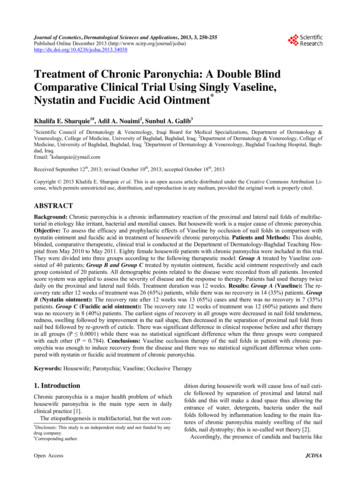
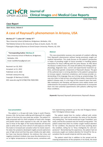
mally invasive endoscopic technique used in patients withtreatment-resistant Raynaud’s [35]. In another study, this wasperformed unilaterally on the left side in eight patients withRaynaud’s, and after surgery on the left hand only, a unilateralimprovement in perfusion was observed in the left hand compared with the right hand [35]. Additionally, the number andduration of attacks in the left hand had decreased over a 2-weekperiod as compared with the right hand with no serious adverseevents occurring in a follow-up period of 10 months [35].Figure 1: Demonstrated here are the patient’s hand with cold stimulation (Left) and then after rewarming (Right).Treatments and therapiesAs the patient referenced, physicians have only commentedon proper dress for weather conditions rather than providinga solution, therapy, or treatment options to his mother whosuffers from the same condition, ultimately leaving the patientfeeling as though seeking treatment would be pointless. Although there is not currently an abundance of treatments ortherapies known for Raynaud’s, it is still important to identifyones that do exist.Alpha blockersAlpha blockers are known for being one of the two mostwidely used treatment options with a high rate of success inmany patients [1]. Alpha blockers work by keeping the norepinephrine from tightening the muscles in the walls of smaller arteries and veins, and thus, the vessels remain open and relaxed,ultimately improving blood flow and lowering blood pressure[32]. One study discovered that use of alpha blockers, with orwithout a combination of a beta blocker, improved the recoveryof finger temperature after being exposed to cold temperatures[32].Calcium channel blockersCalcium channel blockers are the second of the two mostwidely used treatment options with a high rate of success inmany patients, often serving as the first choice of treatment foraffected patients [1]. Calcium channel blockers ultimately worksimilarly to alpha blockers, only by preventing calcium from entering the cells of the heart and arteries [33]. As calcium causesthe heart and arteries to contract stronger, by blocking the calcium, blood vessels are allowed to relax and open [33]. Use ofcalcium channel blockers in patients with Raynaud’s, it has beenobserved that this medication increases peripheral blood flowand reduces levels of pain experienced during episodes [33].Beetroot juiceAs it is known that Raynaud’s phenomenon is characterizedby recurrent transient peripheral vasospasm and lower NitricOxide (NO) bioavailability in the cold, a research team investigated the effect of nitrate-rich beetroot juice supplementationon NO-mediated vasodilation, cutaneous vascular conductanceand skin temperature following local cooling, and systemic antiinflammatory status [34]. After the participants drank the beetroot juice every day for 2 weeks, the research team discoveredthat each participant saw an improvement in thumb blood flow,improved endothelial function and anti-inflammatory status, aswell as a reduction in BP [34].SulodexideSulodexide is a mixture of glycosaminoglycans ( 80% heparan sulfate and 20% dermatan sulfate), which are components of the arterial wall, or basal membrane, and extracellularmatrix, and ultimately influences the function and activity ofheparin-binding proteins [36]. Although calcium antagonists,angiotensin-converting enzyme inhibitors, and α1-blockers arecurrently the most common medications in the pharmacotherapy of Raynaud’s, one study found that sulodexide improved capillary blood flow in patients with Raynaud’s [36]. A significant increase in blood perfusion in the capillary vessels was observedas well as a reduction in the frequency of pain episodes and adecrease of pain intensity during episodes [36].GeneticsVariations in genes may be involved in the process of excessive constriction of small blood vessels in response to coldor stress; however, the exact connection between these genevariations and the abnormal blood vessel response that occursin this disorder is currently unknown [37,43]. The main reasoning for this unknown connection is due to the lack of robustbiomarkers needed to predict vascular outcomes or responsesto therapy in patients with Raynaud’s [38]. It has been observedthat certain cases of Raynaud’s may be familial, but the specific inheritance pattern is still unknown [37,43]. Some studiessuggest that roughly 30% of individuals with a first-degree relative, such as a parent, sibling, or child, who has primary Raynaud’s phenomenon may also have the condition as well [37].Although the exact genetic component has not been clarified,one study suggests that hyperhomocysteinemia resulting frommethylenetetrahydrofolate reductase, or MTHFR, gene mutation may have a role in Raynaud’s etiology [39]. Most individuals observed with this MTHFR gene mutation and hyperhomocysteinemia were, more frequently than not, diagnosed withRaynaud’s phenomenon, making up an estimated 3-20% of theglobal population [39].In cases of secondary Raynaud’s, this exaggerated vasoconstriction could be caused by autoimmune diseases; hypothyroidism; cancers of the blood, bone marrow, or immune system; disease processes that cause obstruction of blood vessels;exposure to certain medicines or chemicals; cigarette smoking;injury or trauma; prolonged repetitive motions such as typing;long-term use of vibrating tools; and more [37,40].Social impactWhen asked about the impact his condition has on his sociallife, the patient attributed the “uncertainty” of when the symptoms will arise as the leading cause of difficulty to plan aroundhis condition; having to always dress warm just in case, alwayshave hand warmers available, and to make sure he has heatedSingle-port thoracoscopic sympathectomygloves. He additionally notes that a “negative or passive” menSingle-port thoracoscopic sympathectomy is a novel, mini- tal state plays a large factor as well, naming stress and/or anxiety some of the worst culprits. The patient notes the irony thatwww.jcimcr.orgPage 5
his condition brings forth these stressors, which in turn, causesthe symptoms to get worse, stating “It’s a constant source ofstress when I’m anywhere the temperature gets even a littlechilly”.5.National Institute of Arthritis and Musculoskeletal and Skin Diseases. Raynaud’s phenomenon. Retrieved ds-phenomenon.2016.He continues to comment by stating “The symptoms mostlyimpact my social life. If I’m out to dinner or a bar with friendswho want to sit outside, I don’t want to be the one to have everyone sit inside since I have an obscure condition that nobodyunderstands. Especially when I’m talking to someone when it’sa little chilly outside, I find that sometimes I can’t focus on theconversations happening around me because I’m so focused onkeeping my hands warm.6.Charkoudian N, Stachenfeld N. Sex hormone effects on autonomic mechanisms of thermoregulation in humans. Auton,Neurosci. 2016; 196: 75–80.7.Vanhoutte PM. Physical factors and regulation of vascularsmooth muscle function, in Handbook of Physiology, eds BohrDF, Somlyo AP, Sparks HV. (Washington, DC: The American Physiological Society), 1980; 443–474.8.Wigley FM, Flavahan NA. Raynaud’s phenomenon. N. Engl. J.Med. 2016; 375: 556–565.9.Herrick AL. The pathogenesis, diagnosis and treatment of Raynaud phenomenon. Nat. Rev. Rheumatol. 2012; 8: 469–479.10.Cordeiro RA, de Andrade RM. Raynaud’s phenomenon in theoccupational context. Revista da Associacao Medica Brasileira.2019; 65: 1314-1320.11.Nicola S, Fornero M, Fusaro E, Peroni C, Priora M, Rolla G, et al.Th1- and Th17-related cytokines in venous and arterial blood ofsclerodermic patients with and without digital ulcers. BioMedResearch International, 2019; 2019: 7908793.12.Rimar D, Rimar O, Rosner I, Rozenbaum M, Kaly L, et al. Nailfold video capillaroscopy: Beyond systemic sclerosis. The IsraelMedical Association Journal. 2019; 21.13.Dinsdale G, Wilkinson S, Wilkinson J, Moore TL, Manning JB,Berks M, et al. State-of-the-art technologies provide new insights linking skin and blood vessel abnormalities in SSc-relateddisorders. Microvascular Research. 2020; 130: 104006.14.Di Benedetto P, Ruscitti P, Liakouli V, Cipriani P, Giacomelli R, etal
Differential diagnosis Based on the patient's constellation of symptoms, his family . the trans-Golgi receptor, Alpha 2C, is translocated to the cell membrane. When . 6. Charkoudian N, Stachenfeld N. Sex hormone effects on auto-nomic mechanisms of thermoregulation in humans. Auton, Neurosci. 2016; 196: 75-80. 7. Vanhoutte PM. Physical .