Transcription
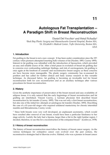
11Autologous Fat Transplantation –A Paradigm Shift in Breast ReconstructionDaniel Del Vecchio1 and Hetal Fichadia21BackBay Plastic Surgery and Massachusetts General Hospital, Boston MA,2St. Elizabeth’s Medical Center, Tufts University, Boston MA,USA1. IntroductionFat grafting to the breast is not a new concept. It has been under consideration since the 19thcentury when pioneers attempted inserting bulk volumes of fat (Neuber, 1893, Czerny 1895).Interest in fat grafting was rekindled with the introduction of liposuction, which providedan easy and reliable source of fat. After a brief pause in evolution of breast fat grafting dueto concerns over confounding radiologic findings, and risk of carcinogenesis, fat grafting isonce again at the forefront of cosmetic and reconstructive breast surgery as these concernsare have become more manageable. The plastic surgery community has re-assessed itsposition and has called for further clinical and basic science research in this versatiletechnique. As discussed below, fat grafting is becoming an invaluable tool for breastreconstruction both for core reconstruction and as an ancillary technique after variousprocedures for breast reconstruction.2. HistoryGiven the aesthetic importance of preservation of the breast mound and easy availability ofadipose tissue, it is only natural that the early beginnings of breast reconstruction and fatgrafting are interlinked. Vincent Czerny, a German physician first described breastreconstruction by fat transfer (Czerny, 1895). This was not only a first breast reconstruction,but also one of the initial few attempts at autologous fat transfer (Neuber, 1893). Describingthe case of a 41-year-old singer who required unilateral mastectomy for chronic interstitialmastitis and fibroadenoma, Czerny wrote:‘’ Since both breasts were very well developed, an unpleasant asymmetry, which wouldhave resulted after removal of one breast, would have been a particular hindrance to herstage activity. Luckily the lady had a lipoma, larger than a fist in the right lumbar region. Idecided, therefore, to use this for a reconstruction of the extirpated breast ‘’ (Goldwyn, 1978)2.1 History of breast reconstructionThe history of breast reconstruction must follow the history of breast cancer surgery. As thevarious techniques for extirpative cancer cure evolved over the past century, thereconstructive strategies had to likewise evolve. Halsted, who was a pioneer in breast cancerwww.intechopen.com
218Breast Reconstruction – Current Techniquessurgery, discouraged initial attempts at breast reconstruction. His teachings kept breastreconstruction from emerging as an option because he believed it could conceal localrecurrence. (Halsted 1894, 1907, McGraw 1980, Losken and Jurkiewicz 2002). Therefore,many of the initial attempts at breast reconstruction took place in Europe. Ombredanne inFrance is credited with using the first muscle flap (a reflected pectoralis minor) to create abreast mound, whereas Tansini is credited with using a latissimus dorsi myocutaneous flap(Losken and Jurkiewicz, 2002). In the pre-antibiotic era, the use of prosthetic materials likepolyvinyl sponges resulted in high complication rates (DeCholnoky, 1963).In the post antibiotic era following World War II, the introduction of prosthetic materialslike silicone implants ushered in a new era in breast reconstruction (Cronin and Gerow,1963), and these received a wider acceptance after use of tissue expansion as an initial stepafter mastectomy (Radovan, 1982). Around the same time, with a better understanding ofthe vascular supply to the skin, microvascular techniques were developed and this resultedin re-introduction of the latissimus dorsi muscle flap for breast reconstruction (Schneider etal, 1977). This became a workhorse flap for a brief period of time, but had shortcomings likeinadequate bulk of the muscle, often necessitating an underlying implant to fullyreconstruct the breast volume. In addition, the donor site scar left on the back wassignificant. In 1982, Carl Hartrampf introduced the transverse rectus myocutaneous flap“TRAM” flap (Hartrampf, 1982). The TRAM flap with its many modifications is one of themost popular methods of autologous breast reconstruction today. Its donor site has anabdominoplasty scar, and there is often adequate soft tissue in the flap to reconstruct mostdefects. A major disadvantage of this flap is the sacrifice of the rectus muscle, which oftenresults in donor site deformities such as muscle weakness, and abdominal wall hernias.With further development of microsurgical techniques, perforator flaps became popular inthe 1990s with development of DIEP and SGAP. Such flaps use the subcutaneous fat andskin of the TRAM flap, but obviate the need for the rectus to be harvested as the vascularpedicle. This procedure demands expertise in microsurgical techniques and is timeconsuming, requiring meticulous dissection of the donor vessels, and still leaves a donor sitescar.2.2 History of breast fat graftingWith the advent of liposuction in the 1980s, large amounts of unwanted fat could beremoved from different body areas using small access incisions and a suction cannula. Inthis setting, fat grafting was re-introduced in the early ‘80s, pioneered by Mel Bircoll, whofirst described a series of fat transplantation for breast augmentation and reconstruction(Bircoll, 1987). Bircoll’s contribution to fat grafting, albeit impressive, was met with aconsiderable amount of criticism from the plastic surgery leadership, with the AmericanSociety of Plastic Surgeons (“ASPS”) releasing a position statement about the procedure in1987. Plastic surgeons were essentially banned from attempting this technique (Snyderman,1987). With all the concerns regarding the unknown risks of carcinogenesis and radiologicchanges with fat grafting, the procedure was never adopted and fell into obscurity.Some Europeans, undeterred by the American position, persisted and continued to push forthe technique, though not for cosmetic augmentation. Emmanuel Delay in Lyon, France hadbegun using fat grafting to the breast for reconstruction as early as 2000 (Delay, 2009) andwww.intechopen.com
Autologous Fat Transplantation – A Paradigm Shift in Breast Reconstruction219Gino Rigotti in Verona, Italy also had a large series that he presented at the European ofAesthetic Plastic Surgeons in 2007 (Rigotti, 2007).At the 2006 meeting of the American Society for Aesthetic Plastic Surgery (ASAPS), Baker etal., presented a series of 20 patients augmented with a combination of external expansionand fat grafting (Baker, 2006). Using serial breast MRI and 3D volumetric analysis Bakerdocumented a 180 ml augmentation with documented volumetric survival of the grafts.None of the women had difficult-to-interpret findings on the mammogram. At the latestupdate of this prospective clinical trial, with over 40 women followed up for at least 6months and for an average of 30 months, there were still no issues with breast imaging ordifficult to interpret masses.In 2007, Coleman published his landmark review of 17 breast augmentation andreconstruction patients who were treated using autologous fat and were followed up withserial photography (Coleman and Saboeiro, 2007). The results were overall successful withmaintenance of volume over 7-12 years of follow up. Coleman used serial grafting sessionsinstead of injecting large volumes in a single session in a pre-expanded recipient breast likeBaker et al.With the growing realization that with optimal technique, fat grafts to the breast havepotential to survive long term and that the radiographic arguments behind the ASPSimposed ban were no longer valid many surgeons across the world have started publishingtheir previously unpublished work (Khouri and Baker, 2002, Rigotti and Marchi, 2007,Gosset et al 2008)3. Physiology of volume maintenance in fat transplantationThe physiology of fat grafting was initially studied scientifically in 1950 by Peer (Peer, 1950),who observed that isogenous fat grafts have a 100% resorption rate whereas autogenous fatgrafts have a 45% resorption rate. It should be noted that Peer’s work preceded liposuctionand he was using blocks of adipose tissue that were excised using open surgical techniques.Peer observed that the larger the graft transplanted, up to a point, the better the survivalseemed to be. He postulated that there may be a critical element of micro-angiogenesis thatoccurs and once it does, perfusion could be re-established to the entire block of tissue. Tissuetherefore needed to survive prior to this angiogenesis event, or the entire block wouldundergo necrosis. Peer postulated this ‘cell-survival theory’ to explain his findings, but thiswork was largely overlooked as the trends in plastic surgery at the time focused on skin andfascial flaps for soft tissue reconstruction.Based partly on the initial work by Peer, there are two main theories explainingmaintenance of volume after adipocytes are transplanted into recipient tissue. The“Diffusion/Angiogenesis” theory postulates that adipocytes survive by oxygen diffusion inthe recipient site during the first 7-14 days following grafting, with eventual microangiogenesis and the formation of a viable blood supply to the grafted cells. In this scenario,the transplanted adipocytes are envisioned as surviving the transplantation event in wholeor in part, and constitute the volume maintenance that is observed clinically. Overcrowdingor excessive interstitial pressure in the recipient site is thought to interfere with diffusion,which leads to cellular death, apoptosis and loss of graft volume. This accounts for the useof successful small graft volume (18 to 34 cc) that has been reported in breast reconstructionwww.intechopen.com
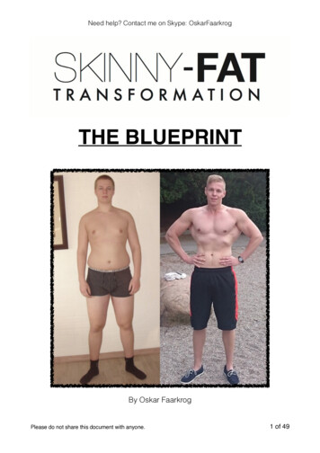
220Breast Reconstruction – Current Techniqueswhere ancillary fat grafting has been used successfully for breast reconstruction of borderzone contour irregularities (Kanchwala, 2008). Smaller volumes of graft compared with therecipient-site volume capacity potentially result in maintenance of a physiologic interstitialpressure environment and a favorable surface-to-volume ratio of graft to recipient,improving oxygen diffusion in the early days after grafting. Just like in a skin graft, earlytrauma or shearing of the graft-recipient interface is thought to damage micro-angiogenesisand decrease graft survival.A competing theory of graft volume maintenance is based on the experimental work ofMorrison who demonstrated adipocyte proliferation and angiogenesis in a perforatedhollow tube filled with a non-viable poly (D, L-lactic-co-glycolic acid) (“PLGA”) spongematrix which was implanted in the groin of rats (Dolderer et al, 2007). In the so-called“Morrison” (or better described as the “Scaffold”) theory of volume maintenance post fattransplantation, all or most of the transplanted adult adipocytes are destined to die. Ratherthan survive to maintain volume, these dead cellular elements act as a non-viable matrix orscaffold, through which macrophages penetrate and through which recipient-site stem cellmediated angiogenesis and adipogenesis occurs. Interestingly, Peer’s original 1950 “cellsurvival theory” postulated that human fat grafts disappeared completely a short time aftertransplantation and noted that “small autogenous multiple grafts had surviving portionsthat one year after transplantation appeared like normal fat tissue.’’Rather than choose between mutually exclusive theories, it is our belief that a third theoryexists to explain volume maintenance after fat transplantation. According to the“Tandem” theory, some transplanted adipocytes survive by diffusion and receive a bloodsupply by eventual micro-angiogenesis, while others do not and act as a scaffold forrecipient site cells to enter and remodel. Thus, both mechanisms are working in tandem inany given system.4. Not all fat grafting is the same – Definition of mega-volume fat graftingEarly applications of fat grafting in breast reconstruction involved injection of smallamounts of fat for contour correction or filling defects left after breast conservation therapy(Losken et al 2011, Rietjens et al 2011). However, there have been reports of complete breastreconstruction after mastectomy being accomplished by fat grafting with good results(Serra-Renom et al, 2011, Babovic, 2010, Fitoussi et al 2009, Delay et al 2010). The techniqueof fat grafting used in the face for nasolabial folds is different from the technique of fatgrafting used to treat chronic wounds, or to reconstruct radiated mastectomy sites. What isapparent is that not all fat grafting can be lumped into one technique, as not all fat graftingis the same. Sometimes fat is required to only provide volume, whereas in other cases its useis more to supply a regenerative effect, as in the case of a chronic wound (Cervelli et. al,2009). Therefore, we propose there are at least four separate categories of fat transplantation:high volume vs. low volume, and regenerative vs. non-regenerative, as depicted in Figure 1,below:We define mega-volume fat grafting as transplantation of over 100 cc of processed fat forcore volume projection replacement. In so doing, our technical strategies will cater to thedemands of this large volume, mainly non-regenerative case.www.intechopen.com
Autologous Fat Transplantation – A Paradigm Shift in Breast Reconstruction221Fig. 1. A Matrix Classification of Fat Grafting. The correct strategy for fat grafting must takeinto consideration the relative volume capacity and regenerative demands of the recipientsite. (Reconstruction case courtesy of Roger Khouri, MD)5. From science to the operating room – Surgical variables in fattransplantationFour clinical factors appear to have a significant effect on the survival of grafted fat cells:1.2.3.4.Fat HarvestingFat ProcessingFat GraftingRole of the Recipient Site5.1 Fat harvestingCannulas: Diameter and Hole Size: Although it has been reported in a histological studythat larger cannula sizes (6mm) harvest fat with better viability than with smaller cannulas(Erdim et al, 2009), viability in this report was measured using cell isolation and countingadipocytes with a haemocytometer. Such an experimental endpoint only represents the firststep of a multi-stage procedure that must consider all stages, with the clinical endpointbeing long-term (six months or greater) volume retention after transplantation into ananimal host.Smaller cannula sizes theoretically create less donor site trauma and allow for removal ofsmaller sized lobules of fat, which may improve flow characteristics and reduce traumaduring re-injection. An important consideration besides cannula size is cannula hole size andnumber of holes. A 12-gauge cannula with 6-8 side holes 2x1 mm in size can extract asignificant amount of fat despite its small caliber.www.intechopen.com
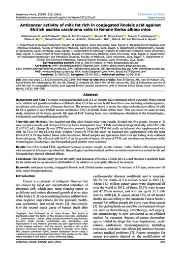
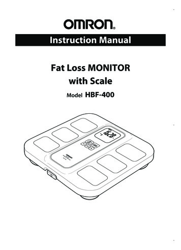
222Breast Reconstruction – Current TechniquesFig. 2. Varying negative pressure and hole size, the volume of fat flowing through cannulas(measured in cc of fat per 10 strokes) is efficient at 6-12 holes, even at lower negativepressures. (Data: courtesy of Roger Khouri, MD)The summation of the surface area of the individual openings on a 12 gauge, 12-holecannula approaches or exceeds the surface area of the opening of a classic 10 mm one-holecannula, once used in the 1980s. The result is better tissue flow with less donor area trauma.Further, each hole selects for lobules of uniform small size, which are more likely to floweasily through the injection cannula during the grafting phase of the procedure, without theneed for further processing or syringe transfers. The hole sizes on the aspiration cannulaapproach the size of the hole on the injection cannula, providing “equalization” of hole sizesfor more efficient fat flow (see below under fat injecting).Negative Pressure: The literature regarding the isolated effects of negative pressure suggeststhat adipocytes can be suctioned below 700 mm Hg without undue trauma (Shiffman, et al,2001). Any claims of syringe suctioning being safer than machine suctioning should becarefully examined. While a standard liposuction machine can generate up to oneatmosphere (760 mm Hg) of negative pressure, a 60 cc syringe connected to an in-linemanometer can also generate nearly one atmosphere of negative pressure (Fig. 3). Withregards to the effect of negative pressure on adipocyte viability, it is likely that absolutepressure and not the source of this pressure is the key variable in adipocyte trauma.Location of Donor Fat: Animal studies have not demonstrated superior donor site fat basedon anatomic location (Ullman et al, 2005), and clinically we have not observed anatomiclocation of the donor fat to be of significance in terms of volume retention. Although reportsin the literature (Padoin et al, 2008) suggest the lower abdomen and inner thighs to be richerin the source of stem cells, this study used C-Kit expression as a proxy measurement forstem cells. C-Kit expression also measures lymphocytes and on this basis the conclusions ofthis paper are in question. In reality, what is more important to consider is the relativeabundance and requirements of donor graft in each individual case, and the surgical planshould aim to avoid or minimize donor site deformities. It may turn out that that adipocytecellular size, which varies in different body regions and also among different patients maybe a more important variable than location per se. Larger cells have a higher likelihood ofmechanical cell membrane damage during extraction, and it may be this variable of cell sizerelative to cannula hole-size that is more important than the specific area on the body usedfor harvest per se.www.intechopen.com
Autologous Fat Transplantation – A Paradigm Shift in Breast Reconstruction223Fig. 3. A 60cc syringe can generate close to one atmosphere (30 in 760 mm Hg) of negativepressure, dispelling the myth that syringe suction is “safer” than machine suction.Another variable of unknown importance in fat harvesting remains the negative impact ofair exposure (Kaufman et al, 2007). Despite its widespread mention, there is a paucity ofscientific data quantifying the effect of air exposure on adipocyte viability (Aboudib et al,1992). Techniques of fat processing range from drying fat on Telfa Rolls (high air exposure),to completely closed systems employing intravenous tubing, three way stopcocks and IVbags for collection.5.2 Fat processingPeer’s cell survival theory of grafted en bloc fat dates back over 50 years and suggests thatthe number of calls transplanted at the time of transplantation may correlate with theultimate fat graft survival volume (Peer, 1950). After Ilouz’s breakthrough application ofliposuction (Ilouz, 1983), fat became available in a fragmented form. The cell survival theoryof solid fat transplantation may have influenced the use of high-speed centrifugation as apotential strategy for effective fat grafting. Historically, the penchant for centrifugation mayhave arisen from the need to graft as much adipocyte biomass as possible into a limitedspace. Although centrifugation can process highly concentrated fat, there are potentialproblems associated with it when this technique is employed in megavolume fat grafting: The cells may be damaged due high G-forces (Kurita et al, 2008)It is a time and labor-consuming processHigh fat concentrations may cause clumping and more difficult flow during re-injectionOne of the most confusing metrics in fat grafting is a lack of standardization when onediscusses “percent yields”. Once fat is lipo-aspirated as donor graft there are an infinitenumber of different concentrations of adipocyte volume relative to non-adipocyte volume(blood, serum, crystalloid) that can be reached prior to grafting the recipient site. Unless thewww.intechopen.com
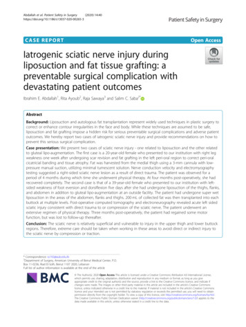
224Breast Reconstruction – Current Techniquesconcentration of adipocytes in the grafted material can be accurately measured and unlessthe process of fat concentration is standardized, one cannot reliably measure the percent ofadipocyte volume that survived grafting. In an effort to move toward an acceptabletechnical standard and to allow for better comparison of volumetric data in clinical series,careful documentation of the following data should be performed in mega-volume fatgrafting patients: Obtain a baseline mammogram of the breastPrior to expansion, objectively document breast volume (MRI, 3D imaging, or both)Document the process used for crystalloid separation (decanting, low speedcentrifugation, high speed centrifugation)Document the volume of processed material grafted in ccObjectively document the post graft volume of the breast at 6 months or more (MRI, 3Dimaging, or both)Our strategy in mega-volume fat grafting is based on the concept of minimizingextracorporeal adipocyte time (“EAT”). Whichever theory of fat volume maintenance onebelieves is true, all should agree that excess time is detrimental for patient safety, cells, andlastly, surgeons. Therefore, we employ the large syringe technique to process 480 cc of fat in2 minutes, which is the most efficient method of fat dehydration currently available.5.3 Fat injecting and shape-modifying techniquesThe selection of an injection cannula used in mega-volume fat grafting follows similarprinciples to those of harvesting. Small gauge cannulas theoretically reduce trauma to thebreast recipient site, which potentially reduces the risks of bleeding, hematoma, andresultant poor graft oxygen diffusion. The hole size of the injection cannula should matchclosely with the hole sizes of the aspiration cannula. By matching hole sizes, the selected sizeof the harvested lobules of fat are more likely to flow easily through the injection cannulawithout blockage or undue resistance (Fig 4).Fig. 4. Equalization of Hole Size. The opening in the 3 mm, 9-hole aspiration cannula (top,center) is nearly equal in size to the hole opening in the 16-gauge blunt tipped side holeinjection cannula (bottom), improving tissue flow on injection.www.intechopen.com
Autologous Fat Transplantation – A Paradigm Shift in Breast Reconstruction225There are currently two methods of injecting fat in a manner that seeks to increasedispersion and surface to volume contact with the recipient site – the “Mapping” techniqueand the “Reverse Liposuction” technique. Despite using these different techniques, theauthors’ independent long-term volume maintenance is essentially the same, eachdemonstrating an average increase in breast volume of 250 cc on average at six months byquantitative volumetric MRI imaging.The Mapping Technique: Donor cells have the highest chance of survival with the techniquethat best ensures an even, three-dimensional dispersion of the fat. The mapping techniqueinvolves the use of small (3 cc-5 cc) syringes handheld and connected directly to a 16 Gaugeblunt curved side-hole cannula. Markings are made in the recipient areas to aid in asystematic, diffuse and even injection of the entire recipient area. 8-10 circum-mammary and4 circum-areolar entry points are usually made with a 14G hypodermic needle (Fig. 5).Through each entry point the 15-20 cm long cannula makes multiple tunnels that fan outradially and injects 1-2 cc of fat only upon axial withdrawal. The cannula is then insertedinto another adjacent entry point and the fanning process is repeated to yield a 3-D weavethat evenly crisscross and covers the recipient space.Fig. 5. Markings for the Mapping Technique of Fat Injection. On the breasts, 8-10 circummammary and 4 circum-areolar needle puncture entry sites with radially fanning tunnelsfrom each site provide a well- diversified insertion of the graftsMultiple levels of graft are deposited, deep from the base of the breast just above thepectoralis fascia, to the subcutaneous space immediately subjacent to the dermis. Directinjection of fat into the dense parenchyma of the breast is never performed. This technique isdeliberate and exact but does take time. In addition, it requires the operator to deploy theplunger and withdraw the needle at the same time. Overall, the Mapping technique may bemore suitable for surgeons beginning mega-volume fat grafting for breast augmentationbecause it is more exact and deliberate. For breast reconstruction, it is clearly beneficial,www.intechopen.com
226Breast Reconstruction – Current Techniquesespecially in scarred areas caused by mastectomy and/or irradiation where a careful anddeliberate graft placement is necessary.The “Reverse Liposuction” Technique: The reverse liposuction technique seeks to evenlydisperse the fat into the subcutaneous and non-breast parenchyma in as efficient a manneras is possible. 6-8 needle insertions are made using a 14-gauge needle on the breast, alongthe infra-mammary fold (“IMF”) and spaced 4-5 cm laterally toward the axilla. Using astraight, 15 cm blunt side- hole, 16 gauge needle loaded directly onto 60 cc syringes, the fatis injected using a controlled “to and fro” liposuction movement with constant lightdepression on the plunger. With every pass of the needle the direction is changed slightly tocreate a fanning pattern of vectors. This is repeated in each different insertion site and atmultiple planar levels from the base of the breast. The axillary insertion is also used to placegraft in the sub-muscular position and this approach is felt to be the safest method ofnavigating the sub pectoral space. The rate of graft insertion in this technique should be 1-2seconds per cc, which results in a 1-2-minute/60 cc of fat rate, or a 300 cc per breast graftingsession performed in 10-15 minutes on each breast. The Reverse Liposuction Technique ismore time efficient and can be utilized in cases where there is no internal breast scarring, nodense adherence of skin to the chest wall, and where the breast is adequately expanded.Hence, this technique might not be the best technique for breast reconstruction in anirradiated breast. Another potential benefit of the reverse liposuction technique is that 60 ccsyringes generate lower maximum pressures than do 5 cc syringes, which generate higherpressure. Therefore, if there is a blockage of flow along the insertion needle due toclumping, this will occur at a lower pressure using a 60 cc syringe, with less potentialdamage to the grafted cells and less likelihood of pushing the blockage through, creating abolus.5.3.1 Breast shape modification using 3 dimensional ligamentous band releaseOnce the fat is grafted, the internal parenchyma is under higher pressure. Contourirregularities due to internal ligamentous tethering can be manifest, especially seen at theinterface between the natural infra-mammary fold and the newly augmented breast mound.A technique first described by Rigotti, who employed a pickle fork to release heavily scarredrecipient site radiated tissue, is used to release subcutaneous ligaments and scars in breastaugmentation and reconstruction with fat grafting. This technique is called threedimensional ligamentous band release, or “Rigottomy”. 3D Ligamentous band release is apowerful technique that can change breast shape. This technique, like meshing a twodimensional skin graft, releases contour deformities of the breast parenchyma in threedimensions. Grafted fat immediately fills space created and the fat keeps distance betweenthe transected scar or band, so the scar or band does not reform.5.4 The role of the recipient site in fat transplantationNegative pressure on the breast prior to fat transplantation creates internal expansion of thebreast parenchyma by drawing in more fluid, creating an edema-like state, and byincreasing the size and caliber of blood vessels. The authors postulate that non-surgical preoperative expansion of the breast recipient site enhances fat grafting results by five maineffects:www.intechopen.com
Autologous Fat Transplantation – A Paradigm Shift in Breast Reconstruction227Bigger potential spaces available for overall volume of graft;Reduces the demand on adipocytes to act as internal expanders, resulting in unduepressure;Augments tension on internal constrictions and scars, so breast shape can be addressed;Variables that are time consuming (e.g. centrifugation) become less demanding;Angiogenesis effect may increase recipient site oxygen tension and lead to better graft take.The VAC device has markedly improved outcomes in many types of difficult openwounds by clearance of bacteria and reduction in fluid volume (Saxena et al, 2007). In openwounds, micromechanical forces such as negative pressure elicit tissue deformation forcesthat stretch individual cells, thereby promoting proliferation in the woundmicroenvironment. The application of micromechanical forces on cells has beendemonstrated as a useful method with which to stimulate wound healing through thepromotion of cell division, angiogenesis, and local elaboration of growth factors (Saxena etal, 2004, 2007). The deformational forces of the VAC device are consistent with thismechanism of action and are similar to the negative pressure exerted on the breast whenBRAVA pre-expansion is used. Pre-expansion to the breast may therefore be more than just“increasing space”. Negative pressure therapy to the breast may demonstrate similar effectsof angiogenesis, cell division, and up-regulation of growth factors in the breast recipient site.For small graft volumes (100 ml or less), pre-expansion is not a pre-requisite as much as it isfor breast augmentation, as it does not result in high interstitial pressures, as the capacity ofthe recipient site can receive such a small volume with relative ease.6. Surgical techniqueFat grafting for breast augmentation or reconstruction does not begin with surgery. Properpatient selection and motivation for use of the external expansion device, and finallyeducation as to the unknown potential risks of fat graft
1Back Bay Plastic Surgery and Massachusetts General Hospital, Bo ston MA, 2St. Elizabeth s Medical Center, Tufts University, Boston MA, USA 1. Introduction Fat grafting to the breast is not a new concept. It has been under consideration since the 19 th century when pioneers attempted inserting bulk volumes of fat (Neuber, 1893, Czerny 1895).