Transcription
PhosphoSens Protein Kinase Assay Instruction ManualINSTRUCTION MANUALTABLE OF CONTENTS1. INTRODUCTION2. PHOSPHOSENS ASSAY PRINCIPLE3. PHOSPHOSENS ASSAYS COVERED BY THIS INSTRUCTION MANUAL4. ASSAY KIT COMPONENTS AND REQUIRED MATERIALS4.1. MATERIALS AND EQUIPMENT NOT INCLUDED4.2. MATERIALS INCLUDED5. PERFORMING THE ASSAY5.1 PREPARING ASSAY REAGENTS5.2 GENERATING A KINASE TITRATION5.3 KINETIC ANALYSIS5.4 DETERMINING KM AND VMAX5.5 DETERMINING INHIBITOR KINETIC PARAMETERS6. TROUBLESHOOTING7. TECHNOLOGY LICENSING8. REFERENCES9. APPENDIX A: PERFORMING A PHOSPHOSENS -RED ASSAYAssayQuant Technologies, Inc. 260 Cedar Hill Street, Marlborough, MA 01752 (508) 804-3155 hello@assayquant.comCopyright 2021, AssayQuant Technologies, Inc. All rights reserved.1
PhosphoSens Protein Kinase Assay Instruction ManualAssayQuant Technologies, Inc. 260 Cedar Hill Street, Marlborough, MA 01752 (508) 804-3155 hello@assayquant.comCopyright 2021, AssayQuant Technologies, Inc. All rights reserved.2
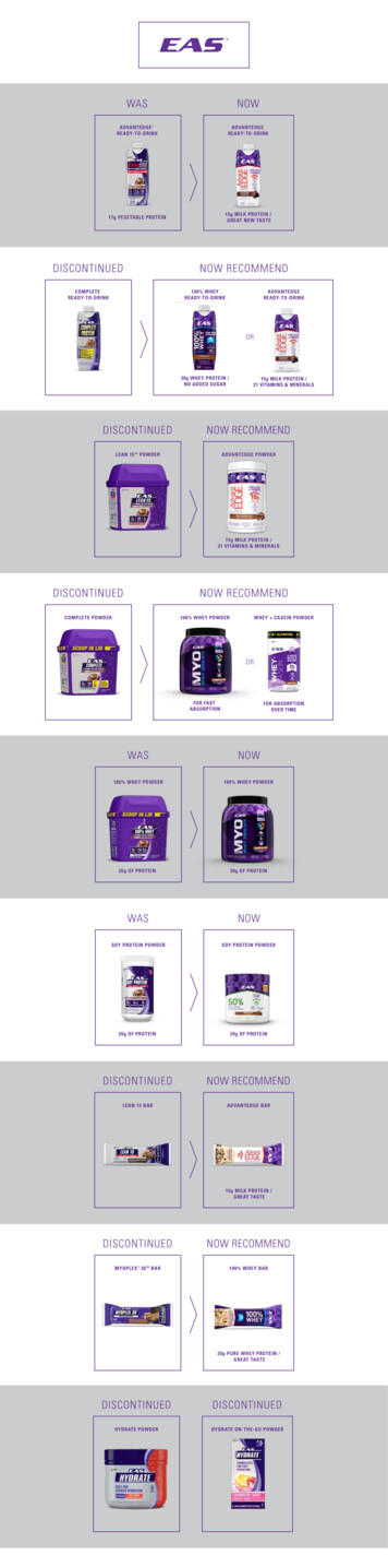
PhosphoSens Protein Kinase Assay Instruction Manual1. INTRODUCTIONProtein kinases play pivotal roles in cellular signaling and regulate many essential biological processes,including cell growth, differentiation, metabolism, proliferation, and programmed cell death. The humangenome encodes for 518 protein kinases, making this one of the largest enzyme families. Many humandiseases are now known to be associated with dysregulated kinase activity. The ability to accurately andprecisely quantify kinase activity and define the effects of compounds that modulate this activity isessential for understanding the complex biology of protein kinases and for the development andmonitoring of effective therapeutic agents against these enzymes (1-4). Precise kinase activitymeasurements are also critical for understanding the effects of kinase mutations on enzyme function tounderstand the relationships between mutations, signal transduction and cellular function (5,6).The PhosphoSens kinase assay platform provides a simple, one-step homogeneous, fluorescence-basedassay for rapid and sensitive detection of serine/threonine and tyrosine kinase activities. PhosphoSens assays directly measure the catalytic activity of target kinases, without the need for antibody reagents,coupling steps or radioisotope labeling to develop the signal. The PhosphoSens technology usesoptimized peptide substrates that provide robust assays with minimal lot-to-lot variation and highaccuracy and precision. PhosphoSens assays are easily performed in continuous kinetic mode, usingcommonly available fluorescence plate readers in 96-, 384- and 1536-well plate formats and a widerange of sample types including recombinant enzymes, immunoprecipitated kinases, crude cell or tissuelysates (7-9) and innovative microfluidic applications extending down to single-cell analysis (10-12). UsingPhosphoSens , kinase activity is measured under optimal conditions including pH, selected metal ioncofactors, and low to physiological (mM) ATP concentrations allowing both ATP competitive and ATPnon-competitive (allosteric) kinase inhibitors to be selected and characterized. The technology issupported by 90 scientific publications with demonstrated utility across applications for studying kinaseactivity regulation, kinase inhibitor screening, and inhibitor mechanism of action analysis including Ki,kinact, residence times and IC50 determinations (13,14).2. PHOSPHOSENS ASSAY PRINCIPLEThe Chelation-Enhanced Fluorescence (ChEF) method for protein kinase sensing, filed under thePhosphoSens trademark, was first introduced by Imperiali and coworkers (15-17). Since that time, theChEF method has been rigorously validated and further developed (8,9,18-21) for optimum performancewith a wide variety of protein kinases. The ChEF sensing mechanism exploits a synthetic α-amino acidwith a side chain bearing an 8-hydroxyquinoline derivative (sulfonamido-oxine, Sox) which uponcoordination to Mg(II), relays information on the phosphorylation state of proximal serine, threonine ortyrosine residues in peptide- and protein-based kinase substrates (Figure 1). In the absence ofphosphorylation, the Sox shows low affinity for Mg(II); upon phosphorylation, Mg(II) affinity is enhanceddue to the advantageous chelate effect, involving the Sox and the introduced phosphate group, andfluorescence is turned on.FIGURE 1: ChEF Mechanism for Direct Protein Kinase Activity SensingAssayQuant Technologies, Inc. 260 Cedar Hill Street, Marlborough, MA 01752 (508) 804-3155 hello@assayquant.comCopyright 2021, AssayQuant Technologies, Inc. All rights reserved.3
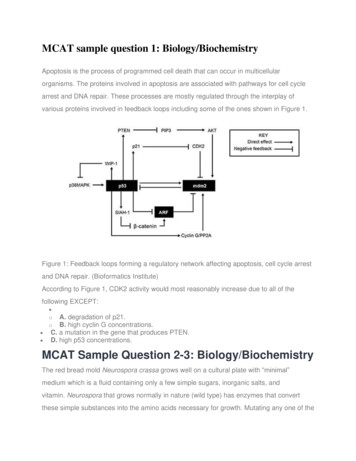
PhosphoSens Protein Kinase Assay Instruction ManualJust Add & ReadFigure 2A illustrates typical fluorescence changes upon phosphorylation of a PhosphoSens peptidesubstrate and Figure 2B shows excitation and emission spectra of a typical phosphorylated PhosphoSens peptide. The fluorescence properties of the Mg(II)-coordinated with the 8-hydroxyquinoline of Sox has anλExMax of 360 nm (358-363 nm) and λEmMax of 492 nm (485-498 nm). Since the fluorescence emissionspectrum is relatively broad (see Figure 2B), fluorescence emission can be monitored between 475-508nm with 7% loss in signal intensity.FIGURE 2: Fluorescence Spectra of PhosphoSens Peptides3. PHOSPHOSENS ASSAYS COVERED BY THIS INSTRUCTION MANUALThis manual can be used as a guide for serine/threonine and tyrosine kinase assays using PhosphoSens or PhosphoSens -Lysate assay kits and stand-alone CSox peptide substrates (available as 96- or 240assays for 96- and 384-well plates, respectively, and bulk net peptide). All kits include reaction buffer,enzyme dilution buffer, ATP, DTT and the CSox peptide substrate. With stand-alone CSox substrates, theuser must provide their own stocks of buffers and other required components. All PhosphoSens assaysare compatible with any purified recombinant protein kinase and with biological samples if the userqualifies the experimental system (see Section 4). PhosphoSens -Lysate assay kits contain CSoxsubstrates that have been optimized for the analysis of target protein kinases in unfractionated cell andtissue lysates. For a complete listing of available CSox-based kinase substrates, as configured in kits or asstand-alone substrates, please refer to www.assayquant.com or contact AssayQuant TechnologiesAssayQuant Technologies, Inc. 260 Cedar Hill Street, Marlborough, MA 01752 (508) 804-3155 hello@assayquant.comCopyright 2021, AssayQuant Technologies, Inc. All rights reserved.4
PhosphoSens Protein Kinase Assay Instruction Manual(info@assayquant.com) for additional information, including custom services for compound testing orthe development of PhosphoSens peptide substrates and assays for new targets of interest.The activity of protein phosphatases, either serine/threonine and tyrosine-directed, can also bemeasured effectively with the PhosphoSens platform by using CSox-based phosphopeptide substrates.This approach provides a physiologically-relevant, homogeneous, continuous and highly-sensitivemeasure of protein phosphatase activity that is described in the PhosphoSens Protein PhosphataseAssay Instruction Manual. In addition to the continuous (kinetic) format, the activity of protein kinasesand phosphatases can be measured in an endpoint format using Europium (III)-coordinated with the 8hydroxyquinoline of Sox to produce a red-shifted signal with λExMax of 360 nm (358-363 nm) and λEmMaxof 620 nm (610-630 nm) that is measured using time-resolved fluorescence with a 100-150 µsec delayand a 300-500 µsec data acquisition time, thereby eliminating any interference due to compoundautofluorescence. The Europium (III) format is described in the PhosphoSens -Red Assay InstructionManual.4. ASSAY KIT COMPONENTS AND REQUIRED MATERIALS4.1. MATERIALS AND EQUIPMENT NOT INCLUDED4.1.1. Recombinant kinase: The PhosphoSens products are compatible with any commercially availableprotein kinase for which a Sox-based substrate has been developed. Kinases providers include, but arenot limited to, BPS Bioscience, Carna Biosciences, EMD-Merck/Millipore, ProQinase, Proteros,SignalChem, and Thermo Fisher/Invitrogen). When choosing a commercially available kinase preparation,an assessment of purity, specific activity (in the supplier’s assay format), the nature and location of coexpression and purification tags (e.g., N- or C-terminal GST, His, or FLAG tags), and the size of theconstruct (full-length or truncated) should be considered. The most rigorous approach is to obtain akinase from multiple sources and compare the activity with the PhosphoSens platform, where thekinetic format allows a quantitative measurement allowing the most appropriate enzyme to be selectedfor further study.4.1.2. Cell and tissue lysate samples: Any PhosphoSens peptide substrate can be used with biologicalsamples if the user qualifies the experimental system. For example, the generic serine/threonine ortyrosine substrates from AssayQuant can be used to measure total serine/threonine or tyrosine kinaseactivity in crude cell and tissue lysates or enriched fractions. Individual substrates can be qualified for usewith crude lysates from appropriately stimulated cell systems. For example, Li and coworkers (7) usedthe Omnia Y7 substrate for highly-selective detection of the Syk tyrosine kinase in crude lysates of IgMstimulated Ramos cells. If required, these experiments can include off-target kinase inhibitors to ensurespecificity of the assay (9). Alternatively, the target kinase can be immunoprecipitated from crude lysatesprior to performing an IP-kinase assay with the PhosphoSens technology; For examples refer to Li (7) forcontrol experiments and plate-IP methods for the Syk tyrosine kinase, Lauchle (22) for MK2, and Chen(23) for GSK3b. Sox-based bipeptide or protein sensors, including both docking and sensing sequences,have been used to specifically and quantitatively measure the kinase activity of individual MAPK familyAssayQuant Technologies, Inc. 260 Cedar Hill Street, Marlborough, MA 01752 (508) 804-3155 hello@assayquant.comCopyright 2021, AssayQuant Technologies, Inc. All rights reserved.5
PhosphoSens Protein Kinase Assay Instruction Manualmembers in crude cell and tissue lysates (9,19,20). In each of the above scenarios, the continuous-readmode of the PhosphoSens technology provides more information about the activation state of theisolated kinase or kinase complex in the presence of other cellular proteins. Indeed, there is considerableinterest in using these more complex systems for drug development to obtain more accurate rank orderof kinase inhibitor potency and for Structure-Activity Relationship (SAR) studies (Li et al., 2009). Thehighly quantitative and precise measure of kinase activity provided by the PhosphoSens technology isan important improvement over conventional western blot analysis with phosphoprotein-specificantibodies, which is at best only semi-quantitative (1,2). Sox-based substrates, either single chain or bipeptide formats, with improved specificity for the targets of interest will be the focus of PhosphoSens Lysate sensors from AssayQuant Technologies (please inquire at info@assayquant.com).4.1.3. Fluorescence microplate reader: Instrument must be capable of reading fluorescence intensity incontinuous (kinetic) mode with an excitation wavelength (λExMax) of 360 nm (358-363 nm) and anemission wavelength (λEmMax) of 492 nm (485-498 nm). Readings can be made at the desired intervalsand duration (e.g., reading every 30 seconds for 60 minutes or every 3 minutes for 150 minutes).PhosphoSens assays can be run on readily available microplate reader instruments including but notlimited to the Tecan Safire 2 , Infinite M1000, Infinite F500, Molecular Device SpectraMax M5, BMGLABTECH PHERAstar, FLUOstar OPTIMA, BioTek FLx800 , Neo, Synergy 2, and Synergy 4, andThermoFisher Varioskan. Contact Technical Support or e-mail us directly at info@assayquant.com forinstrument-specific setup guidelines.4.1.4. Precision pipettes: Precision pipettes with disposable plastic tips to accurately deliver 2–200 μL.4.1.5. Ultrapure deionized water: 18 Ω or higher4.1.6. Plastic tubes or plates: Plastic with low protein-binding properties is used for diluting anddispensing assay components. We recommend USA Scientific (1615-5500) 1.5 ml polypropylene microcentrifuge tubes (DNase, RNase, DNA, and pyrogen free). For larger volumes, use polypropylene and notpolystyrene tubes. A low protein-binding and v-bottom dilution plate is available from Greiner (651201).4.1.7. Heat Block or Water Bath: Set at 30 oC, if this is the temperature at which reactions are to be run.4.1.8. Microtiter plates: White or Black Microtiter plates should be used to minimize light scattering andbackground fluorescence and to reduce well-to-well crosstalk. These plates come in multipleconfigurations, where improved signal and signal/background are obtained with the white plastic noprotein binding (NBS) plates. Excellent performance has been obtained with:Corning, half-area 96-well, white flat bottom polystyrene NBS microplates (3642)Corning, low-volume 384-well, white flat bottom polystyrene NBS microplates (3824)Corning, 1536-well, white flat bottom polystyrene NBS microplates (3729)With the PhosphoSens Platform, we recommend using a 50 µL final reaction volume per well in a halfarea 96-well plate or a 25 µL final reaction volume in a low-volume 384-well plate. PhosphoSens technology also can be used with 1536-well plates, where established assays have been successfullyscaled down to 5 µL for high-throughput applications. With all plate formats, the final reaction volumedetermines the path length, where larger volumes provide higher signals and subsequently greater assayprecision. As a guide, Figure 4 illustrates the effect of assay volume on assay precision (% CVs).AssayQuant Technologies, Inc. 260 Cedar Hill Street, Marlborough, MA 01752 (508) 804-3155 hello@assayquant.comCopyright 2021, AssayQuant Technologies, Inc. All rights reserved.6
PhosphoSens Protein Kinase Assay Instruction Manual4.1.9. Adhesive Seal for Microtiter Plates: To control for evaporation, especially with long kinetic readsor under low humidity conditions, plates should be sealed with an optically-clear adhesive film that stillallows top-reading with minimal light scattering. We have tested many products and recommend PerkinElmer TopSeal-A Plus (6050185) applied with either a roller or a paddle (VWR 60941-118 or 60941-128,respectively).4.2. MATERIALS INCLUDEDEach PhosphoSens kit provides the reagents listed in Table 1 allowing the user to perform 96 kinasereactions at 50 µL final reaction volume per well in a half-area 96-well plate or 192 reactions at 25 µL finalreaction volume in a low-volume 384-well plate. Solutions provided include at least a 10% overfill. Allcomponents are available from AssayQuant Technologies as stand-alone items that can be ordered usingthe Catalog numbers provided below. Components can also be ordered in bulk (please inquire atinfo@assayquant.com). Bulk PhosphoSens peptide substrate is provided as lyophilized powder as mgs ofnet CSx-peptide (please inquire at info@assayquant.com for bulk pricing discounts).TABLE 1: Components provided with each PhosphoSens Kinase Assay Kit:CATALOG T1XEDBNOEGTAAQT1000XEGTACOMPONENTPhosphoSens Substrate, 100XATP solution, 100XDTT solution,1000XReaction Buffer(RB), 10X, No DTTor EGTAEnzyme DilutionBuffer (EDB), 1X,No DTT or EGTAEGTA solution,(1000X)DESCRIPTIONPhosphoSens Cysteine-Sox KinaseSensor peptide substrate, 1 mM100 mM ATP in ultrapure deionizedwater. The pH is not adjusted.1M DTT in ultrapure deionizedwater500 mM HEPES, pH 7.5, 0.1% Brij35, 100 mM MgCl220 mM HEPES, pH 7.5, 0.01% Brij35, 5% Glycerol, 1 mg/mL BovineSerum Albumin0.55 M ethylene glycol-bis(β-aminoethyl ether)-N,N,N′,N′-tetraaceticacid (EGTA), pH 8AMT*60 µL1.5 mLSTORAGE-20 oC or below. Minimizerepeated freeze/thaw cycles-20 oC or below. Minimizerepeated freeze/thaw cycles-20 oC or below. Minimizerepeated freeze/thaw cycles-20 oC or below.1.5 mL-20 oC or below.120 µLRoom temperature or below.120 µL120 µL*AssayQuant adds a 10% overfill to each vial to ensure recovery of the amount listed (for example, 66 µL of the PhosphoSens 100x substrateis dispensed to each CSox-peptide substrate vial)5. PERFORMING A PHOSPHOSENS KINETIC ASSAYThe PhosphoSens assay format is SIMPLE! As a one-step homogeneous assay, you just MIX and READ.There are NO washing or separation steps and NO stop solutions or additional components needed todevelop the fluorescence signal. The assay reaction is initiated typically by addition of a Master Mixcontaining either a protein kinase or the CSox-peptide substrate to a well containing the final componentneeded to start the reaction. This can be done using 96-, 384- or 1536 well plate formats. Assays arecommonly performed at 30 C and fluorescence measurements are recorded in kinetic mode (e.g.,readings every 30 seconds for 30-60 minutes or every 3 minutes for 150 minutes). Alternatively, if a singleAssayQuant Technologies, Inc. 260 Cedar Hill Street, Marlborough, MA 01752 (508) 804-3155 hello@assayquant.comCopyright 2021, AssayQuant Technologies, Inc. All rights reserved.7
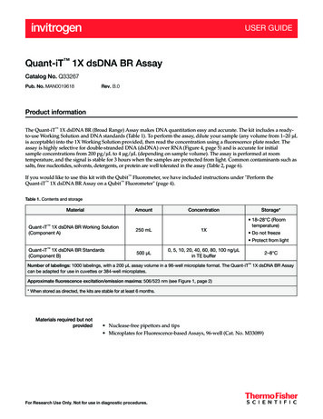
PhosphoSens Protein Kinase Assay Instruction Manualendpoint reading is desired, this can be made after adding a stop solution (an equal volume of 8 M urea),where the signal is stable for at least 24 hours. The λExMax of the chelated Mg(II) with the 8hydroxyquinoline is 360 nm and the λEmMax is 492 nm (485-498 nm). An overview of the PhosphoSens Platform Workflow is shown in Figure 3.FIGURE 3: Summary of the PhosphoSens Protein Kinase Assay Platform WorkflowAn endpoint assay can also be performed using Europium (III) to produce a red-shifted signal with λExMax of 360 nm (358-363 nm) and λEmMax of 620 nm (610-630 nm) that is measured using time-resolvedfluorescence with a 100-150 µsec delay and a 300-500 µsec data acquisition time, thereby eliminating anyinterference due to compound autofluorescence. The Europium (III) format is described in thePhosphoSens -Red Assay Instruction Manual.AssayQuant Technologies, Inc. 260 Cedar Hill Street, Marlborough, MA 01752 (508) 804-3155 hello@assayquant.comCopyright 2021, AssayQuant Technologies, Inc. All rights reserved.8
PhosphoSens Protein Kinase Assay Instruction Manual5.1. PREPARING ASSAY REAGENTSPrior to setting up the individual reactions, prepare the following solutions:5.1.1. PhosphoSens Sox-based Substrate: The concentration of the peptide stock solution has beenaccurately determined by absorption spectroscopy using the extinction coefficient of sulfonamido-oxine(Sox) at 355 nm prior to being lyophilized. With PhosphoSens Kits, the substrate is provided as a 1 mM(100X) solution. With PhosphoSens Bulk peptide, this is supplied as a lyophilized powder. Resuspend thepeptide substrate as indicated on the vial or in the Technical Notes section of the Certificate of Analysisto create the 1 mM (100X) stock. Gently vortex to ensure that all the substrate goes into solution.Caution: Avoid repeated freeze/thaw cycles by aliquoting the 1 mM stock solution into smaller volumesfor storage at -20 C or below until ready for use.5.1.2. Peptide Substrate Solution (10X): Prepare 0.1 mM (10X) Substrate solution by thawing the 1 mM(100X) peptide substrate stock solution, mixing well by vortexing gently, removing an appropriateamount and diluting 10-fold into ultrapure deionized water. To use the entire kit, add 594 µL of water tothe 66 uL of 1 mM substrate provided with the kit to make 660 µL (this is sufficient for 131 x 50 µLreactions for half-area 96-well plate or 261 x 25 µL reactions for low-volume 384-well plates, all at 10µM final substrate). Gently invert tube to mix and perform a 10 second spin in a micro-centrifuge.Alternatively, only make up as as much as is needed for the current experiment.5.1.3. ATP Solution (10X): To run final reactions at 1 mM ATP, prepare 10 mM (10X) ATP solution byadding 50 μL of 100 mM (100X) ATP to 450 μL ultrapure deionized water. Make fresh and discard afteruse. Note: The 100X stock should be aliquoted and stored at -20 oC or below. Minimize repeatedfreeze/thaw cycles.5.1.4. DTT Solutions: Most kinase assays are run in the presence of DTT, although some protein kinasesare more active in the absence of DTT, which must be determined empirically. Prepare 100 mM (100X)DTT solution by adding 5 μL of the 1 M (1000X) DTT provided to 45 μL ultrapure deionized water and useto prepare final EDB in 5.1.6 below. Prepare 10 mM (10X) DTT solution by adding 5 μL of the 1 M (1000X)DTT to 495 μL ultrapure deionized water and use to prepare final reactions per Tables 2-4. Caution:Diluted DTT is readily oxidized so use fresh dilutions.5.1.5. EGTA Solutions: Some kinases show enhanced activity when EGTA is included in the reaction,which must be determined empirically (e.g., the activity of GRK2 is increased by 50% in the presence of0.55 mM EGTA). Including EGTA will also chelate any Zn2 , which can be present as a trace metalcontaminant that can negatively affect assay performance. For these reasons, and with the exception ofworking with kinases activated by Ca2 (EGTA can’t be included because it chelates Ca2 ), we recommendthat EGTA be added to reactions (0.55 mM final) and the EDB (0.1 mM final). We include a 0.55 M stockof EGTA in each PhosphoSens kit for this purpose. Prepare 20 mM EGTA solution by adding 18.2 μL of0.55 M EGTA to 481.8 μL ultrapure deionized water and use to prepare final EDB in 5.1.6 below. Prepare5.5 mM EGTA solution by adding 5 μL of 0.55 M EGTA to 495 μL ultrapure deionized water and use forAssayQuant Technologies, Inc. 260 Cedar Hill Street, Marlborough, MA 01752 (508) 804-3155 hello@assayquant.comCopyright 2021, AssayQuant Technologies, Inc. All rights reserved.9
PhosphoSens Protein Kinase Assay Instruction Manualfinal reactions per Tables 2-4. All data in this manual were generated using EGTA in the final reaction.5.1.6. Enzyme Dilution Buffer (EDB, 1X) with DTT and EGTA: EDB is added to Blank or Background wellsas a "No Enzyme" control. To make EDB with 1 mM DTT final, add 5 μL of 100X DTT prepared in 5.1.4above to 495 μL of the EDB provided in the kit and keep on ice. Caution: Diluted DTT is readily oxidized souse fresh dilutions. To make EDB with 0.1 mM EGTA final, add 2.5 μL of 20 mM EGTA prepared in 5.1.5above to 497.5 μL of the EDB containing DTT or without DTT as provided in the kit. Keep EDB on ice.5.1.7. Kinase Reaction Master Mix: Prepare as listed in Tables 2 through 4.5.1.8. Kinase Stock: Kinases are enzymes and therefore must be maintained on ice. Following steps inTables 2 through 4. Just before use, dilute an appropriate amount of the kinase stock to 5X (10 nM if thefinal desired concentration is 2 nM) in EDB (with or without DTT, depending on the enzyme) and followsteps outlined in Tables 2 through 4. The required volume of 5X kinase in EDB, then is added to assaywells to initiate the reaction (this can be done using automated reagent dispensing if timing is critical).Discard the unused portion of the diluted kinase. Aliquot the remaining undiluted kinase into individuallow-protein-binding storage vials and return to the -80 C freezer, however, indicate that these stockshave been thawed. A limited number of freeze-thaw cycles may be acceptable; however, multiple freezethaw cycles are likely to compromise activity.5.1.9. Final Reaction Conditions: Typical final concentrations of each reaction component are as follows:54 mM HEPES, pH 7.5, 1 mM ATP (or as adjusted as needed), 1.2 mM DTT (optional), 0.55 mM EGTA(optional, but recommended), 0.012% Brij-35, 10 mM MgCl2, 10 µM peptide substrate, 0.05-5 nM kinase(or adjusted as needed) and any additional co-factors or additives (as required).5.2. KINASE TITRATION EXPERIMENT5.2.1. Additional Background: The amount of kinase used in a PhosphoSens assay is dependent on thespecific activity of the kinase towards the optimized CSox-based substrate and must be determinedempirically. Kinases obtained from commercial suppliers can vary widely with respect to their specificactivity and stability, which depends on many factors. In general, 0.1-5 nM of kinase in the final reactionis a good starting concentration, however, a kinase titration experiment will allow you to choose theappropriate amount of kinase for your application (most applications are best run under conditionswhere only 10-15% of the substrate is phosphorylated) and to determine the amount of kinase requiredto completely phosphorylate the peptide substrate to create a phosphopeptide standard curve. With thelatter, the fluorescence signal obtained from the fully phosphorylated peptide, in the presence of Mg(II)and other assay buffer constituents, can be used as the 100% phosphorylation control to determine the% of substrate phosphorylation achieved in an application and to convert RFU/minute to µM/minute forVmax and kcat determinations.5.2.2. Experimental Protocol: The steps and volumes of each component for a kinase titrationexperiment are outlined in Table 2 for a 96-well half-area plate. A dilution scheme is also provided. YouAssayQuant Technologies, Inc. 260 Cedar Hill Street, Marlborough, MA 01752 (508) 804-3155 hello@assayquant.comCopyright 2021, AssayQuant Technologies, Inc. All rights reserved.10
PhosphoSens Protein Kinase Assay Instruction Manualshould only plan to dilute as much kinase as you plan to use for experiments to be run that day as kinasesamples are generally less stable upon dilution. Note: Protocol Tables and dilution schemes for all theexperimental examples provided in this manual are available upon request for 96- and 384-well (lowvolume) plate formats (please inquire at info@assayquant.com).TABLE 2: PROTOCOL 1 – KINASE TITRATION (50 µL assay for a half-area 96-well plate)STEP1234567PROCESSPrepare Master Mix (all componentsexcept Active Kinase) by combiningeach of the components listed to theright. With certain kinases, additionalcomponents may be needed (e.g.,cofactors, glycerol, etc.) and so theamount of water should be adjusted.For 1 RXN:For 40 RXNs:5 µL200 µLReaction Buffer (10X)5 µL200 µLPhosphoSens Substrate (10X)5 µL200 µLATP solution (10X)5 µL200 µLDTT solution (10X)200 µLEGTA solution (5.5 mM or 10X) 5 µL15 µL600 µLUltrapure deionized H2O40 µL1.60 mLTotal volumeEquilibrate Master Mix (all components except kinase) to assay temperature (typically 30 C) by placingthe sealed tube in a heat block or water bath set at the desired temperature for 5 minutes.Equilibrate the assay plate in the plate reader to the desired assay temperature (typically 30 C).Aliquot 40 µL of the Master Mix into each well.Prepare Serially-diluted Kinase as described below.Add 10 µL of each concentration of 5X kinase to designated wells to start the reaction. Mix well. Add 10µL of Enzyme Dilution Buffer (EDB) to "No-kinase" control wells.Incubate at 30 C collecting fluorescence intensity (RFU) readings (λExMax 360 nm/λEmMax 492 nm [485498 nm]) at intervals (e.g., every 30 seconds for 60 minutes or every 3 minutes for 150 minutes, etc.).Kinase titration instructions for Table 2: As outlined below, prepare an 11-point, 2-fold serial dilution ofkinase using 1X EDB. Each dilution in the series should be at 5X the final concentration of kinase in thereaction. This dilution scheme will result in a concentration range that spans 3 log units (e.g., from 20 nMto 20 pM). To generate a titration curve that starts at 20 nM, the first 5X stock needs to be 100 nM. The12th well in a 96-well plate row is designated as a 'No-kinase control'. Replicates (typically in triplicate)are oriented vertically in the plate. The EDB and the protein kinase stocks have glycerol, so make sure allsamples are well mixed.AssayQuant Technologies, Inc. 260 Cedar Hill Street, Marlborough, MA 01752 (508) 804-3155 hello@assayquant.comCopyright 2021, AssayQuant Technologies, Inc. All rights reserved.11
PhosphoSens Protein Kinase Assay Instruction Manual96-well PlateSerial 2-foldDilutions:Tube #Final [Kinase],nM5X [Kinase], 253.131.560.780.390.200.10None38 µL38 µL38 µL38 µL38 µL38 µL38 µL38 µL38 µL38 µL76 µLEnzyme Dilution X µL asSuppliedBuffer, µLStock KinaseY µL38 µL ofTube #138 µL ofTube #238 µL ofTube #338 µL ofTube #438 µL ofTube #538 µL ofTube #638 µL ofTube #738 µL ofTube #8Final Volume76 µL76 µL76 µL76 µL76 µL76 µL76 µL76 µL76 µL38 µL of 38 µL ofNoneTube #9 Tube #1076 µL76 µL76 µLCommercial preparations will vary significantly in their concentration. The first tube takes into account the required dilution of the suppliers kinasepreparation to create Tube #1 (100 nM stock). For example, if the commercial kinase preparation is provided at 1,000 nM, then add 7.6 µL to 68.4µL of Kinase Dilution Buffer to create 76 µL of a 10-fold dilution. You need 38 µL to create each 2-fold dilution and 10 µL for each replicate at eachconcentration (typically triplicate wells, so 30 µL) or 68 µL of the 76 µL total. Tube #12 receives only Kinase Dilution Buffer to use as a "No-kinase"control. In this example, with 11 kinase concentrations plus 1 control (12 conditions), each in triplicate 36 wells (or 38% of a 96-well kit).5.2.3. Data Analysis: Subtract the background fluorescence for each time point from the totalfluorescence signal to obtain corrected Relative Fluorescence Units (RFU) values. Plot the corrected RFUvs. Time. Determine the slope of the initial linear portion of each curve, which is the initial reaction rate(RFU/min or convert to RFU/pmole kinase/min). Reaction rates from linear or non-linear (kinases thatexhibit a lag phase) fit of the data can be generated
PhosphoSens assays can be run on readily available microplate reader instruments including but not limited to the Tecan Safire 2 , Infinite M1000, Infinite F500, Molecular Device SpectraMax M5, BMG LABTECH PHERAstar, FLUOstar OPTIMA, BioTek FLx800 , Neo, Synergy 2, and Synergy 4, and ThermoFisher Varioskan.