Transcription
Kriege et al. BMC Anesthesiology (2018) EARCH ARTICLEOpen AccessA comparison of two hyperangulated videolaryngoscope blades to direct laryngoscopyin a simulated infant airway: a bicentric,comparative, randomized manikin studyMarc Kriege* , Nina Pirlich, Thomas Ott, Eva Wittenmeier and Frank DetteAbstractBackground: In infants, securing the airway is time-critical because of anatomical and physiological differencesrelated to airway management in children less than 1 year old. The aim of this study was to compare the time toventilation using two different hyperangulated video laryngoscope blades with the time to ventilation viaconventional direct laryngoscopy in a normal airway [NA] and in a simulated difficult airway [DA].Methods: This study was a comparative, bicentric, open-label, randomized controlled evaluation. An infant highfidelity simulator (SimBaby ; Laerdal Medical, Stavanger, Norway) was used, and two scenarios were proposed, asfollows: NA and DA evoked with tongue edema and cervical collar. After theoretical and practical briefing, eachparticipant compared in the two airway scenarios the novel King Vision Pediatric aBlade (KV) (Ambu A/S, BadNauheim, Germany) video laryngoscope and the C-MAC D-blade Ped (DB) (Karl Storz SE & Co. KG, Tuttlingen,Germany) video laryngoscope to conventional laryngoscopy using the Miller Blade (MiB) and the Macintosh Blade(MaB) in a random sequence.Results: Eighty physicians (65 AN and 15 PCCM staff) were included. In the NA scenario, the median [IQR] time tosuccessful time to ventilation (TTV) was significantly shorter for the KV at 13 s [12–15 s] than for the MaB at 14.5 s[13–16 s], DB at 14.5 s [13–16] and MiB at 16 s [14–19] (p 0.001). In DA, the KV also shortened TTV to 14 s [13–16],whereas TTV was 23 s with the MaB [20–26], 19 s with the DB [16–21], and 27 s with the MiB [22–31] (p 0.001).There were no differences in first-pass intubation success rates (FPAs) between hyperangulated blades and directlaryngoscopes in NA. In DA, the hyperangulated blades enabled 92 (DB) to 100% (KV) FPAs compared with 65 (MiB)to 76% (MaB) for conventional laryngoscopy (p 0.001).Conclusion: Video laryngoscopes with hyperangulated blades were associated with shorter TTV in normal anddifficult infant airway situations. The higher FPAs of hyperangulated blades in DA may avoid desaturations anddecrease adverse events in pediatric airway management.Keywords: Infant, Airway, Intratracheal intubation, Video laryngoscopy, Pediatric* Correspondence: makriege@uni-mainz.deDepartment of Anesthesiology, University Medical Center of the JohannesGutenberg-University, Langenbeckstraße 1, 55131 Mainz, Germany The Author(s). 2018 Open Access This article is distributed under the terms of the Creative Commons Attribution 4.0International License (http://creativecommons.org/licenses/by/4.0/), which permits unrestricted use, distribution, andreproduction in any medium, provided you give appropriate credit to the original author(s) and the source, provide a link tothe Creative Commons license, and indicate if changes were made. The Creative Commons Public Domain Dedication o/1.0/) applies to the data made available in this article, unless otherwise stated.
Kriege et al. BMC Anesthesiology (2018) 18:119BackgroundSecuring a pediatric airway is a critical skill for theanesthesiologist and pediatric intensive care physician[1]. Problems in airway management can lead to severehypoxemia and are associated with prolonged mechanical ventilation in pediatric intensive care units (PICUs)[2]. Infants are significantly different from older childrenor adults with regard to their anatomical and physiological characteristics. The major anatomical differencesin infants are large occiputs, larger tongues relative totheir pharyngeal space, omega-shaped floppy epiglottises,and, most significantly, more cranially located larynges.Furthermore, the ranges of pathological processes typicallyseen in the pediatric population present unique anatomical or functional difficulties in airway management [3].Consecutively, anesthesiologists and pediatric critical caremedicine choose different airway tools to provide safe andeffective control of the airway. Physiological differencesinclude limited pulmonary reserve due to hemodynamicand respiratory compensation and urgent situations necessitating tracheal intubation place infants at increasedphysiological risk for adverse events. To optimize the timerequired for successful tracheal intubation during electiveand urgent airway management, physicians can choose between direct or indirect laryngoscopy using the videolaryngoscopy technique. Direct laryngoscopy requiresalignment of the oropharyngeal-laryngeal axes, and thesedifferences restrict optimal visualization of the glottis. Themanagement of a difficult airway in infants with a weightof less than 10 kg has always been a challenge [4]. Fortunately, an unanticipated difficult airway is extremely rarein infants; however, the overall incidence of difficult laryngoscopy (Cormack & Lehane Class III) is significantlyhigher in patients 12 months of age (4.7% vs. 0.7%) thanin older children [1, 4].Over the last 10 years, several studies have shown thatvideo laryngoscopy is useful in pediatric anesthesia and critical care medicine [5, 6]. In contrast to conventional directlaryngoscopy, the video laryngoscopy technique enablesvisualization of the glottis without alignment of theoropharyngeal-laryngeal axes. The video laryngoscope bladeoffers field of vision of laryngeal structures similar to aMacintosh or Miller blade and provides 15- to 30-degreeviews to 60- to 90-degree views when used with a hyperangulated blade. Lifting the tongue and laryngeal structuresusing the direct laryngoscope may require a force up to approximately 1.5–3.0 kg [7]; in contrast, the force requiredwhen using a video laryngoscope with a hyperangulatedblade design is only 0.4–1.3 kg, as measured in adult patients [7]. These advantages yield a smaller upward liftingforce with less neck movement and alleviate stimulation ofthe oropharyngeal structures during laryngoscopy [7]. Selection of an adequate blade type may provide advantagesof visualization of the glottis, higher first-pass intubationPage 2 of 9success rate (FPA) and optimization of the time to successful ventilation. The use of hyperangulated blades and conventional laryngoscope blades has not been extensivelycompared in pediatric patients to date, and such studieshave been limited to a few small trials using these devicesin normal pediatric airways in children aged approximatelyless than 1 year of age [6–8].We designed a randomized controlled study involvinga normal airway (NA) and simulated difficult airway(DA) with an infant high-fidelity simulator. We aimed tocompare the performance of the novel King Vision Pediatric aBlade (KV) to that of the C-MAC D-BladePed (DB), both of which use hyperangulated video laryngoscopy blades, and two types of conventional direct laryngoscopes. The purpose of this study was to evaluatewhether KV and DB can improve the time to successfulventilation over that achieved using conventional laryngoscopy in a simulated infant airway. As secondary endpoints, we collected the time to view, time to trachealtube placement, FPA, glottis visualization and degree ofease or difficulty of tracheal intubation based on theLikert scale. We hypothesized that the use of hyperangulated blades would be superior to direct laryngoscopy interms of time to ventilation in NA and DA.MethodsThis study was a bicentric (Department of Anesthesiologyand Center of Pediatric and Adolescent Medicine), comparative randomized study performed in the operatingroom (OR) and PICU at a tertiary university hospital. Theethics committee of the Medical Association of the stateof Rhineland Palatine (Germany) approved this study(Registration No.: 837.384.14 (9623)). The order of devicesused by the participants was determined by computerizedrandomization (http://www.random.org).Selection of participantsIn all, 65 anesthesiologists and 15 practitioners ofpediatric critical care medicine (PCCM) participated inthis study. All were previously trained with video laryngoscopes and had sufficient clinical experience using thedevice (Table 1), with a lack of experience with the novelKing Vision Pediatric aBlade. The expertise of the participating anesthesiologists and faculty of pediatric critical care medicine (PCCM) ranged from “beginner”(residents) to “expert” (consultants). Each physician wasintroduced to the study devices separately via a standardized instructional video by the principal investigator.After an introduction including handling as well the specifics of the device in NA, an intubation procedure wasdemonstrated. Subsequently, the participants had five attempts with each device to familiarize themselves andobtain an adequate learning curve (8) with each devicebefore the evaluation started. As there were four devices
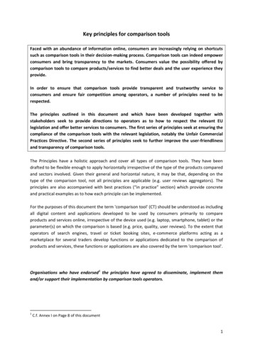
Kriege et al. BMC Anesthesiology (2018) 18:119Page 3 of 9Table 1 Demographics and level of experienceAnesthesiologists (n 65)PCCM staff (n 15)p-value51/11/36/4/50.3330 (6–180 [21–48])36 (6–180 [24–96])0.43 10 applications6 (9%)00.22 50 applications21 (32%)4 (26%)0.67 100 applications26 (40%)2 (13%)0.05 100 applications12 (19%)9 (61%)0.001 5 kg37 (57%)15 (100%)0.001 10 kg62 (95%)15 (100%)0.39 20 kg65 (100%)15 (100%)1.0 1000 254 (6%)5 (33%)0.002 5013 (22%)1 (7%)0.22 10048 (74%)9 (60%)0.28 5000 1002 (3%)4 (27%)0.001 50015 (23%)5 (33%)0.4Provider ncePractice experience (months)Airway management in children ( 6 y)Airway management in infants/childrenExperience in VL (applications)Experience in DL (applications) 100031 (48%)4 (27%)0.14 100017 (26%)2 (13%)0.29Data are presented as medians (range [IQR] and absolute numbers (proportion)and two airway scenarios, each participant performed atotal of 8 intubations.Setting and interventionsThe study was conducted in an antechamber of the ORPICU with the high-fidelity simulator positioned at thehead of a stretcher (Fig. 1). Participants were allowed to adjust the stretcher or chair to a comfortable height. Apediatric high-fidelity Simulator SimBaby (Laerdal Medical, Stavanger, Norway) was built to resemble a 65-cm longhealthy baby and was capable of creating easy and difficultairway situations. The SimBaby is a realistic airway that includes crying and gurgling sounds, cyanosis, tongue edema,pharyngeal obstruction, chest excursion, and breath sounds.In relation to a real infant derived from growth charts ofthe WHO, this length corresponds to that of a 6- to8-month-old infant when looking at the area between the5th and 95th growth percentile [9, 10]. We evaluated thedevices under two airway scenarios as follows: (i) normal(NA) and (ii) difficult intubation condition (DA) createdthrough tongue edema and a cervical collar (Fig. 1; SAM Splint , SAM Medical Products, Wilsonville, OR, USA) toFig. 1 Standardized position of the SimBaby with a shoulder roll toelevate the shoulders and a soft donut to stabilize the head
Kriege et al. BMC Anesthesiology (2018) 18:119reduce the mouth opening from 2.6 mm before to 1.5 mmafter adjustment of an extrication collar to inhibit neckmovement.We chose to compare two video laryngoscopes thatuse a hyperangulated blade with two conventional directlaryngoscopes. The King Vision Pediatric aBlade (KV)(Ambu GmbH, Bad Nauheim, Germany) is a novelvideo laryngoscope used in a clinical setting in pediatricpatients and has not been evaluated under various airway conditions. The KV has a built-in video screen andis available in three pediatric sizes, #1–3 ( #2 has achanneled and standard disposable blade with a 37.8-degree angulated blade). In this study, we used a KV #2standard blade (Fig. 2). The C-MAC D-Blade Ped (DB)(Karl Storz , Tuttlingen, Germany) is a compact systemconsisting of a monitor, electronic module and interchangeable video laryngoscope blade. The DB has an increased curvature with a 75-degree blade angulation.The manufacturer recommends its use in infants orsmall children weighing 9.8 to 22 kg. The DB was specifically developed to manage anatomically difficult airwayconditions such as those encountered in Down syndrome or Pierre-Robin syndrome. The conventional direct laryngoscopes included the Miller straight blade #1(MiB) and a Macintosh curved blade #2 (MaB) with astandard laryngoscope handle for pediatric patients. AllFig. 2 In order from top to bottom: KV #2 standard blade, DB,Macintosh blade #2 and Miller blade #1Page 4 of 9sizes of the blades were tested prior to the study; theyare shown in Fig. 2.Per the recommendation of the manufacturer of theSimBaby, all intubations were performed with a 3.5-mminternal diameter cuffed endotracheal tube (ET). Duringintubation using one of the video laryngoscopes, a malleable stylet was inserted into the ET, which was angulatedinto a hockey-stick shape (distal end of ET angulated90 ). When using a conventional direct laryngoscope,the rigidity of the ET was provided by a malleable stylet(Mallinckrodt satin slip, 2.6 mm). The SimBaby wasplaced in a standardized position, and a shoulder rolland a soft donut-shaped foam headrest were used tosupport the head in all cases (Fig. 1).Methods of measurementsDemographic analysis was collected for all participantsconcerning the clinical level of airway management experience with children 5 years of age, the time since theirmost recent intubation in each age group (newborns, infants, and small children) and experience with video laryngoscopy (VL) and conventional direct laryngoscopy in realpatients [11]. Experienced users (consultants) were defined as those with performance experience in pediatrictracheal intubation exceeding 100 infants. Inexperiencedusers were defined as those with experience in less than100 tracheal intubations in infants [6].The time interval between the blades passing the gumsto confirmation of the best glottic view was recorded asthe time to best view (TTBV). The participant announced the Cormack & Lehane Classification (C&L)and the percentage of glottic opening (POGO) scoreonce the best glottic exposure was obtained [12, 13]. External laryngeal manipulations (ELMs) [14], such as theBURP (backwards, upwards and rightwards pressure)maneuver, can be applied to improve the view of theglottis to achieve a C&L I or II. The time to place (TTP)the ET was defined as the time when the first blackmark on the ET was threaded between the vocal cords.The time to ventilation (TTV) in seconds (s) was defined as the time from when the blade tip passed thegums of the SimBaby to the point of confirmation of thefirst visible chest rise of the simulator. Time was measured using a stopwatch. Study personnel were positioned on the right site of the SimBaby.Failed intubation was defined as follows: a) an elapsedintubation time of more than 40 s in accordance withthe current recommendations and in the time period ofpreviously published data [6, 15, 16]; b) failed trachealplacement of the ET (i.e., esophageal); and c) removal ofthe device/repositioning from the oral cavity without advancing the ET. If the first attempt failed, the providermade a second laryngoscopy attempt with the same device. A total of two laryngoscopy attempts were allowed.
Kriege et al. BMC Anesthesiology (2018) 18:119If DL failed, the clinician changed to a prescribed rescuetechnique with KV. If VL failed after two attempts, theclinician was advised to proceed with DL. The limitationof two intubation attempts and choice of an alternativetechnique has been recommended in several studies andwas chosen in accordance with clinical standards [17].After each intubation, the physician was asked to ratethe degree of difficulty of intubation using a 5-pointLikert scale (1 very easy to 5 very difficult) for eachblade [18].Outcome measuresOur primary study objective was to determine whetherthere was a difference in TTV, demonstrated by bilaterallung inflation of the manikin with positive pressure viabag-valve ventilation, between the KV and the DB, MiBand MaB. Secondary outcomes were the TTBV and theTTP, visualization of laryngeal structures using the C&LClassification and the POGO score, FPA, the use ofELMs and the degree of difficulty.Statistical analysisA statistical analysis of the data was subsequently undertaken. The Shapiro-Wilk test was used to test the assumption of normal distribution (p 0.1). Normallydistributed data are presented as the means (SD) andwere analyzed using an independent t-test for unequalvariances. Non-normally distributed interval and ordinaldata are reported as medians (interquartile range [IQR]);results were compared among groups using the Wilcoxon-Mann-Whitney test. Categorical variables are presented as counts; results were evaluated using thechi-square test. TTV data were compared betweengroups using the log-rank test. All recorded data weredocumented using a controlled case report form.Devices were randomized to treatment groups usingthe GraphPad QuickCalcs Web site: http://www.graphpad.com/quickcalcs/randmenu (accessed January 2015). GraphPad Prism (Vers. 6.0 for MAC; GraphPad Software, SanDiego, CA, USA) was used for all statistical analyses. Differences in TTV were considered statistically significant if thep-value was less than 0.05.ResultsCharacteristics of study subjectsFrom July to August 2017, a total of 80 physicians (anesthesiologists, n 65; PCCM staff, n 15) eligible to participate in this study were included (Table 1). These 80participants performed 640 intubation attempts. The faculty of PCCM had more experience in airway management in children less than 6 years of age (p 0.001) andinfants with a weight 5 kg (p 0.001). Otherwise,the anesthesiologists had more overall experience inVL ( 25 applications prior to the study; p 0.002)Page 5 of 9and DL ( 100 applications prior the study; p 0.001)compared to PCCM. Demographics and the level ofexperience of all subjects are presented in Table 1.Main resultsFor the primary outcome, the median duration for TTVwas shorter for the KV at 13 s [12–15] than for the MaBat 14.5 s [13–16], the DB at 14.5 s [13–16], and the MiBat 16 s [14–19]; p 0.001) in the NA scenario. In DA,the KV similarly significantly shortened TTV to 14 s[13–16], whereas TTV was 23 s using the MaB [20–26],19 s using the DB [16–21], and 27 s using the MiB [22–31]; p 0.001). Both groups (anesthesiologists andPCCM staff) were similar with respect to the time requiredto obtain TTV with all four devices (p 0.5). Furthermore,TTV was shorter when using the hyperangulated blades forboth experienced and inexperienced physicians in the NA/DA scenario (p 0.001). Study outcomes for the variousblades are outlined in Table 2 for the NA scenario and inTable 3 for the DA scenario. Figure 3 present theKaplan-Meier plots of the TTV with all four devices in theNA scenario and DA scenario, respectively.The overall intubation success rates were higher forthe KV 80/80 (100%) in both airway scenarios performedby anesthesiologists and PCCM staff (p 0.001). Thenumber of attempts with each device is listed in Table 2for the NA scenario and in Table 3 for the DA scenario.A proportion of 8/320 (2.5%) participants had 2 failedattempts with the DL in the DA scenario (p 0.001). Allwere successfully intubated with the KV. The reasonfor a second attempt in the NA scenario was a timeout 40 s in 3/320 (0.9%) cases, and the reasons fora second attempt in the DA scenario were a timeoutin 28/320 (9%) of the trials and removal of the bladein 17/320 (5.3%) of the trials (p 0.1).In the NA scenario, we obtained a better view of theglottis with hyperangulated blades than with the conventional laryngoscopes (p 0.001). In the DA scenario, theKV enabled a better glottic view (C&L I) 80/80 (100%)than was achieved using the DB 74/80 (92%), MaB 61/80(76%) and MiB 52/80 (65%; p 0.001).After completing the series of simulated intubations,the participants rated the degree of difficulty in the NAscenario as lower for the DB than for the MaB, MiB andKV (p 0.001). In contrast, in the DA scenario, the KVwas rated easier to use compared with the DB, MaB orMiB (p 0.001).DiscussionIn the present study, a shorter TTV was achieved usingvideo laryngoscopes with a hyperangulated blade in agroup of 640 intubation attempts in an infant DA. Inparticular, TTV was shorter in both airway situationswhen applying the novel KV. The FPA, visualization of
Kriege et al. BMC Anesthesiology (2018) 18:119Page 6 of 9Table 2 Study results by laryngoscope blades in the NA scenariop-values of Pairwise DifferencesOutcomesMaBDBTTBV5.5 [5–7]5 [4–6.5]TTP10.25 [9–11.5] 10 [9–11.5]TTV14.5 [13–16]MiBKV7 [5–8]5 [3.5–6]DB vs. MaB DB vs. MiB KV vs. MaB KV vs. MiB KV vs. DBTime a Sequences (s)12 [10–14.5] 9.5 [8.5–11]14.5 [13–16] 16 [14–19]13 [12–15]0.01 0.001 0.001 0.0010.020.85 0.0010.02 0.0010.0020.750.0010.002 0.001 0.001 0.990.150.31No. attemptsb1st80/80 (100%)79/80 (98%) 78/80 (97%) 80/80 (100%) 0.310.562nd01/80 (2%)2/80 (3%)00.56Failed0000C&L 1/2/3b67/13/078/2/057/23/079/1/00.007 0.0010.001 0.001 0.99POGO (%)a90 [90–100]100 [100]95 [80–100]100 [100] 0.001 0.001 0.001 0.0010.840.02 0.0010.090.0020.320.44 0.001 0.0010.9 0.001Glottic viewbELM5/80 (6%)0/80 (0%)11/80 (14%) 1/80 (1%)BURP006/11 (54%)1/1 (100%)Neck extension5/5 (100%)07/11 (63%)01 [1–1]1 [1–2]1 [1–2]Degree of a difficulty (1–5) 1 [1–2]aMedians and [IQR] are shown. Pairwise differences among devices were evaluated using the signed-rank testbAbsolute numbers (proportion) are shown. Pairwise differences among devices were evaluated using the chi-square testThe demographics of the participants were comparableto those in other studies investigating the MaB, DB orMiB [6, 10, 12, 20–22] in pediatric airways. In contrast toother studies comparing experienced and inexperiencedpractitioners [6, 9–11, 19, 21, 22], this study demonstratedthat TTV was similar between the groups. However, inthis study, there were no institutional differences in FPAor TTV among the blades or airway scenarios. This likelythe glottis and subjective assessment in this comparativestudy were superior to those of conventional directlaryngoscopy. To date, only one study compared thenovel KV with the traditional MiB in elective surgeriesperformed in children 2 y of age [19]. This study isone of the first studies comparing the KV with anotherhyperangulated blade and direct laryngoscopy in a simulated infant airway.Table 3 Study results by laryngoscope blades in the DA scenariop-values of Pairwise DifferencesOutcomesMaBDBMiBKVDB vs. MaB DB vs. MiB KV vs. MaB KV vs. MiB KV vs. DBTTBV11 [8.5–14]8 [6.5–9]13.5 [9–15.5]6.5 [5–7] 0.001 0.001 0.001 0.001 0.001TTP17.5 [15–20] 14 [11–15]20.5 [16.5–24] 10 [9.5–12] 0.001 0.001 0.001 0.001 0.001TTV23 20–26]27 [22–31] 0.001 0.001 0.001 0.001 0.001 0.001 0.0010.01Time a Sequences (s)19 [16–21]14 [13–16]bNo. attempts1st61/80 (76%) 74/80 (92%) 52/80 (65%)80/80 (100%) 0.004 0.0012nd16/80 (20%) 6/80 (8%)23/80 (29%)00.290.26Failed3/80 (4%)05/80 (6%)019/60/161/19/07/70/377/3/0 0.001 0.001 0.001 0.001 0.001POGO (%)40 [30–60]90 [90–100] 40 [30–60]100 [90–100] 0.001 0.001 0.001 0.001 0.001ELMb3/80 (4%)1/80 (1%)4/80 (5%)0/80 (0%)0.310.170.080.040.32BURP0000Neck extension3/3 (100%)1/1 (100%)4/4 (100%)1 [1–2]3 [3–3] 0.001 0.001 0.001 0.0010.001Glottic viewC&L 1/2/3baDegree ofa difficulty (1–5) 2 [2–3]a1 [1–1]Medians and [IQR] are shown. Pairwise differences among devices were evaluated using the signed-rank testbAbsolute numbers (proportion) are shown. Pairwise differences among devices were evaluated using the chi-square test
Kriege et al. BMC Anesthesiology (2018) 18:119Page 7 of 9Fig. 3 Kaplan-Meier plots of the time to ventilation for all four devices in (left) the normal airway situation and (right) a difficult airwayreflects a familiarity with the direct and video laryngoscopy techniques. Furthermore, every participant only had5 attempts with the novel KV or DB prior to the study.A number of new devices have been introduced tofacilitate tracheal intubation in pediatric patients with normal and difficult airways, with reports describing varyingsuccess rates and experience in pediatric airway management [10, 11, 22]. In these experiments, manikins were usedin various simulation scenarios, including tracheal intubation. In addition to their use in training, they have alsobeen used in various clinical research projects to demonstrate the efficacy of one device over another. SimBaby hasbeen used to teach clinical and decision-making skills whilemimicking patient care scenarios [9, 10, 23]. Tracheal intubation using a video laryngoscope might be easier to learnthan conventional direct laryngoscopy [8, 9]. The successrates vary considerably, and the proportion of patients inwhom the glottic opening can be visualized but the ETcannot be inserted into the trachea is high [20, 21, 24]. Several study results have noted that intubation time is longerwhen using a video laryngoscope with a hyperangulatedblade [6, 9, 21]. Surprisingly, TTV was significantly shorterfor the novel KV blade in the NA (1.5 s) and DA (13 s) scenarios than for the other devices, which influence the occurrence of systemic complications (e.g., hypoxemia). TTV waslonger for the MaB and MiB than for hyperangulated bladesrelative to the results of studies looking at two conventionalblades; however, the values were within the range of published data [6, 9, 23]. In two studies that evaluated childrenaged 2 years of age, TTV was shorter for the MiB [19, 22]than for the KV. Straight blades are often recommended foruse in neonates and infants to lift the relatively large andfloppy epiglottis [22]. These blades avoid a situation inwhich the hyoid bone cannot be displaced forward and theepiglottis continues to obstruct a view of the larynx. In summary, a good laryngeal view with the intubating device didnot equate with ease of intubation. Otherwise, a curvedMaB provides more room in the oropharynx to maneuverthe ET. The time to visualization of the glottis and successful ventilation were also comparable to the results reportedin another study [19]. However, the measurements differedbetween these studies in which the practitioner took hold ofthe handle of the device until the cuff of the ET was inflated[9, 20, 24] or until confirmation of the first inflection of theend-tidal capnogram on the anesthesia respirator [6, 19]. Inthree studies, time measurement stopped when successfullung expansion occurred [9, 20, 21]. All intubations weresuccessfully performed using the KV on FPAs. ComparingTTV or FPA using the DB is difficult because the literatureis quite sparse with regard to the use of the DB in pediatricpatients or in a suitable manikin. The prolonged intubationtime and lower success rate in DB and conventional directlaryngoscopes might be explained as follows: first, the anterior view angle of 55 degrees achieved using the KV differsfrom the half-moon shaped DB (embedded optical lens withaperture angle of 80 ); furthermore, the length differs between the two hyperangulated blades (KV, 7 cm vs. DB,10 cm). Additionally, it is generally thought that the position of the head has an influence on the laryngeal view[6]. We used a standardized position for the SimBaby inwhich a shoulder roll elevated the shoulders and a smalldonut was used to support the head. The goal was to alignthe oral, pharyngeal, and tracheal axes to facilitate trachealintubation.As a secondary outcome, visualization of the glottis wassignificantly easier with VL in both scenarios. These dataare consistent with previously published data in simulatedpediatric patients in whom the laryngeal view was found tobe better with VL [6, 9, 10, 19]. In contrast to other studiescomparing hyperangulated blades with DL, we found ahigher FPA and overall success rate when using the KV orDB [11, 15, 21]. In fact, a higher FPA was shown with theKV in DA compared to the first study that evaluated theKV in a normal pediatric airway [19]. These data are comparable to that in study with manikins where the resultsvary with the operator’s prior experience and familiaritywith the equipment, institutional preferences, and how wellthe manikin simulates a real patient [11, 20]. Finally, whenconsidering the number of ELMs, which were slightlyhigher with direct laryngoscopy, and the participants’
Kriege et al. BMC Anesthesiology (2018) 18:119subjective difficulty scores, which were also higher for thesedevices, our data agree with those reported on the management of adult patients [18]. This fact may be derived fromthe absence of prior experience of the subjects with the DBand KV or with the specific characteristics of the conventional blades and the infant manikin’s airway anatomy.Page 8 of 9Additional fileAdditional file 1: Availibility data. (XLSX 99 kb)AcknowledgementsThe authors wish to thank Gerrit Toenges (Institute of Medical Biostatistics,Epidemiology and Informatics, University Medical Center of the JohannesGutenberg University, Mainz, Germany).Study limitationsOur study has several limitations. First, we used two different conventional and two different hyperangulated videolaryngoscopy blade sizes in an infant simulator. We cannotassure that the simulated clinical conditions truly reproducethe real patient’s conditions. Second, this comparative studymight have been limited by the heterogeneous experiencesof the providers (e.g., experiences in anesthesia practice orpediatric critical care medicine and in video laryngoscopytechniques). We feel, however, that this approach is moreclinically relevant than are the study results, reflecting theexperience of a small team of a
tween direct or indirect laryngoscopy using the video laryngoscopy technique. Direct laryngoscopy requires alignment of the oropharyngeal-laryngeal axes, and these differences restrict optimal visualization of the glottis. The management of a difficult airway in infants with a weight of less than 10 kg has always been a challenge [4]. Fortu-