Transcription
Prevention and Management ofAcute and Chronic WoundsFederal Bureau of PrisonsClinical Practice GuidelinesMarch 2014Clinical guidelines are made available to the public for informational purposes only. TheFederal Bureau of Prisons (FBOP) does not warrant these guidelines for any other purpose, andassumes no responsibility for any injury or damage resulting from the reliance thereof. Propermedical practice necessitates that all cases are evaluated on an individual basis and that treatmentdecisions are patient-specific. Consult the FBOP Health Management Resources Web page todetermine the date of the most recent update to this document:http://www.bop.gov/resources/health care mngmt.jsp
Federal Bureau of PrisonsClinical Practice GuidelinesPrevention and Management of Acute and Chronic WoundsMarch 2014Table of ContentsPURPOSE AND USE OF THESE GUIDELINES .1BASIC SUPPORTIVE WOUND CARE ALGORITHM .2TABLES FOR BASIC WOUND CARE ALGORITHM.3TABLE 1.1: Wound History.3TABLE 1.2: Wound Assessment .3TABLE 1.3: Basic Vascular Exam for Wounds on the Lower Extremities .3TABLE 2: Wound Type and Mechanism of Injury .4TABLE 3: Wound Color, Goals, and Treatment for Healing a Wound .5TABLE 4: Assessment of 2-Week Healing Goal .6TABLE 5: Topical Antiseptics with Low Cytotoxic Profiles for EmpiricTreatment of Critical Colonization .6STEPS 1–4 OF BASIC WOUND CARE ALGORITHM .7STEP 1A: Perform a Basic Initial Wound Assessment. .7Wound History .7Wound Assessment .7Basic Vascular Exam for Wounds on the Lower Extremities .9STEP1B: Identify the Type of Wound. . 10STEP 2A: Alleviate the Mechanism of Injury for the Wound Type. 11STEP 2B: Establish Treatment Plan and Healing Goal Based on the Wound Bed Color(s). . 11Key Principles of Topical Therapy . 11Therapy and Healing Goals Based on Wound Bed Color . 12Black Wounds . 12Mostly Mixed Color Wounds: Red/Pink/Yellow/Brown/Black . 13Red or Pink Wounds . 15STEP 3: Evaluate 2-Week Healing Goal. . 16Healing Goal for Mixed Color Wounds: Slough Reduction of 25% in 2 Weeks . 16Healing Goal for Red/Pink Wounds: Size Reduction of 25% in 2 Weeks . 16STEP 4: Review Wounds Not Meeting Healing Goals. . 18i
Federal Bureau of PrisonsClinical Practice GuidelinesPrevention and Management of Acute and Chronic WoundsMarch 2014COMMON WOUND TYPES . 20Pressure Ulcerations . 20Risk Factors. 20Mechanism of Injury . 20Characteristics . 20Diagnostic Tests . 21Interventions to Alleviate Mechanism of Injury . 22Prevention strategies for the spinal cord injury population: . 23Specific Topical Treatment Recommendations . 24Arterial Insufficiency Wounds . 24Risk Factors. 24Mechanism of Injury . 24Characteristics . 25Diagnostic Tests . 26Interventions to Alleviate Mechanism of Injury . 27Specific Topical Treatment Recommendations . 28Venous Insufficiency Wounds . 28Risk Factors. 28Mechanism of Injury . 28Characteristics . 28Diagnostic Tests . 29Interventions to Alleviate Mechanism of Injury . 30Specific Topical Treatment Recommendations . 33Neuropathic Disease (diabetic foot ulcers) . 33Risk Factors. 33Mechanism of Injury . 33Characteristics . 34Diagnostic Tests . 35Interventions to Alleviate Mechanism of Injury . 36Specific Topical Treatment Recommendations . 38Mixed Lower Extremity Disease . 38ii
Federal Bureau of PrisonsClinical Practice GuidelinesPrevention and Management of Acute and Chronic WoundsMarch 2014GENERAL CONCEPTS OF WOUND HEALING SCIENCE . 40General Healing Physiology . 40General Therapy Goals: Healing, Stabilization, and Palliation . 41Colonized versus Infected Wounds . 42Wound Culture Techniques . 44Use of Antibiotics for Wound Care: Topical and Systemic . 45Use of Topical Antiseptics. 45Nutrition Recommendations for Wound Healing . 46USE OF ADVANCED THERAPIES AND SPECIALTY EQUIPMENT . 47Negative Pressure Wound Therapy (NPWT) . 47Clinitron and Other Special Sleep Surfaces and Specialty Beds . 48Hyperbaric Oxygen (HBO) Therapy . 48REFERENCES AND RESOURCES . 49APPENDIX 1: PHASES OF HEALING. 51Hemostasis Phase . 51Inflammatory Phase . 51Proliferative Phase . 52Remodeling Phase . 53APPENDIX 2: BASIC DRESSING TYPES. 54Saline-Moistened Gauze . 54Hydrogels . 54Hydrocolloids . 54Alginates and Hydrofibers . 55Foam Dressings . 55Transparent Film Dressings . 56APPENDIX 3: DURABLE MEDICAL EQUIPMENT (DME) RESOURCES . 57APPENDIX 4: PATIENT EDUCATION HANDOUTS (ENGLISH) . 58APPENDIX 5: PATIENT EDUCATION HANDOUTS (SPANISH) . 67iii
Federal Bureau of PrisonsClinical Practice GuidelinesPrevention and Management of Acute and Chronic WoundsMarch 2014PURPOSE AND USE OF THESE GUIDELINESThe Bureau of Prisons (BOP) Clinical Practice Guidelines for the Prevention and Managementof Acute and Chronic Wounds reflect the collaborative work of a multidisciplinary taskforceincluding dietitians, midlevel providers, nurses, pharmacists, physical therapists, and physicians,as well as health care professionals with advanced certification in wound care. This document provides guidance on the prevention and treatment of common types ofwounds, including pressure ulcers, diabetic foot ulcers, venous insufficiency ulcers, andarterial insufficiency ulcers. It is not intended to provide guidance about all types of wounds or skin ulcerations.Instead, guidance regarding timely consultation is provided. It is also not intended for the management of acute traumatic wounds, such as crushinjuries, or skin lesions that are primarily dermatologic in nature, e.g., rashes, MRSAlesions, and hidradenitis suppurativa (HS). The focus of these guidelines is to optimize treatment outcomes in the correctionalenvironment. The algorithm that is provided to help practitioners meet these goals isorganized around the concept of “basic supportive wound care”—a phrase coined by thegroup to encompass the principles that best support the body to heal itself. The algorithm isnot intended to replace a thorough evaluation of the patient’s health care history and needs. Guidance regarding durable medical equipment (DME), consultation, and referral isincluded. Terminology: Although the term antimicrobial is used in wound care to identify topical agents that arenot antibiotics, for the sake of clarity, these guidelines use the term antiseptic. Eschar is a term normally used for dry necrotic tissue of various colors. In theseguidelines, however, the phrase dry necrotic tissue is used in lieu of this term. Slough isa term normally used for moist yellow necrotic tissue, which is more amenable than drynecrotic tissue to various forms of debridement.1
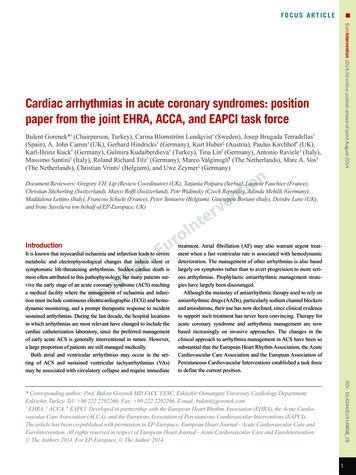
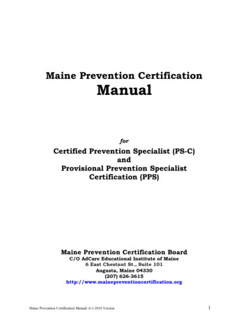
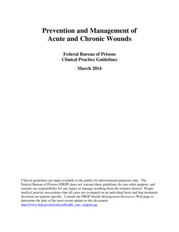
Federal Bureau of PrisonsClinical Practice GuidelinesPrevention and Management of Acute and Chronic WoundsMarch 2014BASIC SUPPORTIVE WOUND CARE ALGORITHMPatient Presents with WoundSTEP 2STEP 1A. Alleviate the mechanism of injury foridentified wound type. See TABLE 2A. Perform a Basic Initial Wound Assessment: Wound History Wound Assessment Basic Vascular Exam for Wounds on LowerExtremities See TABLES 1.1 –1.3B. Establish treatment plan and healinggoal based on the wound bed color(s). See TABLE 3B. Identify type of wound:For common wound types, see Table 2 and thespecific sections on: Pressure Ulcerations Arterial Insufficiency Venous Insufficiency Neuropathic Disease (diabetic foot ulcers)Meets 2-wkgoalSTEP 3Evaluate 2-week healing goal: Re-assess wound. See TABLE 1.2 25% slough or size reduction goal met? See TABLE 4IMPORTANT NOTE: Consider prompt referralfor pressure ulcers thatpresent with cellulitis. Consider immediatereferral for any foot or toewound that presents withcellulitis, abscess,gangrene, or deepulceration.Does NOT meet2-wk goalSTEP 4Review wounds not meeting healing goals: Wound type/mechanism of injury and basic interventions Patient adherence to basic interventions Consider empiric treatment with topical antiseptics with lowcytotoxic profiles for critical colonization See TABLE 5 Consider additional diagnostic studies: Biopsy for tissue culture and/or pathology Serum ESR/CRP & imaging if suspect osteomyelitis Vascular study/consult if suspect arterial disease Consider consultation with wound care specialist andRegistered Dietitian if healing goals not met after 4 weeks.2
Federal Bureau of PrisonsClinical Practice GuidelinesPrevention and Management of Acute and Chronic WoundsMarch 2014TABLES FOR BASIC WOUND CARE ALGORITHMTABLE 1.1: W OUND HISTORYOnsetWhen did it first occur? Is it recurrent? What is the patient’s description of the cause ofthe wound?Prior Treatment andDiagnostic WorkDressings? Antibiotic use? Offloading or prevention strategies to alleviate mechanism ofinjury? Diagnostic tests? Previous consultations/referrals?Past PainPast pain and pain level related to the wound(s)? Interventions tried for relief?Effectiveness of interventions?TABLE 1.2: W OUND ASSESSMENTParametersDescriptorsLocationAnatomical siteSize/VolumeL x W x D (cm) (L x W for wounds without depth)Always measure in centimeters (cm) the longest measure for each axis: Length: 12–6 o’clock measure in cm Width: 9–3 o’clock measure in cm Depth: deepest point in cm If no depth, document “no appreciable depth.” If wound covered with slough/dry necrotic tissue, document as “indeterminate.” Note any tunneling or undermining.Wound BedEstimate percentage of colors (e.g., black, brown, yellow, grey, red, green).Drainage Surrounding Skin Intact or not intact? Color? Is there a palpable temperature change? How does it feel to palpation: supple (normal), soft (fluctuant),or hard (indurated)? Does it blanch?Current PainLocation, scale, quality, qualities, onset, duration, exacerbating/relieving factors,commentsBarriers to HealingIntrinsic: Ability to comprehend and understand instructions Any physical limitations or mobility issues that may affect healing Willingness to be an active participant in care and treatmentExtrinsic: Equipment not available Institution specific limitations: building, housing, terrain challenges Correctional/security challengesAmount (none, scant, moderate, or copious)ColorOdor (none, mild, moderate, or strong).Moisture balance (surrounding skin is not wet; dressing is not adheredto wound bed). Is the drainage well contained by the dressing?For more details, see discussion of Wound Assessment under Step 1A.TABLE 1.3: BASIC V ASCULAR EXAM FOR W OUNDS ON THE LOWER EXTREMITIESAppearance of Skin Color/discoloration Edema Distribution of hairVascular Assessment Skin temperature Capillary refill (in seconds) Palpation of dorsalis pedis andpost-tibial pulses Presence of dependent rubor Pallor elevation testFor more details, see discussion of Basic Vascular Exam under Step 1A.3
Federal Bureau of PrisonsClinical Practice GuidelinesPrevention and Management of Acute and Chronic WoundsMarch 2014TABLE 2: WOUND TYPE AND MECHANISM OF INJURYWound TypeMechanism of InjuryBasic InterventionsPressureUlcerationsPressure over ulcerationOffload or limit pressure over the area around theulcer (sitting and/or lying). Sleep surface andwheelchair cushion must be in accordance with CMScriteria. Provide a shower cushion, when needed. If cellulitis present, consider prompt referral fortreatment. If ulcer tunnels near the bone, review STEP 4 ofalgorithm: Suspect osteomyelitis.ArterialInsufficiencyDecreased arterial bloodflow in limbLimit constriction and elevation of affected area.Keep warm and protect from injury (e.g., thick socks,extra layer of clothing). External heating devices arenot recommended due to potential for burns. Providepadding over the ulcer (e.g., extra layers of gauze). If ulcers are on toes or heels, may need awheelchair and wound shoe to offload wounds. If cellulitis, abscess, gangrene, or deep ulcerationis present, consider immediate referral fortreatment and amputation prevention.VenousInsufficiencyVenous congestion/edemaShort term, consider elasticated tubular bandagesize F (e.g., Medigrip or Tubigrip ); use ACEwraps and TED hose if no other alternatives areavailable or acceptable to the patient. Ambulate totolerance. Consider consultation (e.g., with physicaltherapist; wound or vascular specialist) for safe,long-term compression.NeuropathicDisease(diabetic footulcers)Change in foot sensationand/or structure (poorfitting footwear)Maintain good glycemic control. Recommend limitedweight bearing on affected foot; may need awheelchair and wound-healing shoe (not the sameas a diabetic shoe) to offload wounds. Callousdebridement by qualified provider may be needed.Once resolved, recommend medical footwear andfoot care for life. If cellulitis, abscess, gangrene, or deep ulcerationis present, consider immediate referral fortreatment and amputation prevention.Skin InfectionMRSA or other infectionsSee the BOP Clinical Practice Guidelines forManagement of MRSA Infections and forAntimicrobial Stewardship Guidance. If there is aresidual wound after treatment of the infection, thenfollow the Basic Supportive Wound Care Algorithm inconjunction with these guidelines.KEY: critical decision points; potential life or limb threatening issueFor more in-depth review of interventions to alleviate specific mechanisms of injury, see the sectionsunder Common Wound Types which provide more detailed discussions.4
Federal Bureau of PrisonsClinical Practice GuidelinesPrevention and Management of Acute and Chronic WoundsMarch 2014TABLE 3: WOUND COLOR, GOALS, AND TREATMENT FOR HEALING A WOUNDWound Bed Color*Mostly black tissueTreatment**2-Week Healing GoalHealing see “mostly mixed colors” in next row belowStabilization Povidone-Iodine and gauze 1–2times/day.Discuss therapy goal withprovider: healing vs.stabilization See General Therapy Goals: Healing, Stabilization,and Palliation in Section 2 under General Concepts. If black tissue on heels or toes, see caution withblack heels and toes under STEP 2B discussion. For alternatives to iodine, see section on Use ofTopical Antiseptics. If signs of cellulitis on foot or toes, considerimmediate referral.Mostly mixed colors:red/pink/yellow/brown/black25% reduction in yellowsloughRinse with normal saline (NS), and apply NS-moistenedgauze to wound. Cover with gauze and/or ABD, securewith tape OR consider collagenase (Santyl ) 1–2 timesa day, nickel thickness to entire wound. If yellow/ brown/black tissue is dry, considerremoving the dry roof or cross-hatching—by qualifiedprovider—to optimize therapy. See the descriptionof conservative sharp debridement. Healing goals are not calculated until most of the dryyellow/brown/black tissue is removed.Mostly red or pink25% reduction in sizeRinse with normal saline (NS), and apply NS-moistened,gauze to wound. Cover with gauze and/or ABD, securewith tapeHealed/intact skinRemains healed x 2 weeksProtect the wound bed and apply OTC topicalmoisturizer if dry. Continue interventions used to alleviate mechanismof injury and consider long-term interventions formechanism of injury.KEY: critical decision points; potential life or limb threatening issue* For a more in-depth review and examples, see discussion under STEP 2B: Establish Treatment Plan andHealing Goal, Based on the Wound Bed Color(s).** Unless otherwise indicated, dressings are to be changed daily or less often. At each dressing change, thewound is assessed for moisture balance and containment of drainage. Moisture balance is adjusted by addingor subtracting the amount of saline used when applying the dressing. If moisture balance is difficult to establishwith normal saline and gauze, consider using an alternative dressing type listed in Appendix 2, Basic DressingTypes. Notify the provider if unable to establish good moisture balance. For a more thorough discussion, seeKey Principles of Topical Therapy under STEP 2B.5
Federal Bureau of PrisonsClinical Practice GuidelinesPrevention and Management of Acute and Chronic WoundsMarch 2014TABLE 4: ASSESSMENT OF 2-WEEK HEALING GOALWound Re-assessment CriteriaSee Table 1.2 for criteria.Percentage of Slough ReductionOnly for yellow slough: 1 – (current % wound slough/previous % wound slough) % slough reduction %Percentage Size Reduction Calculation1 – (new size/previous size) % size reductionNote that there are some minor differences for the following: Wound without depth Wound with depth Comparing a wound which previously had measurable depth and now does notFor examples of slough reduction and size reduction calculations, see Step 3: Evaluate 2-Week Healing Goal.TABLE 5: TOPICAL ANTISEPTICS WITH LOW CYTOTOXIC PROFILES FOR EMPIRIC TREATMENT OFCRITICAL COLONIZATIONAgentSilvadene 1% cream(silver sulfadiazine)Iodasorb gel 0.9%ointment(cadexomer iodine)FrequencySpecial Instructions2–3 timesa dayNickel thickness to entire wound; avoid letting cream dry out.daily toevery 3 daysNickel thickness to entire wound; changes color as it absorbsdrainage. Light yellow signals time to change. Caution: Colorchange should not be confused for pus.Dose limit: 50g per application or 150 g per week. Caution if patient has sulfa allergy. Caution if patient has iodine allergy. Contraindicated for patients with thyroid disorders,and for pregnant or breastfeeding patients0.25% acetic acid solution(1/4 strength acetic acid)1–2 timesa dayDeliver on moistened gauze and adjust dressing for moisturebalance (not too wet, not too dry). Undiluted solution can cause serious acid-related injury totissue and bone.KEY: potential life or limb threatening issueTreat with one of the above antiseptics for two weeks. If healing goals are met, then return to basictreatment listed in Table 3. If no improvement, consider a different agent listed above in this table.For more in-depth review, see Use of Antibiotics for Wound Care: Topical and Systemic and Use of TopicalAntiseptics.6
Federal Bureau of PrisonsClinical Practice GuidelinesPrevention and Management of Acute and Chronic WoundsMarch 2014STEPS 1–4 OF BASIC WOUND CARE ALGORITHMSTEP 1A: PERFORM A BASIC INITIAL WOUND ASSESSMENT.When performing the initial evaluation of a wound, conduct (1) a wound history and (2) a woundassessment. If the wound is located on the lower extremity, also perform a (3) basic vascularexam for wounds on lower extremities.WOUND HISTORYDetermine from the patient the onset, prior treatments and diagnostic studies, and a review ofpast pain related to the wound. See TABLE 1.1 for more details.WOUND ASSESSMENT The following discussion is summarized in TABLE 1.2.1. Location: Describe the portion of the body affected. Be clear and concise.2. Size/Volume: Determine the length x width x depth (L x W x D) in centimeters.There are a variety of methods to measure the size or volume of a wound. The key is touse the same pattern of measurement to evaluate wound size so that comparisons overtime are valid. The ruler method is simple and available in almost all health care settings. Thewound is visualized as a clock, with 12 o’clock oriented towards the patient’s headand 6 o’clock oriented towards the patient’s feet. Length is measured first, from 12 to6 o’clock. Width is measured next, from 9 to 3 o’clock. Depth is then measured fromthe deepest point of the wound upward to the wound edge. The gentle use of acotton-tipped applicator can be helpful in measuring depth. If the wound bed contains yellow, brown, or black tissue, then depth cannot bedetermined and should be labeled as “indeterminate.” A tunnel or area of undermining is also measured using the clock method: A tunnel is a narrow channel that extends from the edge of the wound into thetissue laterally. For instance, picturing the wound as a clock with 12 o’clockoriented towards the patient’s head, a tunnel at 2 o’clock that extends 3 cm wouldbe documented as “tunneling, 3 cm at 2 o’clock.” A wound can have multipletunnels. Undermining, when a section of tissue is missing under the edge of the wound, isidentified by the starting and ending points, and measured where the underminingextends the farthest. For example, a section of missing tissue that starts at4 o’clock and ends at 8 o’clock, and is 2.3 cm at its furthest measurement, wouldbe documented as “undermining from 4 to 8 o’clock, 2.3 cm. A wound can havemultiple areas of undermining.7
Federal Bureau of PrisonsClinical Practice GuidelinesPrevention and Management of Acute and Chronic WoundsMarch 20143. Wound Bed: Describe the wound bed colors in percentages. Estimation is acceptable.For instance, a wound that is all red would be documented as “100% red.” A wound withmultiple colors might be documented, for example, as “50% red, 30% yellow, and 20%brown.”4. Drainage: Describe the amount of drainage (none, scant, moderate, or copious), color,and the presence of odor (none, mild, moderate, or strong). Also document if there isgood moisture balance; in other words, determine if the wound is not too dry or too wet.A wound is too dry when the dressing adheres to the wound bed. A wound is too wetwhen drainage makes the surrounding skin wet and fragile, otherwise known asmaceration. If moisture balance is difficult to establish with normal saline and gauze,consider using an alternative dressing type listed in Appendix 2, Basic Dressing Types.Note: The drainage amount, color, and odor can indicate increased numbers ofmicroorganisms living on the wound bed (referred to as critical colonization), butthese parameters alone are not diagnostic. The failure to meet healing goalsshould be the primary reason to select topical antiseptic treatment for criticalcolonization. Initial antiseptic treatment options are listed in TABLE 5. Additionalantiseptic treatment options are listed in Use of Topical Antiseptics.5. Surrounding Skin: The condition of the surrounding skin up to 5 cm from the edge ofthe wound is important to evaluate. Assessment features include whether or not the skinis intact, the color, palpable temperature change, and palpable feel such as normal(supple), soft (fluctuant), or hard (indurated).It is normal to find a 1 cm or less band of discolored, warmer, indurated tissue around thewound. It often indicates increased blood flow to support the body’s efforts toautolytically debride any black tissue or yellow slough, and to support granulation tissueformation. A band wider than 1 cm may indicate cellulitis. A white blood count ishelpful to differentiate between cellulitis and an appropriate, localized inflammatoryresponse.Note: In wounds caused by venous insufficiency, the band of erythema is often muchwider than 1 cm. Most often, this is associated with the exaggerated cutaneousinflammatory response common to these types of wounds. A white blood count isespecially helpful in differentiating this condition from cellulitis.6. Current Pain: It is important to evaluate and document a snapshot of the patient’s painassociated with the wound over the last 2 weeks. Assessment characteristics are alignedwith those found in the Bureau Electronic Medical Record (BEMR), and summarized inTABLE 1.2. Proper assessment and subsequent interventions to control pain is an integralpart of wound care. Consequences of Uncontrolled Pain: Pain can significantly affect a patient’s qualityof life, sleep cycle, and psychosocial status. In addition, inadequately treated acutepain can lead to the development of chronic disabling neuropathic pain.Inadequate treatment of pain can also lead to poor wound healing and increasedinfection rates. Acute pain increases catecholamine release, and leads to peripheralvasoconstriction and decreased perfusion of blood to the affected area. The decrease8
Federal Bureau of PrisonsClinical Practice GuidelinesPrevention and Management of Acute and Chronic WoundsMarch 2014in available oxygen inhibits the oxidati
Arterial Insufficiency Venous Insufficiency Neuropathic Disease (diabetic foot ulcers) Patient Presents with Wound STEP 2 A. Alleviate the mechanism of injury for identified wound type. See TABLE 2 B. Establish treatment plan and healing goal based on the wound bed color(s). See TABLE 3 STEP 3 Evaluate 2-week healing goal: Re-assess wound.