Transcription
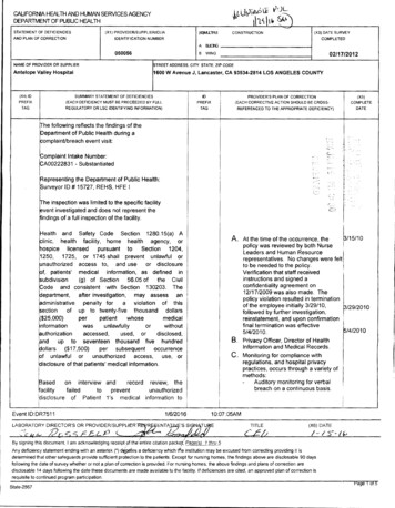
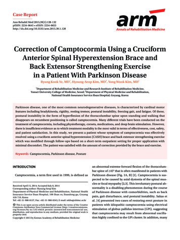
Case ReportAnn Rehabil Med 2015;39(1):128-132pISSN: 2234-0645 eISSN: 28Annals of Rehabilitation MedicineCorrection of Camptocormia Using a CruciformAnterior Spinal Hyperextension Brace andBack Extensor Strengthening Exercisein a Patient With Parkinson DiseaseByung Kook Ye, MD1, Hyoung-Seop Kim, MD2, Yong Wook Kim, MD11Department of Rehabilitation Medicine and Research Institute of Rehabilitation Medicine,Yonsei University College of Medicine, Seoul; 2Department of Physical Medicine and Rehabilitation,National Health Insurance Service Ilsan Hospital, Goyang, KoreaParkinson disease, one of the most common neurodegenerative diseases, is characterized by cardinal motorfeatures including bradykinesia, rigidity, resting tremor, postural instability, freezing gait, and fatigue. Of these,postural instability in the form of hyperflexion of the thoracolumbar spine upon standing and walking thatdisappears on recumbent positioning is called camptocormia. Many different trials have been conducted on thetreatment of camptocormia, including physiotherapy, corsets, medications, and deep brain stimulation. However,there is insufficient evidence as to which treatment modality is the most valid in terms of effectiveness, cost, safety,and patient satisfaction. In this study, we present a patient whose symptom of camptocormia was effectivelyresolved using a cruciform anterior spinal hyperextension (CASH) brace and back extensor strengthening exercisewhich was modified through follow-ups based on a short-term outpatient setting for proper application withminimal discomfort. The patient was satisfied with the amount of correction provided by the brace and exercise.Keywords Camptocormia, Parkinson disease, PostureINTRODUCTIONCamptocormia, a term first used in 1999, is defined asReceived April 15, 2014; Accepted July 8, 2014Corresponding author: Hyoung-Seop KimDepartment of Physical Medicine and Rehabilitation, National HealthInsurance Service Ilsan Hospital, 100 Ilsan-ro, Ilsandong-gu, Goyang410-719, KoreaTel: 82-31-900-0137, Fax: 82-31-900-0343, E-mail: rekhs@nhimc.or.krThis is an open-access article distributed under the terms of the CreativeCommons Attribution Non-Commercial License (http://creativecommons.org/licenses/by-nc/3.0) which permits unrestricted noncommercial use,distribution, and reproduction in any medium, provided the original work isproperly cited.Copyright 2015 by Korean Academy of Rehabilitation Medicinean abnormal extreme forward flexion of the thoracolumbar spine of 45 that is often manifested in patients withParkinson disease (Fig. 1A, B) [1]. Camptocormia is suspected to be caused by axial dystonia of the spinal muscles or focal myopathy [2,3]. This involuntary postural abnormality is a disabling phenomenon during the courseof Parkinson disease with comorbidities, such as backpain, gait disturbance, and postural instability. Sakas etal. [4] presented two cases of restoring erect posture inpatients with idiopathic camptocormia using electricalstimulation of globus pallidus internus (GPi), suggestingthat camptocormia may result from abnormal oscillation highly confined to the GPi cluster. In addition, many
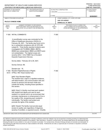
Resolved Camptocormia Using Cruciform Anterior Spinal Hyperextension BraceBAtypes of treatments have been implemented, includingpharmacological therapy as well as physiotherapy andcorsets. However, no effective treatment has been widelyacknowledged yet [5]. Azher and Jankovic [6] reported agood response of levodopa in the motor symptoms of 11patients with Parkinsonism, but with no effect on theircamptocormia. Meanwhile, there are several studies withpromising results of orthosis effect in patients with osteoporotic kyphosis. It was reported that 31 patients withspinal osteoporosis who wore a weighted kypho-orthosisfor six months showed a 11% decrease in their angle ofkyphosis [7]. In two studies published by Sinaki et al. [8]in 2005, groups of patients with spinal osteoporosis wearing a weighted kypho-orthosis with exercise showed animprovement in their kyphotic posture, body balance,gait velocity, and cadence. However, the pathogenesis ofcamptocormia in spinal osteoporosis is quite differentfrom that of Parkinsonism. Studies described above areconducted on patients with osteoporotic camptocormia.Here, we present a case of camptocormia that was effectively corrected in a patient diagnosed with Parkinsondisease using a cruciform anterior spinal hyperextension(CASH) brace and back extensor strengthening exercises.CASE REPORTThe patient was a 70-year-old man who initially visitedthe Department of Neurology at a tertiary training hospital on May 9, 2009 for weakness accompanied by tremorCFig. 1. (A) Hyperflexion of thethoracolumbar spine observedon the initial outpatient visit. (B)Hyperflexion of the thoracolumbar spine resolved by recumbentpositioning. (C) No evidence ofcompression fracture seen onplain X-ray (whole spine lateralview, supine position).in both legs that began in early 2009. He was diagnosedwith Parkinson disease with regular follow-up at an outpatient setting for medication management. Symptomsof involuntary truncal flexion first began in June 2011without response to medication dosage control. He wasreferred to the Department of Physical Medicine and Rehabilitation of our hospital on October 12, 2011. As therewas sustained lumbar flexion during anti-gravity activities but none on recumbent positioning, camptocormia,an involuntary abnormal forward flexion of thoracolumbar spine and anterocollis, was suspected (Fig. 1).The ambulatory ability of the patient was measured by‘Functional Ambulation Category’ (FAC) with category3 and by ‘time up and go test’ at 16 seconds. The patientrefused muscle enzyme level, MRI, and electromyography studies for economic reasons. Moreover, the patient showed no symptomatic response to the followingmedication: Requip (ropinirole 8 mg, once a day, daily,for March 3, 2011 November 16, 2011) and Madopar(levodopa 200 mg and benserazide 50 mg, three times aday, daily, for March 7, 2011 present). Application of abackpack loaded with two standard bricks at 3.8 kg (center of gravity was to located on the level of inferior angleof the scapulae and length of the string was adjusted fortotal contact in order to induce the extension of the trunksimilar to a weighted kypho-orthosis) was intolerable dueto heavy weight (Fig. 2). Therefore, a CASH brace was applied on October 19, 2011 in order to provide a resistivelimitation to the forward flexion of the thoracolumbarwww.e-arm.org129
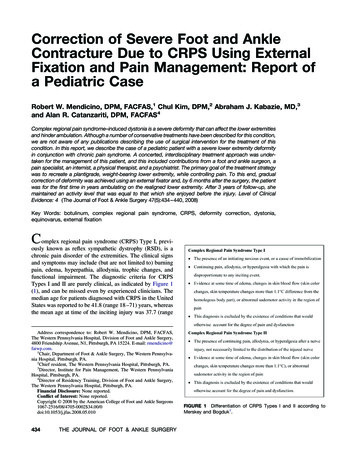
Byung Kook Ye, et al.Fig. 2. Extension of the thoracolumbar spine partiallyachieved with application of a heavily loaded backpack,but the weight was intolerable to the patient.Aspine (Fig. 3). The patient was instructed to perform dailyback and neck extensor strengthening exercises for 30minutes. Through many short-term outpatient visits, theCASH brace was modified to fit the patient without discomfort from any improper pressure (Fig. 4). After fourmonths, the patient was able to walk independently withthe support of CASH brace through holding an involuntary flexion of the thoracolumbar spine. His ambulatory130www.e-arm.orgFig. 3. Camptocormia was still observed after the initialapplication of the cruciform anterior spinal hyperextension brace.BFig. 4. (A, B) Correction of camptocormia after a serial of outpatient-based follow-ups and modifications.ability was also improved. The FAC changed from category 3 to category 5. The time up and go test improved from16 seconds to 9 seconds. The patient has been followedup for 5 months. He remains satisfied with the effect ofthe brace for correcting the uncomfortable postural deformity. In the end, he was able to maintain a good posture without the brace.
Resolved Camptocormia Using Cruciform Anterior Spinal Hyperextension BraceDISCUSSIONThe pathogenesis of camptocormia is not yet wellunderstood. The underlying pathophysiology of camptocormia is suggested to be of either central or peripheralorigins, axial dystonia, or focal myopathy of the spinalmuscles [1,3]. Forward bending of the spine results froma weakness of the paravertebral muscles or over-activityof the abdominal muscles. However, no evidence is available to support either factor in the development of camptocormia.As surgical approaches consisting of deep brain stimulation (DBS) of GPi have corrected the truncal and cervical flexion, the central nervous system involvement,specifically GPi, is the most probable central anatomicallesion for camptocormia. Although interfastigial cerebellum and bilateral midbrain tegmentum play importantroles in the maintenance of erect posture and bipedalism, restoration of erect posture has only been achievedby DBS of the GPi, further suggesting its involvement incamptocormia [4]. However, despite the rapid and dramatic effects of DBS in correcting camptocormia, it islimited due to its major surgical procedure with risks thatmight accompany as any other major neurosurgeries. Inaddition, of 15 cases that have been successfully treatedwith DBS, the average age of patient at onset was 40.57years, and that of surgery was 49.07 years. Therefore,most patients are relatively young. Of these patients, primary patients with a greater number of motor symptoms,including dystonia, showed better results. There was onlyone case of successful treatment among patients abovethe age of 70 years, which is an age group with relativecontraindication of DBS [9]. Antiparkinson medicationsincluding levodopa, dopamine agonists, amantadine andanticholinergics are infrequently mildly beneficial andmostly ineffective [5,10]. Antispastic medications alsohave no obvious benefit. A botulinum toxin injection ismildly effective in four out of nine patients for up to threemonths [6]. It has been suggested that camptocormiamay be caused by myopathic changes [3] due to evidences, such as myopathic electromyographic findings andmyopathic histologic changes. Similar to camptocormia,anterocollis, the dropped head in patients with Parkinsonism, is also closely related with myopathic changes ofneck extensor muscles [2]. The number of patients mentioned in the two studies above was 11 with average ageof 71.45 years, which is relatively older than those in theDBS group. Because of those differences, age is assumedto be a key factor in pathophysiology. Furthermore, therelative contraindication of DBS involves the age groupabove 70 years, and the absolute contraindication of DBSis associated with dementia which explains the cause ofthe difference.Although physiotherapy accompanied by a corset andbrace is feasible, it is often unsatisfactory to patients because corset and braces are uncomfortable due to inappropriate prescription, fitting, or donning. For that reason, we have followed our patient in short-term courseson an outpatient basis and made modifications based onhis feedback. In our case, the patient complained of painaround the sternum. To avoid sternal area pain and discomfort caused by rubbing between the brace and sternum, it is helpful to perform back extensor strengtheningexercises. Although the patient complained of discomfortin the pubic area in this period, the symptom disappeared immediately as the trunk flexion was improved.Our patient is currently able to wear the brace comfortably throughout the day without complaints of excesspressure on any specific site that may lead to pain or discomfort with effective correction of camptocormia during standing and ambulation. Although the effects maybe less dramatic than those achieved by DBS, it seemsvalid to apply a brace before undergoing such proceduresconsidering its safety and cost.In conclusion, despite previous reports on the mediocre effects of applying brace to correct camptocormiain patients with Parkinsonism, we suggest that a CASHbrace with back extensor strengthening exercise can bean effective and satisfactory method of treatment if serialshort-term follow-ups with frequent modifications to thebrace are provided. However, it may be a limitation thatwe report only one case. Therefore, case series or a largetrial should be considered in further studies.CONFLICT OF INTERESTNo potential conflict of interest relevant to this articlewas reported.REFERENCES1. Doherty KM, van de Warrenburg BP, Peralta MC, Si www.e-arm.org131
Byung Kook Ye, et al.2.3.4.5.6.lveira-Moriyama L, Azulay JP, Gershanik OS, et al.Postural deformities in Parkinson’s disease. LancetNeurol 2011;10:538-49.Askmark H, Eeg-Olofsson K, Johansson A, Nilsson P,Olsson Y, Aquilonius S. Parkinsonism and neck extensor myopathy: a new syndrome or coincidental findings? Arch Neurol 2001;58:232-7.Schabitz WR, Glatz K, Schuhan C, Sommer C, BergerC, Schwaninger M, et al. Severe forward flexion of thetrunk in Parkinson’s disease: focal myopathy of theparaspinal muscles mimicking camptocormia. MovDisord 2003;18:408-14.Sakas DE, Panourias IG, Stavrinou LC, Boviatsis EJ,Themistocleous M, Stathis P, et al. Restoration of erectposture in idiopathic camptocormia by electrical sti mulation of the globus pallidus internus. J Neurosurg2010;113:1246-50.Melamed E, Djaldetti R. Camptocormia in Parkinson’sdisease. J Neurol 2006;253 Suppl 7:VII14-16.Azher SN, Jankovic J. Camptocormia: pathogenesis,132www.e-arm.orgclassification, and response to therapy. Neurology2005;65:355-9.7. Pfeifer M, Begerow B, Minne HW. Effects of a new spinal orthosis on posture, trunk strength, and quality oflife in women with postmenopausal osteoporosis: arandomized trial. Am J Phys Med Rehabil 2004;83:17786.8. Sinaki M, Brey RH, Hughes CA, Larson DR, KaufmanKR. Significant reduction in risk of falls and back painin osteoporotic-kyphotic women through a SpinalProprioceptive Extension Exercise Dynamic (SPEED)program. Mayo Clin Proc 2005;80:849-55.9. Reese R, Knudsen K, Falk D, Mehdorn HM, Deuschl G,Volkmann J. Motor outcome of dystonic camptocormia treated with pallidal neurostimulation. Parkinsonism Relat Disord 2014;20:176-9.10. Bloch F, Houeto JL, Tezenas du Montcel S, BonnevilleF, Etchepare F, Welter ML, et al. Parkinson’s diseasewith camptocormia. J Neurol Neurosurg Psychiatry2006;77:1223-8.
the brace for correcting the uncomfortable postural de-formity. In the end, he was able to maintain a good pos-ture without the brace. Fig. 2. Extension of the thoracolumbar spine partially achieved with application of a heavily loaded backpack, but the weight was intolerable to the patient. Fig. 3. Camptocormia was still observed after the initial