Transcription
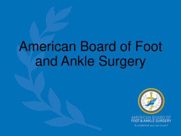
Correction of Severe Foot and AnkleContracture Due to CRPS Using ExternalFixation and Pain Management: Report ofa Pediatric CaseRobert W. Mendicino, DPM, FACFAS,1 Chul Kim, DPM,2 Abraham J. Kabazie, MD,3and Alan R. Catanzariti, DPM, FACFAS4Complex regional pain syndrome–induced dystonia is a severe deformity that can affect the lower extremitiesand hinder ambulation. Although a number of conservative treatments have been described for this condition,we are not aware of any publications describing the use of surgical intervention for the treatment of thiscondition. In this report, we describe the case of a pediatric patient with a severe lower extremity deformityin conjunction with chronic pain syndrome. A concerted, interdisciplinary treatment approach was undertaken for the management of this patient, and this included contributions from a foot and ankle surgeon, apain specialist, an internist, a physical therapist, and a psychiatrist. The primary goal of the treatment strategywas to recreate a plantigrade, weight-bearing lower extremity, while controlling pain. To this end, gradualcorrection of deformity was achieved using an external fixator and, by 6 months after the surgery, the patientwas for the first time in years ambulating on the realigned lower extremity. After 3 years of follow-up, shemaintained an activity level that was equal to that which she enjoyed before the injury. Level of ClinicalEvidence: 4 (The Journal of Foot & Ankle Surgery 47(5):434 – 440, 2008)Key Words: botulinum, complex regional pain syndrome, CRPS, deformity correction, dystonia,equinovarus, external fixationComplex regional pain syndrome (CRPS) Type I, previously known as reflex sympathetic dystrophy (RSD), is achronic pain disorder of the extremities. The clinical signsand symptoms may include (but are not limited to) burningpain, edema, hyperpathia, allodynia, trophic changes, andfunctional impairment. The diagnostic criteria for CRPSTypes I and II are purely clinical, as indicated by Figure 1(1), and can be missed even by experienced clinicians. Themedian age for patients diagnosed with CRPS in the UnitedStates was reported to be 41.8 (range 18 –71) years, whereasthe mean age at time of the inciting injury was 37.7 (rangeAddress correspondence to: Robert W. Mendicino, DPM, FACFAS,The Western Pennsylvania Hospital, Division of Foot and Ankle Surgery,4800 Friendship Avenue, N1, Pittsburgh, PA 15224. E-mail: rmendicino@faiwp.com.1Chair, Department of Foot & Ankle Surgery, The Western Pennsylvania Hospital, Pittsburgh, PA.2Chief resident, The Western Pennsylvania Hospital, Pittsburgh, PA.3Director, Institute for Pain Management, The Western PennsylvaniaHospital, Pittsburgh, PA.4Director of Residency Training, Division of Foot and Ankle Surgery,The Western Pennsylvania Hospital, Pittsburgh, PA.Financial Disclosure: None reported.Conflict of Interest: None reported.Copyright 2008 by the American College of Foot and Ankle Surgeons1067-2516/08/4705-0002 34.00/0doi:10.1053/j.jfas.2008.05.010434THE JOURNAL OF FOOT & ANKLE SURGERYFIGURE 1 Differentiation of CRPS Types I and II according toMerskey and Bogduk1.
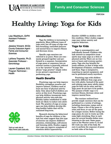
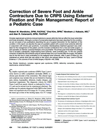
FIGURE 2 Clinical presentation shows the triplanar equinovarus position of the foot and ankle. A. Posteromedial view. B. Anterolateral view.14 – 64) years, and the female to male ratio was 2.3:1 (2– 4).One of the debilitating symptoms of CRPS is dystonia. Inthe lower extremities, an equinovarus deformity of the footand ankle is the most common manifestation of CRPSinduced musculoskeletal contracture (5). Because of theprogressive nature of this symptom, immediate treatment isusually necessary to prevent the development of severedeformity. Treatment options for CRPS-induced dystoniainclude serial casting, aggressive physical therapy, sympathetic ganglion blocks, and adjunctive botulinum toxin injections (6 –16). Following a systematic review of the Medline, Excerpta Medica, and the Cumulative Index of Nursingand Allied Health databases, we were unable to identify areference to the use of gradual correction of the deformityby means of surgical intervention. The patient underwenttendon lengthening, botulinum toxin injections, nerve releases, external fixation application, and spinal cord stimulator implantation. The purpose of this report is to describethe case of a pediatric patient with CRPS-induced dystonia,who was treated surgically with the use of external fixationin order to gradually correct a severe equinovarus contracture. This article also discusses the importance of a multidisciplinary approach to this disease process, including perioperative pain management provided by pain medicinespecialists, as well as contributions from a physical therapist, neurosurgeon, and psychiatrist.Case ReportA 14-year-old girl presented to The Foot and AnkleInstitute of Western Pennsylvania on a referral from a painmanagement specialist at the Western Pennsylvania Hospital. The patient developed a progressive equinovarus deformity of the left lower extremity after she sustained an anklesprain while diving into third base while playing softball.Within 4 months, she had developed severe equinovarusdeformity and was unable to ambulate with weight on herleft lower extremity. During an approximately 1.5-year period before presentation to our practice, the patient hadundergone serial casting, physical therapy that includedcontrast baths, and she used an ankle-foot orthosis, underthe guidance of the previous treating physician. She had alsobeen manipulated under general anesthesia and casted in aneffort to reduce the deformity, however this caused such asevere exacerbation of her pain that she had to be admittedinto intensive care and the cast removed. Because of thesevere nature of the pain syndrome and the resultant equi-VOLUME 47, NUMBER 5, SEPTEMBER/OCTOBER 2008435
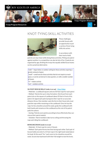
FIGURE 3 Radiographs of the left ankle demonstrate the severity of the deformity and multiple joints involved in the contracture. A. Anteriorview. B. Mortise view.novarus deformity, serious consideration was given to below-the-knee amputation. At the time of the referral to ourpractice, the patient was under the care of a physiatrist,neurologist, psychiatrist, pain management specialist, and aphysical therapist. Moreover, she was receiving lumbarsympathetic ganglion blocks under fluoroscopic guidance,and these resulted in limited periods of partial pain relief.The physical examination revealed a left lower extremitythat was considerably cooler than the contralateral side, andthe skin on the left side demonstrated diminished digital hairgrowth and atrophy. She also exhibited allodynia and hyperesthesia involving the left foot, ankle, and leg. Musculoskeletal examination demonstrated a rigid equinovaruscontracture involving the mid and hindfoot, as well as theankle, with little to no range of motion due to antalgicguarding. Furthermore, the posterior muscle group overpowered the anterior and lateral leg muscles (Figure 2).Radiographic images supported the clinical findings withevidence of an equinovarus deformity of the hindfoot andankle and an adducted position of the forefoot (Figure 3).Additional nerve conduction velocity and electromyographic studies showed no evidence of any other neurological conditions.436THE JOURNAL OF FOOT & ANKLE SURGERYSurgical treatment was carefully planned with cooperation between the foot and ankle surgeon, pain specialist,internal medicine physician, physical therapist, and the psychiatrist. The patient was admitted the day before surgeryand had an indwelling epidural catheter placed, throughwhich ropivacaine and fentanyl were administered under theguidance of the pain medicine service. This was maintainedthroughout the patient’s hospital course. Left lower extremity reconstructive surgery was undertaken the followingday.Because of the nature of the contracture and associateddysfunction of the anterior and lateral muscle groups, common peroneal nerve and posterior tibial nerve decompressions were performed. Additionally, botulinum toxin wasinjected into all of the posterior leg muscles in the region ortheir respective neuromuscular junctions, in an attempt torelieve spasticity and to assist in the subsequent gradualcorrection of the deformity. The gastrosoleus contracturewas the most severe, and the Achilles tendon was lengthened by means of an open technique, although the ankle wasleft in a position of slight equinus in an effort to avoidplacing excessive traction on the musculature of the deepposterior compartment of the leg. An Ilizarov apparatus
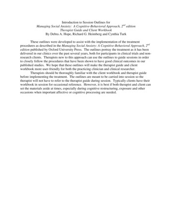
FIGURE 4 Immediate postoperative views of the left lower extremity demonstrate the frame designed to addres the severity of the deformityand the multiple segments that needed to be addressed during the correction. Note that use of pin fixation of the digits helped to preventcontractures distally during the correction. A. Anterior view. B. Posterior view.consisting of a distal tibial block, a half ring at the calcaneus, and a 5/8 ring over the forefoot, was then applied.These were held with the appropriate smooth wire fixation,and then tensioned utilizing Ilizarov tensioning principles.The rings were connected with universal hinges and lengthening rods in an unconstrained fashion so that slow, postoperative adjustments could be made to correct the deformity (Figure 4).The patient’s early postoperative course included continuous infusion of the epidural block, and daily adjustment of theexternal fixation frame to correct the equinovarus deformity.Immediately following application of the external fixationframe, the pain management specialists continued to administer anesthesia throught the epidural. With the adequate resolveof the acute post-operative pain, a neurosurgeon implanted aspinal stimulator in an effort to provide long lasting pain relief.Satisfactory correction was gradually achieved over the next 6weeks at the following rate: medial posterior 8 mm/day,central posterior 5 mm/day, lateral posterior 3 mm/day,lateral foot 2 mm/day, and medial foot 5 mm/day. Thelateral foot distraction was performed to prevent crushingthe cuboid. After the deformity was fully corrected, theframe was left static for an additional 6 weeks, at the end ofwhich the frame was removed in the operating room andadditional botulinum toxin injections were administered toall of the posterior leg muscles. In total, the frame was onfor 3 months. Upon removal of the external fixator, thepatient was casted in the corrected position for an additional2 weeks. The patient began aggressive physical therapy,including ankle range of motion, leg muscle strengthening, and stretching, once the cast was removed. For 6months, she also used an ankle-foot orthosis that blockedplantarflexion and aided in the prevention of recurrentcontracture. The remainder of the postoperative courseproceeded without incident, and after more than 3 yearsof postoperative follow-up, she was ambulating well withno pain in the extremity (Figure 5). She also continued toperform physical therapy modalities in an attempt toprevent recurrent ankle and foot contractures, and shekept biannual visits with her pain specialist for neuromodulation therapy, and with our service for surveillanceof her left lower extremity.VOLUME 47, NUMBER 5, SEPTEMBER/OCTOBER 2008437
DiscussionFIGURE 5 Follow-up clincial non weightbearing views demonstrate the plantigrade position of the foot and the realignment of thedigits. A. Anterior view. B. Medial view.438THE JOURNAL OF FOOT & ANKLE SURGERYDystonia is a well-documented clinical entity that mayhinder ambulation and, particularly in pediatric patientswith CRPS, may lead to progressive equinovarus deformitydespite adequate pain control. Although a wide range oftreatment options exist for the nonsurgical management ofmusculoskeletal contracture (6 –15), including serial casting, physical therapy/manipulation, and injection of botulinum toxin, these methods may not be adequate for somesevere deformities. Moreover, acute surgical correction ofthese deformities can be problematic because of perioperative pain issues that can be very difficult to manage. Gradualcorrection using an external fixation device, combined withadjunctive procedures such as isolated tendon lengtheningand/or botulinum injections, may enable the establishmentof a plantigrade foot in a less acutely traumatic fashion and,as such, may be less stressful to the patient and minimize therisk of exacerbation of the pain syndrome (17–29).Furthermore, the use of prophylactic neurovascular releases can also be useful adjunct procedures to the surgicalcorrection of severe deformity of the lower extremity. Therationale behind this concept hinges on the idea that prophylactic removal of any anatomical structures that maylead to strangulation or impingement of neighboring anatomical structures will decrease the likelihood of subsequentneurological damage and/or ischemia as the deformity isgradually corrected. This concept is particularly applicableto the posterior tibial nerve and its branches in the tarsaltunnel, and the common peroneal nerve near the head of thefibula, in association with the correction of a wide range oflower extremity deformities (20 –23). In the case describedin this report, we implemented this concept by means ofprophylactic release of the neurological components of thetarsal tunnel.Injection of botulinum toxin is a common adjunct intervention used for the treatment of skeletal muscle spasticity(12–16). We used this treatment in an effort to alleviatecontractures of the posterior leg musculature, which appeared to be overwhelming the other muscular compartments of the leg. Botulinum toxin, however, does not usually cause complete resolution of all spasticity, and this wasthe case in our patient. As such, we felt that an openlengthening of the Achilles tendon would be a beneficialadjunct to the relief of posterior leg contracture in thisparticular patient. To avoid placing a sudden and excessivetensile load on the musculature of the deep posterior compartment of the leg, a process that could potentially exacerbate CRPS (27), the Achilles tendon was left in slightequinus. It was felt that the small amount of residual equinus would be adequately corrected gradually by means ofadjustment of the external fixation frame.The external fixation was mounted in the standard blockconstruct with smooth wires. However, the segments were
connected with universal hinges and lengthening rods sothat gradual correction could be obtained over a period of 6weeks, after which the device was maintained static for anadditional 6 weeks. Overall, the frame was left on forapproximately 3 months in the patient we described. Ingeneral, when correcting a contracture, the external fixatoris left on for a total of twice the amount of time it takes tocorrect the deformity. Typically, the first half of this periodof external fixation is used to obtain the desired position ofcorrection, and the second half is used to allow the tissuesto adapt to the new alignment. In certain cases, it may bedesirable to slightly overcorrect the extremity, however thiswas not the case with the patient described in this report. Asan example of this time sequence, if it would take 2 weeksto correct a deformity, then the fixator would be left in placefor a total of 4 weeks. If it takes 10 weeks to obtain thecorrected position, then the fixator would be left in place forand additional 10 weeks (24 –28). As a general rule, deformity correction without osteotomy and/or arthrodesis conveys a high rate of recurrence. For this reason, we feel thatpostcorrectional maintenance using appropriate orthosesand bracing, as well as ongoing physical and occupationaltherapy, is just as important as the surgical correction itself.For the patient described in this report, aggressive physicaltherapy was employed for almost a full year followingremoval of the external fixation.Although the epidural block achieved pain relief throughout the perioperative period, our patient ultimately requiredlong-term management with the use of a spinal cord stimulator. The successful use of spinal column stimulation inpatients with CRPS is well documented (30, 31). In the casedescribed in this report, it was not until after the neurosurgeon implanted the spinal column stimulator that the patientwas capable of undergoing manipulation, physical therapy,and rehabilitation. Moreover, as was the case with ourpatient, ongoing physical therapy and the periodic use oflumbar sympathetic ganglion blockade can also be helpfulin the postoperative phase.The importance of a multidisciplinary approach to thiscomplex clinical problem cannot be overstated. Pain management by pain specialists, surgery by a qualified foot andankle surgeon, aggressive physical therapy, and evaluationof the psychiatric health of the patient, need to be coordinated to obtain therapeutic success (29, 32–34). This concept is supported by Singh et al (33), who reported anincrease in functional outcome in CRPS patients who hadsimultaneous treatment by the pain management physician,psychologist, physical therapist, and occupational therapist.Additionally, we feel that strong family support is alsoimportant, especially in pediatric patients suffering withdystonia secondary to CRPS. Our experience with the patient described in this report leads us to believe that athorough understanding of the complexity of the disorder,as well as an appreciation of the treatment plan, on the partof the parents is essential to achieving a successful outcome.In conclusion, this case demonstrated that CRPS-induceddystonia could be treated using an external fixation devicefor gradual correction. However, perioperative pain management, physical therapy, and the expertise of other medical specialists was also important in regard to this patient’ssuccessful outcome. We believe that the successful management of this type of patient requires constant communication with providers from other medical disciplines, and thatsuch treatment is best undertaken at a tertiary care facilitythat has the capacity to provide the appropriate level ofexpertise.References1. Merskey H, Bogduk N. Classification Chronic Pain, IASP Press,Seattle, 1994.2. Allen G, Galer BS, Schwartz L. Epidemiological review of 134 patients with CRPS assessed in a chronic pain clinic. Pain 80:539 –544,1999.3. Atkins J. Complex regional pain syndrome. J Bone Joint Surg Br858:1100 –1106, 2003.4. Pappagallo M, Rosenberg AD. Epidemiology, pathophysiology, andmanagement of complex regional pain syndrome. Pain Pract 1:11–20,2001.5. Kirkpatrick AF. Reflex Sympathetic Dystrophy/Complex RegionalPain Syndrome – Clinical Practice Guidelines, ed 3, InternationalResearch Foundation for RSD/CRPS, Tampa, Florida, 2006.6. Lee KJ, Kirchner JS. Complex regional pain syndrome and chronicpain management in the lower extremity. Foot Ankle Clin 7:409 – 419,2002.7. Nambi-Joseph P, Stanton-Hicks M, Sferra JJ. Interventional modalitiesin the treatment of complex regional pain syndrome. Foot Ankle Clin9:405– 417, 2004.8. Nelson DV, Stacey BR. Interventional therapies in the management ofcomplex regional pain syndrome. Clin J Pain 22:438 – 442, 2006.9. Berthelot JM. Current management of reflex sympathetic dystrophysyndrome (complex regional pain syndrome type I). Joint Bone Spine73:495– 499, 2006.10. Walia KS, Muser DE, Raza SS, Griech T, Khan YN. A managementof early CRPS I caused by ankle sprain: a case report. Pain Pract4(4):303–306, 2004.11. Wilder RT. Management of pediatric patients with complex regionalpain syndrome. Clin J Pain 22(5):443– 448, 2006.12. Jacks LK, Michels DM, Smith BP, Koman LA, Shilt J. Clinicalusefulness of botulinum toxin in the lower extremity. Foot Ankle Clin9:339 –348, 2004.13. Lim EC, Ong BK, Seet RC. Botulinum toxin-A injections for spastictoe clawing. Parkins Relat Disord 12(1):43– 47, 2006.14. Falso M, Fiaschi A, Manganotti P. Pedobarometric evaluation ofequinus foot disorder after injection of botulinum toxin A in childrenwith cerebral palsy: a pilot study. Dev Med Child Neurol 47(6):396 –402, 2005.15. Cioni M, Esquenazi A, Hirai B. Effects of botulinum toxin-A on gaitvelocity, step length, and base of support of patients with dynamicequinovarus foot. Am J Phys Med Rehabil 85(7):600 – 606, 2006.16. Mancini F, Sandrini G, Moglia A, Nappi G, Pacchetti C. A randomized, double-blind, dose-ranging study to evaluate efficacy and safetyof three doses of botulinum toxin type A (Botox) for the treatment ofspastic foot. Neurol Sci 26(1):26 –31, 2005.VOLUME 47, NUMBER 5, SEPTEMBER/OCTOBER 2008439
17. Scales BA, Kowalczyk J. Complex regional pain syndrome in theambulatory surgical care setting. J Perianesth Nurs 17(4):251–264,2002.18. Ducic I, Maloney CJ, Barrett SL, Dellon AL. Perioperative epiduralblockade in the management of post-traumatic complex pain syndromeof the lower extremity. Orthopedics 26(6):641– 644, 2003.19. Moufawad S, Malak O, Mekhail NA. Epidural infusion of opiates andlocal anesthetics for complex regional pain syndrome. Pain Pract2(2):81– 86, 2002.20. Daniels TR, Lau JT, Hearn TC. The effects of foot position and loadon tibial nerve tension. Foot Ankle Int 19(2):73–78, 1998.21. Barker AR, Rosson GD, Dellon AL. Pressure changes in the medialand lateral plantar and tarsal tunnels related to ankle position: acadaver study. Foot Ankle Int 28(2):250 –254, 2007.22. Mendicino SS, Mendicino RW. The tarsal tunnel syndrome and itssurgical decompression. Clin Podiatr Med Surg 8(3):501–512, 1991.23. Lamm BM, Paley D, Testani M, Herzenberg JE. Tarsal tunnel decompression in leg lengthening and deformity correction of the foot andankle. J Foot Ankle Surg 46(3):201–206, 2007.24. Paley D, Lamm BM. Correction of the cavus foot using externalfixation. Foot Ankle Clin 9(3):611– 624, 2004.25. Ferreira RC, Costo MT, Frizzo GG. Correction of neglected clubfootusing the Ilizarov external fixator. Foot Ankle Int 27(4):266 –273,2006.26. Kucukkaya M, Kabukcuoglu Y, Kuzgun U. Management of neuromuscular foot deformities with the Ilizarov method. Foot Ankle Int23(2):135–141, 2002.440THE JOURNAL OF FOOT & ANKLE SURGERY27. Tsuchiya H, Sakurakichi K, Uehara K. Gradual closed correction ofequinus contracture using the Ilizarov apparatus. J Orthop Sci 8:802–806, 2003.28. Calhoun JH, Evans EB, Herndon DN. Techniques for the managementof burn contractures with the Ilizarov fixator. Clin Orthop Rel Res280:117–124, 1992.29. Schwend RM, Drennan JC. Cavus foot deformity in children. J AmAcad Orthop Surg 11(3):201–211, 2003.30. Turner JA, Loeser JD, Deyo RA. Spinal cord stimulation for patientswith failed back surgery syndrome or complex regional pain syndrome: a systematic review of effectiveness and complications. Pain108:137–147, 2004.31. Kemler MA, de Vet H. Spinal cord stimulation for chronic reflexsympathetic dystrophy—five year follow-up. N Eng J Med 354(22):2394 –2396, 2006.32. Townsend CO, Bruce BK, Hooten WM, Rome JD. The role of mentalhealth professionals in multidisciplinary pain rehabilitation programs.J Clin Psychology 62(11):1433–1443, 2006.33. Singh G, Willen SN, Boswell MV, Janata JW, Chelimsky TC. Thevalue of interdisciplinary pain management in complex regional painsyndrome type I: a prospective outcome study. Pain Physician 7:203–209, 2004.34. Stanton-Hicks MD, Burton AW, Bruehl SP, Carr DB, Harden RN,Hassenbusch SJ, Lubenow TR, Oakley JC, Racz GB, Raj PP,Rauck RL, Rezai AR. An updated interdisciplinary clinical pathway for CRPS: report of an expert panel. Pain Pract 2(1):1–16,2002.
Correction of Severe Foot and Ankle Contracture Due to CRPS Using External Fixation and Pain Management: Report of a Pediatric Case Robert W. Mendicino, DPM, FACFAS,1 Chul Kim, DPM,2 Abraham J. Kabazie, MD,3 and Alan R. Catanzariti, DPM, FACFAS4 Complex regional pain syndrome-induced dystonia is a severe deformity that can affect the lower extremities