Transcription
Network Modeling Analysis in Health Informatics and Bioinformatics (2021) GINAL ARTICLEIn silico studies of selected multi‑drug targeting against 3CLproand nsp12 RNA‑dependent RNA‑polymerase proteins of SARS‑CoV‑2and SARS‑CoVInemesit A. Udofia1 · Kofoworola O. Gbayo1 · Oluwakemi A. Oloba‑Whenu1 · Taofeek B. Ogunbayo1 ·Chukwuemeka Isanbor1Received: 31 October 2020 / Revised: 25 January 2021 / Accepted: 12 March 2021 / Published online: 25 March 2021 The Author(s), under exclusive licence to Springer-Verlag GmbH Austria, part of Springer Nature 2021AbstractAn outbreak of a cluster of viral pneumonia cases, subsequently identified as coronavirus disease 2019 (COVID-19), due toa novel SARS-CoV-2 necessitates an urgent need for a vaccine to prevent infection or an approved medication for a cure. Inour in silico molecular docking study, a total of 173 compounds, including FDA-approved antiviral drugs, with good ADMEdescriptors, and some other nucleotide analogues were screened. The results show that these compounds demonstrate strongbinding affinity for the residues at the active sites of RNA-dependent RNA-polymerase (RdRp) modelled structures andChymotrypsin-like cysteine protease (3CLpro) of the HCoV proteins. Free energies (ΔG’s) of binding for SARS-CoV-2and SARS-CoV RdRp range from – 5.4 to – 8.8 kcal/mol and – 4.9 to – 8.7 kcal/mol, respectively. Also, SARS-CoV-2 andSARS-CoV 3CLpro gave ΔG values ranging from 5.1 to 8.4 kcal/mol and 5.5 to 8.6 kcal/mol, respectively. Interesting results are obtained for ivermectin, an antiparasitic agent with broad spectrum activity, which gave the highest bindingenergy value ( 8.8 kcal/mol) against the 3CLpro of SARS-CoV-2 and RdRps of both SARS-CoV and SARS-CoV-2. Thereason for such high binding energy values is probably due to the presence of hydroxy, methoxy and sugar moieties in itsstructure. The stability of the protein–ligand complexes of polymerase inhibitors considered in this investigation, such asSofosbuvir, Remdesivir, Tenofovir, Ribavirin, Galidesivir, 5c3, 5h1 and 7a1, show strong to moderate hydrogen bondingand hydrophobic interactions (π–π stacked, π–π T-shaped, π-sigma and π-alkyl). The stability provided from such interactions translate into greater antiviral activity or inhibitory effect of the ligands. Assessment of the average free energies ofbinding of the FDA approved drugs are highly comparable for conformers of a particular inhibitor, indicating similar modesof binding within the pockets.Keywords COVID-19 · Molecular docking · Antiviral drugs · Binding energy · Ligand binding interaction1 IntroductionCoronaviruses are a large group of RNA viruses that causerespiratory diseases and even hepatitis (Masters 2006; Spaanet al. 1988). They are widely distributed among mammalsand birds (Masters 2006). There are many identified types ofcoronaviruses, which are about 25 species and are broadlyclassified into four groups—Alpha, Beta, Gamma and Deltacoronaviruses. This classification is based on their genetic* Chukwuemeka Isanborcisanbor@unilag.edu.ng1Department of Chemistry, University of Lagos, Akoka,Lagos, Nigeriaand serological relationship (Corman et al. 2018). There arenow seven coronaviruses known to infect humans—HCoVOC43, -229E, -NL63, -HKU1, SARS-CoV, MERS-CoVand now SARS-CoV-2 (Andersen et al. 2020; Corman et al.2018). Its action is similar to SARS-CoV (Severe Acute Respiratory Syndrome—Coronavirus) and MERS-CoV (MiddleEast Respiratory Syndrome—Coronavirus) coronaviruses(Andersen et al. 2020). According to World Health Organization (WHO), most people infected with SARS-CoV-2, thevirus that causes COVID-19, will experience mild to moderate respiratory illness and will intensify in older people andthose with underlying medical problems such as cardiovascular disease, diabetes and cancer (WHO 2020).Survival of SARS-CoV-2 outside the living systemdepends on the medium of exposure, contact time and13Vol.:(0123456789)
22 Page 2 of 12Network Modeling Analysis in Health Informatics and Bioinformatics (2021) 10:22temperature (van Doremalen et al. 2020). Studies with cryogenic electron microscope show that SARS-CoV-2 bindsto the cell membrane of angiotensin-converting enzyme2(ACE2) (Walls et al. 2020; Yang et al. 2013). This virusis enveloped RNA virus with a single, positive-strand RNAgenome, which is reported to contain about 29,891–29,903nucleotides (Walls et al. 2020). The genome carries twolarge replicase polyproteins that undergo proteolytic processing into a set of mature non-structural proteins (nsp) thatfunction during viral replication, including RdRp, 3CLpro,Papain-like proteinase (PL2pro) and a superfamily 1-likehelicase (HEL1) (Cheng et al. 2005).Andersen et al. (2020) have identified key genomic features of SARS-CoV-2 that are adapted for entry into humanhost—the structure is optimized for binding to the humanreceptor cell known as angiotensin-converting enzyme2(ACE2). Structurally, coronaviruses contain four main proteins in their genomes, namely S (spike protein), M (membrane protein), E (envelope protein), and N (nucleocapsidprotein) and all have been identified in the SARS-CoV-2proteome. The N protein is present in the nucleocapsid, Mand E are the transmembrane proteins, while the S protein isthe spike protein that is responsible for gaining entry into thehost cell (Hasan & Hossain, 2020). Its Spike glycoproteinconsists of two connected halves, S1–S2, which have to beseparated by the furin enzyme on ACE2 cell of the human,by the insertion of 12 nucleotides (Walls et al. 2020).SARS-CoV-2, therefore, selectively binds through S2protein to the surface of ACE2 and from there been able togain access to the human cell (Huang et al. 2006; Li et al.2003; Prabakaran et al. 2004). MERS-CoV, SARS-CoVand SARS-CoV-2 are suggested to have originated frombats. However, while MERS-CoV have their reservoir indromedary camels, SARS-CoV have their reservoir in bats.MERS-CoV, SARS-CoV and SARS-CoV-2 belong the samegenus, the β-coronavirus. From this genus, the followingSARS-CoV are reported to be endemic in humans: HCoVOC43, HCoVHKU1, HCoV-NL63, and HCoV-229E (Chanand Chan 2013).Viral polymerases are essential for viral genome replication and are targets for antiviral drug development (Chenget al. 2005; Martin et al. 2017; Patil et al. 2019; Tomar et al.2019). Since RdRp is an important protease that catalyzesthe replication of RNA from RNA template, it is an attractive therapeutic target (Cheng et al. 2005). In HIV-1 RT, theinhibitors either bind to the hydrophobic pocket close to thepolymerase or to the hydrophobic pocket on the surface ofthe thumb.As scientists try to come up with vaccines or treatments for COVID-19, it becomes necessary to assess thebinding interactions of some potential anti-viral agentsagainst SARS-CoV-2 and SARS-CoV. One class of medicines that have held enormous therapeutic ability is the13nucleotide-based (Flierl et al. 2015). They have often beenmodified to improve their stability and reduce degradationthrough, particularly, phosphorothioate backbone modification of nucleotide-based drugs (Erion et al. 2006; Flierl et al.2015).Some antiviral medications approved by the Food andDrug Administration (FDA) have been tested, while others have been suggested to inhibit SARS-CoV-2 (Gao et al.2020a, b; Touret and de Lamballerie 2020). Ju et al. (2020a,b) note that coronaviruses replicate in the same way as hepatitis C virus and describe novel strategies to design andsynthesize nucleotide analogues, viral polymerase inhibitors. These nucleotides show potent polymerase inhibitoryeffect when the active forms of Sofosbuvir and Alovudineare incorporated to terminate further nucleotide extension byRdRp in the polymerase reaction (Ju et al. 2020a, b). Also,Remdesivir, adenosine nucleotide analogue, is reported toact as a broad-spectrum antiviral drug. However, its mechanism of action is not well known (Ju et al. 2020a, b; Wanget al. 2020a, b).This in silico study, therefore, assesses the binding affinity of nucleotide analogues designed by Ju et al. (2020a, b)and compares their binding energies and interactions withselected antiviral drugs approved by the FDA. Other drugmolecules considered in this work are Quinine (effectiveagainst Plasmodium falciparum, the parasite that causesmalaria), Thymoquinone, Ivermectin, antivirals drugs suchas Oseltamivir, Sofosbuvir, Tenofovir and Peramivir (Caoet al. 2014). In addition, other compounds that have alsobeen studied include; hydroxychloroquine, ribavirin, remdesivir, favipiravir and galidesivir. These have been selected,because recent reports that show their activity againstviruses and possibly SARS-CoV-2 and SARS-CoV (Colsonet al. 2020; Li and De Clercq 2020; Wang et al. 2020a, b).2 Materials and methods2.1 Preparation of RdRp templatesThe “FASTA” (Pearson 1994) query sequences of SARSCoV-2 and SARS-CoV nucleotide genes were obtained fromthe NCBI (National Centre for Biotechnology Information)repository via https://w ww.n cbi.n lm.n ih.g ov. Their referencesequence numbers are NC 045512.2 and NC 004718.3 forSARS-CoV-2 and SARS-CoV nucleotides, respectively. Theelectronic crystallographic structures of SARS-HCoV RdRp(PDB Code: 6NUR, Chain A, resolution 3.1 Å) and SARSCoV-2RdRp (PDB Code: 6M71, Chain A, resolution 2.9 Å)were used as templates to develop the homology models forSARS-CoV-2 and SARS-CoV RdRp, respectively. With thequery sequence and templates in hand, homology modelswere developed for SARS-CoV and SARS-CoV-2 via the
Network Modeling Analysis in Health Informatics and Bioinformatics (2021) 10:22SWISS-MODEL webserver (Waterhouse et al. 2018). Furthermore, the integrity of the obtained models was verifiedby structure validation on SAVES v5.0 (Beg et al. 2018;Lüthy et al. 1992) server of the University of California,Los Angeles.2.2 Preparation of 3CLpro templatesIn another molecular docking experiment, to study the interaction of the inhibitors with chymotrypsin-like cysteine protease (3CLpro), crystal structures of SARS-CoV-2 (PDBcode: 6LU7, resolution 2.16 Å) and SARS-CoV (PDB code:1UJ1, resolution 1.9 Å) were used. All the PDB files used inthis work were downloaded in.pdb format from the ProteinData Bank at http:// www. rcsb. org.2.3 Molecular docking protocolMolecular docking was carried out using Iterated LocalSearch global optimizer implemented in AutoDock Vina(Trott and Olson 2010) with the help of a graphical userinterface program, PyRx (Dallakyan and Olson 2015). FreePage 3 of 12 22water molecules were deleted from the crystallographicstructures of 3CLpro proteins, while the RdRp modelstructures of both SARS-CoV and SARS-CoV-2 wereused as obtained from SWISS-MODEL webserver. All theprotein structures were examined for gabs and hydrogenadded before docking simulation. Throughout the dockingexperiment, the protein structures were kept rigid, whilethe torsions or degrees of freedom for the ligands wereallowed full rotations. Conformational space for dockingsimulation was sampled following the literature reportsof known active sites for 1UJ1 and 6LU7 3CLpro proteins(Jin et al. 2020; Yang et al. 2003). The ligand-bindingsites for SARS-CoV and SARS-CoV-2 RdRps are shownin Fig. 3, similar to the reports of (Gao et al. 2020a, b;Kirchdoerfer and Ward 2019). A total of 173 inhibitorsare considered in this molecular docking study and theirstructural representations are given in Figs. 1 and 2. Thegrid dimensions that were used are contained in Supplementary Information (SI)1. The visualization tools used inthis work are UCSF Chimera (Pettersen et al. 2004) andDiscovery Studio 2017 R2.Fig. 1 Non FDA approvednucleotide analogues. 1–4. a R1 methyl (CH3), b R1 ethyl (CH2CH3), c R1 allyl (CH2CH CH2), d R1 propyl (CH2CH2CH3),), e R1 methoxymethyl (CH2OCH3), f R1 propargy (CH2CCH),g R1 methylthiomethyl (CH2SCH3), h R1 azidomethyl (CH2–N3). 5–8. a1 R2 H, R1 a; a2 R2 OH, R1 a; a3 R2 F, R1 a; a4 R2 OCH3, R1 a. b1 R2 H, R1 b; b2 R2 OH, R1 b; b3 R2 F, R1 b; b4 R2 OCH3, R1 b.c1 R2 H, R1 c; c2 R2 OH, R1 c; c3 R2 F, R1 c; c4 R2 OCH3, R1 c. d1 R2 H, R1 d; d2 R2 OH, R1 d; d3 R2 F, R1 d; d4 R2 OCH3, R1 d. e1 R2 H, R1 e; e2 R2 OH, R1 e; e3 R2 F, R1 e; e4 R2 OCH3, R1 e.f1 R2 H, R1 f; f2 R2 OH, R1 f; f3 R2 F, R1 f; f4 R2 OCH3, R1 f. g1 R2 H, R1 g; g2 R2 OH, R1 g; g3 R2 F, R1 g; g4 R2 OCH3, R1 g. h1 R2 H, R1 h; h2 R2 OH, R1 h; h3 R2 F, R1 h; h4 R2 OCH3, R1 h(Ju et al. 2020a, ba)13
22 Page 4 of 12Network Modeling Analysis in Health Informatics and Bioinformatics (2021) 10:22Fig. 2 Structures of approved antiviral drugs that are also used here for molecular docking, with hyroxychloroquine and quinine, antimalarialdrugs where their mechanism of action affect viral RNA polymerase (Ben-Zvi et al. 2012; Fox 1996)2.4 Preparation of inhibitor structures3 Results and discussionFull geometry optimizations of the molecular structuresof the inhibitors (ligands), without symmetry constraintswere performed. Stationary points on the potential energysurface were characterized, ascertained to have no imaginary frequency. All calculation Density Functional Theory(DFT) calculations were performed using Gaussian 16suite of programs by means of resources provided by SEAGrid (Pamidighantam et al. 2016) facilities. DFT calculations were performed at the B3LYP (Lee et al. 1988)/631G(d) level of theory.3.1 Structure validation13The model structures obtained from the homology modeling using SWISS-MODEL (Biasini et al. 2014) weresubjected to a series of evaluations and validation usingMolProbity (Williams et al. 2018) and the different modules of SAVES v5.0; VERIFY, ERRAT and PROCHECK.For SARS-CoV-2 and SARS-CoV, 97.50% and 96.05% ofthe respective Ramachandran plots are in favoured regionsand their corresponding MolProbity values are 1.03 and
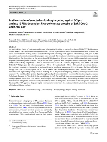
Network Modeling Analysis in Health Informatics and Bioinformatics (2021) 10:22Page 5 of 12 22Fig. 3 Aligned model structuresof SARS-CoV-2 (red) withSARS-CoV (blue) RdRp showing the four catalytic domains;palm, thumb, finger andN-terminal (color figure online)0.66. The Ramachandran plots are presented in Fig. 7and 8 of supplementary information (SI2). VERIFY wasalso employed in validation; for SARS-CoV-2 (89.66%of the residues have averaged 3D-1D score 0.2) andfor SARS-CoV (89.20% of the residues have averaged3D-1D score 0.2). ERRAT gave an overall quality factorfor SARS-CoV-2 and SARS-CoV as 95.905 and 95.594,respectively. The sequence alignment of SARS-CoV3CLpro (PDB Code: 6LU7) and SARS-CoV-2 3CLpro(PBD Code: 1UJ1) gave percent identity of 93.79%(Fig. 9a of SI2) and the sequence alignment of SARS-CoVRdRp with SARS-CoV-2 RdRp shows 95.89% identity, inFig. 9b of SI2.3.2 Aligning the homology modelThe model structures and their respective templates werealigned, Fig. 6 of the SI2, with SARS-CoV-2 and SARSCoV giving RMSDs of 0.144 and 0.069 Å. Similarly, thealigned structures displayed in Fig. 3 indicate that SARSCoV-2 and SARS-CoV RdRp are closely related, havingRMSD of 0.501 Å, with 762 conserved residues, with95.89% identity. These structures also show features that areconserved due to similar protein conformations, particularlyin the four critical regions—Thumb, Finger, N-terminal andPalm. The active sites of the SARS-CoV and SARS-CoV-2RdRp proteins are conserved and are formed by conservedpolymerase motifs located around the palm domain. However, they are structurally different around the N-terminal,as indicated by the “less conserved N-terminal” in Fig. 3.3.3 Analysis of docking resultsCOACH meta-server (Yang et al. 2013) identifies the consensus binding residues for SARS-CoV-2 RdRp model toinclude the following VAL477, PHE482, TYR485, ARG571,ARG572, GLN575, ARG642, LYS643, HIS644, ASN697,CYS699, GLY714. The residues that are within 5.0 Å of theactive site of the template, 6NUR, are CYS647, CYS489,ILE307, LEU302, CYS301, THR293 ASN200. During thedocking experiment, the entire active site conformationalspace was sampled to examine the kind of interactionsthat might occur around the active site and other pockets.Similarly, COACH also identifies the consensus residuesof ligand binding to include; LYS426, ILE466, LEU470,TYR515, MET519, ASP523, ASP525, ALA526, VAL535,ILE539, ASN543, ALA547, SER561, VAL587, ILE589,LYS593, PHE594, HIS650, PHE652, TYR653, ARG654,LEU655, 657, 658, CYS659, GLN661, GLU729, CYS730,LEU731, and SER768.3.4 Gibb’s free energies of bindingGenerally, the free energies of binding are calculated asa sum of four energy terms, namely, intermolecular freeenergy (dominated by enthalpic contribution from steric andelectrostatic interaction upon complex formation), energy ofsolvation, free energy associated with the motion of ligand,protein and ligand–protein complex and free energy due toconformational changes (Raha & Merz, 2005). However,AutoDock Vina scoring function was inspired by the scoring function implemented in X-score and is given in Eq. 1:13
22 Page 6 of 12Network Modeling Analysis in Health Informatics and Bioinformatics (2021) 10:22ΔGbind ΔGvdw ΔGH - bond ΔGdeformation ΔGhydrophobic ΔG0 ,(1)where ΔGvdw takes care of free energy contribution due tovan der Waals interaction between the ligands and the proteins; ΔGH bond accounts for the hydrogen bonding betweenthe ligand and the protein; ΔGdeformation accounts for the freeenergy of deformation during docking; ΔGhydrophobic accountsfor the hydrophobic interaction during simulation and ΔG0 isthe regression constant that includes translational and rotational entropy loss in binding (Trott and Olson 2010; Wanget al. 2002).During each docking process, nine conformational modeswere generated and the best conformers, those with the lowest free energies of binding, are presented in Table 1. Fromthe results, it is easy to see that the free energies of bindingare strongly negative which indicates that the inhibitors bindtightly to the active residues of proteins used in the study.The difference in the standard deviations of the nine conformational modes of each inhibitor is not much, indicating thatthe binding affinity of each conformer does not differ greatlyfrom others. Similarly, the mean free energies of binding arehighly comparable for conformers of a particular inhibitor,as can be seen in SI1.Generally, 3CLpro of SARS-CoV-2 show higher bindingenergies compared to the other protein structures with thesame inhibitors. We use here the result of Remdesivir aspositive control since it has been chosen by the WHO forclinical trials to develop medication for SARS-CoV-2. For3CLpro protein of SARS-CoV-2, the binding energies forthe ligands are higher than values for Remdesivir and Ivermectin, with values 7.4 and 8.4 kcal/mol, respectively.Similar comparison is done for its SARS-CoV counterpart.Except for Sofosbuvir and Ivermectin, with binding energyof 8.2 and 8.1 kcal/mol, respectively, the rest of theinhibitors give binding energies higher than Remdesivir. Toinvestigate the possible reasons why ivermectin bind wellthan Remdesivir, we examined the kinds of interactions thatassist in protein–ligand complexes. SARS-CoV-2-Ivermectincomplex shows mainly carbon-hydrogen bonding at 3.5 Åwhile in the protein-Remdesivir complex, 6 hydrogen bonds(HB’s) are formed at a range of 2.2–3.48 Å and 3 hydrophobic interactions. Since hydrophobic interactions are relatively stronger than other weak intermolecular forces, thistranslates into better binding for Ivermectin ( 8.4 kcal/mol)compared to Remedesivir (between 7.9 and 7.0 kcal/mol) and other FDA-approved nucleotide prodrugs wheninteracted with 3CLpro and RdRp of SARS-CoV and RdRpof SARS-CoV-2. However, Remdesivir binds stronger withSARS-CoV RdRp protein, with an affinity of 8.7 kcal/molthan Ivermectin with a binding affinity of 8.4 kcal/mol.Analysis of the bound complexes shows that electrostaticinteraction plays a major role in the binding of Remdesivirwith ASP218 and LYS50 residues. Asp218, known as thenidovirus conserved residue, is reported to play a criticalrole in the virus activity (Kirchdoerfer and Ward 2019). Thenumber of HB’s and other hydrophobic interactions (suchas pi-alkyl) increase in both SARS-CoV RdRp—RemdesivirTable 1 FDA-approved antiviral inhibitors, their target protein and free energies of binding and the average free energies of binding of the nineconformational modes of each inineRemdesivirRibavirinVitamin rHydroxychloroquineThymoquinoneIvermectin– 5.6– 5.2– 6.7– 7.4– 6.2– 5.1– 5.8– 6.7– 5.4– 6.1– 7.2– 5.2– 5.00 0.35– 4.67 0.25– 5.90 0.26– 6.59 0.42– 5.82 0.22– 4.80 0.23– 5.62 0.18– 6.10 0.27– 5.08 0.18– 5.53 0.31– 6.81 0.24– 4.89 0.19– 5.4– 5.02 0.12 – 5.6– 8.4– 7.78 0.41 – 8.113Average Gof �� 5.6– 5.5– 6.6– 7.9– 6.4– 5.3– 6.0– 6.3– 5.7– 6.3– 8.2– 6.0SARS-CovRdRpAverage Gof inhibitorconformersSARS-– 5.7– 4.9– 7.9– 8.7– 6.4– 5.2– 6.4– 6.3– 6.1– 6.2– 8.1– 6.7– 5.27 0.21– 4.71 0.14– 7.03 0.44– 7.99 0.35– 5.80 0.31– 4.93 0.19– 6.02 0.22– 5.90 0.24– 5.58 0.295.86 0.21– 7.63 0.34– 5.96 0.36– 7.44 0.31 – 6.0– 5.24 0.35 – 5.4– 4.67 0.25– 0.34 – 8.8– 8.31 0.34 – 8.4– 8.03 0.25Average Gof inhibitorconformers3CLpro– 5.18 0.27– 4.76 0.34– 6.18 0.22– 7.28 0.27– 5.83 0.33– 5.04 0.19– 5.73 0.18– 5.81 0.21– 5.21 0.27– 7.37 0.41– 5.66 0.34– 5.03 0.26SARS-Cov-2RdRp– 5.4– 5.6– 7.3– 7.0– 6.3– 5.9– 6.5– 6.7– 5.4– 7.2– 7.7– 6.2Average Gof inhibitorconformersSARS-Cov-2– 5.20 0.14– 5.12 0.21– 6.61 0.296.42 0.27– 5.89 0.23– 5.33 0.28– 5.99 0.24– 6.16 0.31– 5.04 0.20– 6.04 0.56– 6.86 0.35– 5.87 0.15
Network Modeling Analysis in Health Informatics and Bioinformatics (2021) 10:22and SARS-CoV RdRp—Ivermectin complexes. Binding energy of Vitamin C is relatively higher when compared to other inhibitors. This is expected as Vitamin C isa known immune system booster against virus attack andnot an antiviral drug. In general, the binding energies ofthe RdRp homologues are highly comparable, with SARSCoV showing slightly lower values, with few exceptions likeIvermectin and Galidesivir. However, Quinine with value of 7.3 kcal/mol binds to SARS-CoV-2 better than Remdesivir, so also Peramivir, 7.2 kcal/mol, Sofosbuvir, – 7.7 kcal/mol and Ivermectin, 8.8 kcal/mol. It is interesting to notethat our result is in excellent agreement with a recent report(Caly et al. 2020) that Ivermectin, an FDA-approved antiparasitic, previously shown to have broad-spectrum antiviral activity in vitro, is an inhibitor of the causative virus(SARS-CoV-2).With a single addition to Vero-h SLAMcells 2 h post-infection with SARS-CoV-2 is able to effect 5000-fold reduction in viral RNA at 48 h. Ivermectin is,therefore, currently in the frontline as one of the drugs beingconsidered as possible treatment for COVID-19 infection(Chaccour et al. 2020).Antiviral drugs that are polymerase inhibitors such asSofosbuvir, Remdesivir, Tenofovir, Ribavirin and Galidesivir are purine nucleotide prodrugs that are considered inthis investigation. Syntheses of their analogues used in thiswork, Fig. 1, have been described by Ju et al. (2020a, b). Theanalogue show very promising inhibitory potentials whenconsidering their binding energies. Generally, the bindingenergies of the inhibitors including the nucleotide analogues,range between 5.4 and 8.4 kcal/mol for SARS-CoV-2RdRp, 4.9 and 8.7 kcal/mol for SARS-CoV RdRp, 5.5and 8.6 kcal/mol for SARS-CoV 3CLpro and 5.1 and 8.4 kcal/mol for SARS-CoV-2 3CLpro. However, Ivermectin recorded the highest binding affinity, with a ΔG of 8.8 kcal/mol, while Thymoquinone gave one of the lowestbinding affinities, with a value of 5.4 kcal/mol.3.5 Chemistry of binding of the HCoV structureswith inhibitorsSeveral kinds of chemical interactions mediate the differences in the calculated free energies of binding betweenpocket amino acid residues and the molecular inhibitorsthat are used in this investigation. Some of the interactionsinclude electrostatic interactions (HB), hydrophobic andhalogen bonding. HB in this 3CLpro SARS-CoV-2-ligandbinding interaction is mainly due to the presence of amidegroups in the organic backbone of the protein, which actas sites for H-bond donor and acceptor. Pertinent to note isthat the various non-covalent interactions that occur, apartfrom HB, within the protein–ligand complex in our studyare weak but play a major role in determining the stabilityof the system. Electrostatic interactions that are observedPage 7 of 12 22are generally charge (attraction and repulsion) and π-cation.The hydrophobic interactions that are observed are π-sigma,π-πstacking, π–π T-shaped and π-alkyl.3.5.1 3CLpro of SARS‑CoV‑2—ligand‑binding interactionsNon-covalent interactions are the major chemical interaction observed, particularly HB between the ligands andprotein residues. HB is important in protein–ligand interaction, because it leads to the structure stability (Panigrahi2008). The classification scheme for the H-bonding interactions described in this work is Jeffrey’s (Jeffrey 1997). Theyare divided into three, namely, strong and mostly covalent(2.2–2.5 Å), moderate and electrostatic (2.5–3.2 Å) andweak and electrostatic (3.2–4.0). These hydrogen bonds,therefore, are formed between an electronegative atom anda hydrogen atom bonded to a second electronegative atom(Henderleiter et al. 2001). In protein, the H-bonding aremainly within the moderate region and shifts to either sidedue to the kind of moiety that is present in the environmentof the protein.The nature of the chemical interactions of the ligands(Figs. 1 and 2) with the proteins of HCoVs that are investigated here are now discussed. Sheet 1 of SI1 shows thatapart from the HB interactions that are generally observed,other interactions further contribute to a decrease in theenergy of binding of the complex. This can be observedin Remdesivir and Sofosbuvir, among the FDA approvedantiviral drugs and also in the non-FDA approved nucleotide analogues (Ju et al. 2020a, b), Fig. 1, (5–8). They arehere described as non-FDA approved drugs since they arerecently synthesized and have not even undergone clinicaltrials. 5c3, 5h1 and 7a1 show strong HB which occur withina range of 2.20–3.02 Å, and give a low binding energy,ΔG – 8.4 kcal/mol. While 5c3 and 7a1 are π-stacked withHIS41 (active residue) over a distance of 4.33 and 4.41 Å,respectively, 5h1 rather interacts with HIS41 via a non-conventional C-H bonding within 3.5 Å. The spatial distanceof the benzene rings of both 5c3 and 7a1 from HIS41′s islarge enough to reduce the electrostatic interactions betweenthe π-electrons of the interacting benzene rings (Fu et al.2016). As expected of such interaction, the bond type isweak and acidic behavior will dominate (Spiwok 2017).π-sulphur is another non-covalent interaction that occurswhere the π-electron cloud of MET165 interacts with thelone pair of electron cloud of the sulphur atom. Remdesivir,which is chosen as reference here, show π-orbital interaction with MET165 (active residue) at 5.74 Å. Pertinent isthe π–π stacked interaction between the electron cloud ofthe benzene ring in HIS41 and the central aromatic ring ofRemdesivir at a distance of 4.00 Å. This is 0.34 Å higherthan 3.66 Å, for which an equivalent value would be enoughto cause 7.5 eV rise in energy of the system (Fu et al. 2016).13
22 Page 8 of 12Network Modeling Analysis in Health Informatics and Bioinformatics (2021) 10:22This buttresses the fact that π-π stacking at large distancesstabilizes the protein–ligand complex by reducing the repulsive electrostatic interaction. Strong HB interactions that areobserved lie between 2.12 and 2.87 Å. In addition to the HBwith GLY143, GLN189 and 142, Remdesivir made hydrophobic contacts with HIS41, LEU27 and MET49. The weakattractive charge interaction is due to P-atom of the nucleoside phosphoramidite group and ASP289 (OD2, 4.3 Å), anacidic residue, which results in minimal stability at site ofinteraction (Bajji and Davis 2000) and the conventional HBare due to ARG131 (HH2, 2.80 Å) as donors to the oxobenzenoid group of the Sofosbuvir.Quinine and Galidesivir, with the same binding score( 6.7 kcal/mol) are dominated mainly by HB but Galidesivir shows π-π T-shaped hydrophobic interaction due to theπ-electron cloud of its pyrryl ring with HIS41 in the activesite, as shown in Fig. 4. This contributes to the extra stabilityof the protein–ligand complex (Martinez and Iverson 2012).Oseltamivir interacts with the residues of the pocket,howbeit, through HB with LYS137 (HZ2, 2.11 Å; HZ23,2.63 Å) and LEU287 (O, 2.35 Å). Even though conventionalHB interaction was not observed, most of the interactionsobserved are non-covalent, electrostatic except non-conventional C-H (CG2, 3.91 Å) bonding with ILE106.3.5.1.1 CLpro of SARS‑CoV‑ligand‑binding interac‑tions Generally, AutoDock Vina binding scores for 3CLproof SARS-CoV are slightly better than 3CLpro of SARSCoV-2. From sheet 2 of SI1, the free energy of bindingranges from 5.5 to 8.6 kcal/mol. The structures ofHCoV 3CLpros have been described clearly elsewhere,(Chen et al. 2020; Cheng et al. 2005) hence, only their salient features would be highlighted here. 3CLpro structuresof SARS-CoV and SARS-CoV-2 are dimers with threedomains, I, II and III as shown in Fig. 5, for HCoV-2–RemFig. 4 Docked Galidesivirmolecule showing π-π T-shapedinteraction with HIS41active residue of SARS-CoV-23CLpro13desivir complex. The catalytic mechanism does not followthe triad seen in other chymotrypsin protease, comprisingSER-HIS-ASP (Shi et al.
Search global optimizer implemented in AutoDock Vina (Trott and Olson 2010) with the help of a graphical user interface program, PyRx (Dallakyan and Olson 2015). Free water molecules were deleted from the crystallographic structures of 3CLpro proteins, while the RdRp model structures of both SARS-CoV and SARS-CoV-2 were