Transcription
6/14/2016THE LOWER EXTREMITIESAMA GUIDES CHAPTER 17Tim MussackMarlene PhillipsBradford & Barthel, LLPAMA Analysis and Ratings DivisionBradford & BarthelAMA Analysis and Ratings Division Tim Mussack(916) 569-0790tmussack@bradfordbarthel.com Marlene Phillips(909) 476-0552mphillips@bradfordbarthel.com l-form/www.bradfordbarthel.com21
6/14/2016www.bradfordbarthel.com3Most Frequently Used Chapters Chapters 1 & 2 --- The ‘Constitution’– From page 17, in the Introduction to Chapter 2, the Practical Application ofthe Guides:“Two physicians, following the methods of the Guides to evaluate the samepatient, should report similar results and reach similar conclusions.Moreover, if the clinical findings are fully described, any knowledgeableobserver may check the findings with the Guides criteria.” The Almaraz Guzman en banc decision of 9/3/2009:“ by requiring use of the AMA Guides to determine impairment, theLegislature furthered its expressly stated goal of achieving “consistency,uniformity, and objectivity.” Chapter 15 --- Spine Chapter 16 --- Upper Extremity (UE) Chapter 17 --- Lower Extremity (LE)www.bradfordbarthel.com42
6/14/2016The Lower Extremities Lower extremity impairment values Combining– Impairment– PD (after adjustment) Methods of Evaluationwww.bradfordbarthel.com5Chapter 17 – The Lower Extremitiesthere are errata 17.1 Principles of Assessment(p. 524-525) 17.2 Methods of Assessment (p. 525-554) 17.3 LE Impairment Evaluation ProcedureSummary and Examples (p. 555-560) Impairment evaluations are performed after theinjured worker attains MMIwww.bradfordbarthel.com63
6/14/2016www.bradfordbarthel.com7‘Regional’ Impairment AMA Guides is not jurisdictionally specific for evaluatingPermanent Disability The California Schedule for Rating gives additional instructions for rating PermanentDisabilities.Section 1 of the rating schedule is Introduction and Instructions.From page 1-4:"The impairment number identifies the body part, organ system and/ or nature of theinjury.""Under Section 2 of the Permanent Disability Rating Schedule, an appropriate impairmentnumber can be found for most impairments."On page 1-5:"A single injury can result in multiple impairments of several parts of the body. Forexample, an injury to the arm could result in limited elbow range of motion and shoulderinstability."On page 1-11 of the Schedule:".'adjusting' refers to adjusting an AMA impairment rating for diminished future earningcapacity, occupation, and age.""Impairments of an individual extremity are adjusted and combined at the whole personlevel with other impairments of the same extremity."84
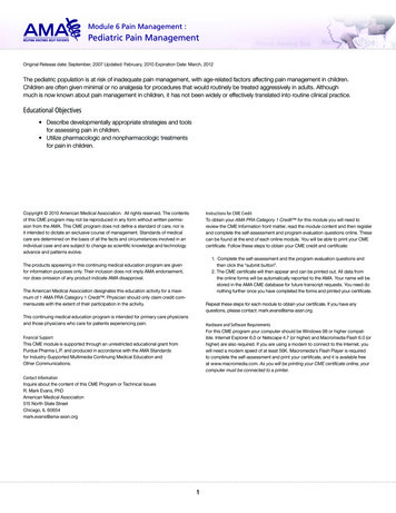
6/14/2016Impairment Values 100% LE 40% WPI [LE % x .4 WPI %]– Table 17-3 – page 527 100% Foot 70% LE [Foot % x .7 LE %]www.bradfordbarthel.com9Table 17-3 – page 527Conversion Table – LE to WPIwww.bradfordbarthel.com105
6/14/2016Combine or Add, and CVC Numbers that are put together for evaluation of impairment/ PDmust be either added or combined. When to combine: COMBINE – for most situations -- unless specific instructions stateto ADD impairment values. The effect/ purpose of combining isthat it prevents the combined value from exceeding 100.A B(1 – A) [where A and B are decimal equivalents] When to add: The most notable exception to combining impairments is with theevaluation of range of motion impairment for the same part of thebody (for example, right ankle motion) [hand evaluation hasunique methodology]www.bradfordbarthel.com11Combine or Add, and CVC How to combine:Page 1-11 of the 2005 PDRS:Multiple impairments such as those involving a single part of an extremity, e.g.two impairment involving a shoulder such as shoulder instability and limitedrange of motion, are combined at the upper extremity level, then converted towhole person impairment and adjusted before being combined with other partsof the same extremity.Impairments with disability numbers in the 16.01 and 17.01 series are convertedto whole person impairment and adjusted before being combined with any otherimpairment of the same extremity.Impairments of an individual extremity are adjusted and combined at the wholeperson [PD] level with other impairments of the same extremity before beingcombined with impairments of other body parts. For example, an impairment ofthe left knee and ankle would be combined before further combination with animpairment of the opposing leg or the back.www.bradfordbarthel.com126
6/14/2016www.bradfordbarthel.com13Chapter 17 13 Methods of Assessmentwww.bradfordbarthel.com147
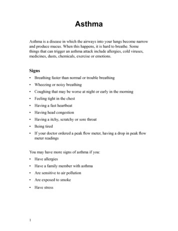
6/14/2016Methods Used to EvaluateImpairments of the Lower Extremitieswww.bradfordbarthel.com15Table 17-2, page 526“After all potentially impairing conditions havebeen identified and correct ratings recorded select the most specific method(s) andrecord the estimated impairment for each.”“explain in writing why a particularmethod(s) was chosen.”(p. 526)www.bradfordbarthel.com168
6/14/2016Table 17-2 “Typically, one method will adequatelycharacterize the impairment ” “Avoid combining methods that rate thesame condition.” “If more than one method can be used, themethod that provides the higher ratingshould be adopted.” (page om189
6/14/2016Interpolation When a Table gives a range for objectivefindings, and a correlating range forimpairment, use interpolation to provide theappropriate value (as shown in Example 1715, with leg shortening)www.bradfordbarthel.com19Method #1Limb Length Discrepancy X-rays strongly recommendedRepeat 3 times3“averaged to reduce measurement error”P. 528www.bradfordbarthel.com2010
6/14/2016Table 17-4 (p. 528)Impairment Due to Limb Discrepancywww.bradfordbarthel.com21Shortening due to: Overriding, Malalignment, or Fracture deformities0-1.25 cm (0-1/2 in.) 5% LE1.25-2.5 cm (1/2-1 in.) 10% LE2.5-3.75 cm (1-1 ½ in.) 15% LE3.75-5.0 cm (1 ½ - 2 in.) 20% LE“combine with other functional sequela ”(p 528)www.bradfordbarthel.com2211
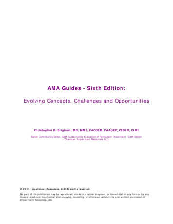
6/14/2016Method #2Gait Derangement Why is it being used?Should use more specific methodCorrelating objective findingsRead Table 17-5 (p. 529) carefullyNo combiningwww.bradfordbarthel.com23Table 17-5, p. 529 www.bradfordbarthel.com2412
6/14/2016 “Except as otherwise noted Table 17-5 [is] forfull-time gait derangements of persons whoare dependent on assistive devices.” Not for “abnormalities based only onsubjective factors, such as pain or suddengiving-way, as with an individual with lowback discomfort who chooses to use a cane toassist in walking.”www.bradfordbarthel.com25 “Whenever possible, the evaluator should use a morespecific method. When the gait method is used, awritten rationale should be included in the report. Thelower limb impairment percents shown in Table 17-5stand alone and are not combined with any otherimpairment evaluation method.” As also expressed in the Comment section for Example17-1 on page 528 of the AMA Guides, “Although the individual has a limp (gait abnormality),gait derangement should be used only when no othermethod is available to rate the person.”www.bradfordbarthel.com2613
6/14/2016Method #3Muscle Atrophy (Unilateral) At thigh 10 cm above patella Calf at “max level” Must compare measurement to opposite,uninjured LE Combine thigh and calf atrophywww.bradfordbarthel.com27Atrophy Measurements Compare measurement to opposite member Difference in circumference might be:– Swelling– Varicose veins– Opposite member injuredwww.bradfordbarthel.com2814
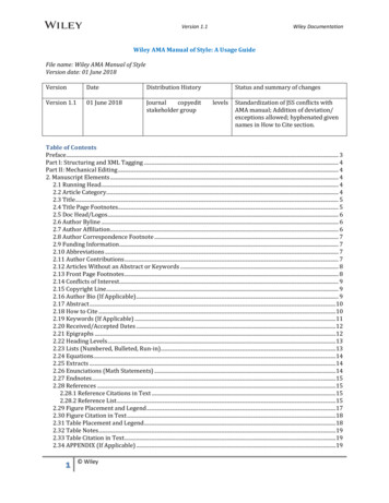
6/14/2016Table 17-6 (p. 530)Impairment Due to UnilateralLeg Muscle AtrophyMildwww.bradfordbarthel.com29Example Right tibia fractureMMIPain free walkingRight thigh atrophy 2 cmRight calf atrophy 1 cmwww.bradfordbarthel.com3015
6/14/2016Impairment?Thigh 2 cm 8% LE 3% WPICalf 1 cm 3% LE 1% WPICombine: 8% C 3% 11 LEConvert: 11% LE 4% WPIwww.bradfordbarthel.com31Method #4Manual Muscle Testing Use Table 17-7 &17-8(pages 531- 532) Note typo - Hip abduction,Grade 3 37% LEwww.bradfordbarthel.com3216
6/14/2016Table 17-7 (p. 531)Criteria for Grades of Muscle Functionof the Lower Extremitywww.bradfordbarthel.com33Manual Muscle Testing (cont’d)Cautions – page 531:“depends on the examinee’s cooperation”“should be concordant with other signs andmedical evidence”– More than one grade between examiners– More than one grade from exam to exam– Pain– Fear of pain– Attributed to deficit of a peripheral nervewww.bradfordbarthel.com3417
6/14/2016Table 17-8, p. 532Combine – Example 17-5www.bradfordbarthel.com35Method #5Range of Motion(pages 533-538)www.bradfordbarthel.com3618
6/14/2016 Use ROM only “If it is clear restricted [ROM]has an organic basis ” Obtain 3 measurements; use greatest Add ROM impairments in jointwww.bradfordbarthel.com37Figure 17-1 (p. 534)Using a Goniometer to Measure Flexion of the Right Hipwww.bradfordbarthel.com3819
6/14/2016Figure 17-2 (p. 534)Neutral Position, Abduction, Adductionof the Right Hipwww.bradfordbarthel.com39Figure 17-3 (p. 535)Measuring Internal and External Hip Rotationwww.bradfordbarthel.com4020
6/14/2016Figure 17- 4 (p. 535)Measuring Knee Flexionwww.bradfordbarthel.com41Figure 17-5 (p. 535)www.bradfordbarthel.com4221
6/14/2016Range of Motion (page 537) Table 17-9Table 17-10Table 17-11Table 17-12Table 17-13Table 17-14- Hip- Knee- Ankle- Hindfoot- Ankle/Hindfoot- Toes (see hel.com4422
6/14/2016Example:Ankle flexion of 6 ? 6% WPI (15% LE) (Table 17-11, p. 537)Ankle extension of 5 ? 3% WPI (7% LE) (Table 17-11)1 cm calf atrophy? 1% WPI (Table 17-6, p. 530)www.bradfordbarthel.com45Impairment?Add flexion extension 15% LE 7% LE 22% LE 9% WPIDo not use atrophy (1% WPI)R: 1) Table 17-2 prohibits combining2) ROM is more generouswww.bradfordbarthel.com4623
6/14/2016Range of Motion (continued)Invalid if: Class inconsistency between 2 observersClass inconsistency between examsPainFear of painwww.bradfordbarthel.com47Method #6Ankylosis (joint immobility)See text for “optimal position” valuesHip 50% LE 20% WPI (p. 538)Knee 67% LE 27% WPI (p. 540)Ankle 14% Foot 10% LE 4% WPI (p. 541)Foot 14% Foot 10% LE 4% WPI (p. 542)Toes – see Table 17-30 (page 543)www.bradfordbarthel.com4824
6/14/2016Ankylosis (continued)1. Determine value for “optimal position” (text)2. Use Tables (pages 538-543) for deviation values(malposition increases impairment)3. Add multiple malpositions for same joint4. Combine ankylosis of different joints5. “The baseline rating for ankylosis in a neutralposition is used only once for each joint.”49www.bradfordbarthel.comBody PartMotionTablePagehipinternal rot17-16539hipext -20540kneevalgus17-21540kneeflex17-22540kneeint or ext17-23540malrotationwww.bradfordbarthel.com5025
6/14/2016Body PartankleMotionplantar flex ankleankleankleint malrotationext malrotationtibia-os calcis angletoesankylosiswww.bradfordbarthel.com51Method #7Arthritiswww.bradfordbarthel.com5226
6/14/2016Arthritis (cont’d) Use x-rays (“standing if possible”) with Table17-31 (page 544) ( ) normal cartilage intervals Compare uninjured opposite memberwww.bradfordbarthel.com53Table 17-31, p. 544www.bradfordbarthel.com5427
6/14/2016Arthritis (cont’d)Table 17-31 Footnote:if (i) direct trauma; (ii) patellofemoral pain(between knee cap and thigh bone/femur), and(iii) “crepitation” on physical exam; (iv) no jointspace narrowing- 2% WPI (5% LE) The knee has a medial and lateral compartment– only the more significant loss is used(can be combined with patellofemoral arthritis)www.bradfordbarthel.com55Example Right tibia fracture 10 years ago Over years, increase knee pain, occasionalswelling Standing x-ray – 2 mm cartilage intervalwww.bradfordbarthel.com5628
6/14/2016Impairment?Cartilage interval (knee) 2 mmTable?Table 17-31 (p. 544)Likely amount of cartilage loss?“normal” 4 mmWPI?8% WPI (20% LE)www.bradfordbarthel.com57Method #8Amputations See Table 17-32 (page 545)www.bradfordbarthel.com5829
6/14/2016Table 17-32, p. 545www.bradfordbarthel.com59Method #9Diagnoses-Based Estimates – Table 17-33(used in 70-80% of LE cases)Covers 9 al Shaft FractureKneeMalalignment of Tibial Shaft FractureAnkleHindfootMidfoot DeformityForefoot Deformitywww.bradfordbarthel.com6030
hel.com6231
6/14/2016Hip and Knee Replacements requirethe use of 2 TablesHip Replacement:Table 17-34 and 17-33 (p. 548, 546-547)Knee Replacement:Table 17-35 and 17-33 (p. 549, 546-547)All others:Table 17-33 (p. 546-547)www.bradfordbarthel.com63DRE Hip Replacement Add points from Categories a-e (Table 17-34,p. 548)– Note that the primary factor is pain Apply to Table 17-33 (p. 546)(Good, Fair, Poor Results)www.bradfordbarthel.com6432
6/14/2016Table 17-34, p. 548www.bradfordbarthel.com65Table 17-35, p. 549 www.bradfordbarthel.com6633
6/14/2016Total Hip and/ or Knee Replacement Good Results, 85-100 pts.15(37) Fair Results, 50-84 pts.20(50) Poor Results, less than 50 pts. 30(75)www.bradfordbarthel.com67Method #10Skin Loss(p. 550)www.bradfordbarthel.com6834
6/14/2016Use Table 17-36 (p. 550)Full-thickness skin loss: impairment “evenwhen the areas are successfully covered with[a] skin graft.”Chronic osteomyelitis (bone infection*) too* Usually a bacterial infection of bone/marrow.Resulting inflammation can lead to reductionof blood supply to bone.www.bradfordbarthel.com69Table 17-36 (p. 550)www.bradfordbarthel.com7035
6/14/2016Method #11Peripheral Nerve Injuries(p. 550-552)Table 17-37 - maximum impairment valuesAssessed sensory/ motor deficits are appliedCombine with other LE methods except:muscle weaknessatrophygait (p. 552)www.bradfordbarthel.com71Dysesthesia Impairment of sensitivity, especially to touch.Altered feelings, such as burning, wetness,electric shock, pins and needles, itching(p. 600)www.bradfordbarthel.com7236
6/14/2016Table 17-37 (p. 552)www.bradfordbarthel.com73Method #12Causalgia & CRPS(p. 553)www.bradfordbarthel.com7437
6/14/2016CRPS I (RSD)CRPS II (causalgia)“characterized by pain, swelling, stiffness,discoloration skeletal demineralization”“may follow a sprain, fracture or nerve orvascular injury”www.bradfordbarthel.com75CRPS I (RSD)CRPS II (causalgia)Use Chapter 13 (“Central & Peripheral NervousSystem”):Section 13.8 (p. 343-344)Section 13.5 (p. 336)www.bradfordbarthel.com7638
6/14/2016“to rate [causalgia and RSD] , diagnosis iskey ” Diagnosis: rely on clinical findings, threephase bone scan, x-rays, laser Dopplerflowmetry, sudomotor reflex Table 16-16 (p. 496)www.bradfordbarthel.com77Table 13-15 (p. 336)www.bradfordbarthel.com7839
6/14/2016Method #13Vascular Disorders(p. 553-554)www.bradfordbarthel.com79Table 17-38, p. 554)www.bradfordbarthel.com8040
6/14/2016 Intermittent claudication: cramping or aching in thecalves (sometimes the thighs or buttocks) caused bywalking; relieved by rest; a manifestation ofatherosclerosis (blockage of an artery). It is called“intermittent” because of the pattern of pain onlywith walking. Edema: abnormal buildup of fluid in the ankles, feetand legs. Common causes:– Prolonged standing– Long airplane flights or automobile rides– Menstrual periods (for some women)– Pregnancy– Being overweight– Increased age– Injury or traumawww.bradfordbarthel.com81Summary Multiple potential methods for impairmentevaluation Some can be combined; others cannot Combine impairments at the LE impairmentlevel before converting to WPI Cautions regarding the use of GaitDerangement; Strength evaluationwww.bradfordbarthel.com8241
6/14/2016Almaraz/ Guzman Almaraz/ Guzman rating not automaticMust be substantial evidenceWithin four corners of AMA GuidesPhysician rationale required2015 DWC Educational ConferenceEvidence and reasoningwww.bradfordbarthel.com83Tim Mussack(916) 569-0790Marlene Phillips(909) fordbarthel.comwww.bradfordbarthel.com8442
AMA G UIDES CHAPTER 17 Tim Mussack Marlene Phillips Bradford & Barthel, LLP AMA Analysis and Ratings Division Bradford & Barthel AMA Analysis and Ratings Division Tim Mussack (916) 569-0790 tmussack@bradfordbarthel.com Marlene Phillips (909) 476-0552 mphillips@bradfordbarthel.com ratings@bradfordbarthel.com