Transcription
Arch Clin Med Case Rep 2022; 6 (1): 91-104DOI: 10.26502/acmcr.96550455Case ReportImpact of free Fall Impact versus Treadmill Physical Exercise Programs:Which is Most Osteogenic?Aveline PC1, Lespessailles E3,4, Best TM2, Cesaro A1, Toumi H3,4I3MTO, University d’Orléans, Orléans 45000, France12UHealth Sports Medicine Institute, Department of Orthopedics, Division of Sports Medicine, U of Miami, USADepartement de Rhumatologie, Centre Hospitalier d’Orleans, Orléans, France3Plateforme Recherché Innovation Medicale Mutualisée d’Orléans, CHR, Orleans, France4Corresponding Author: Hechmi Toumi. Departement de Rhumatologie, Centre Hospitalier d’Orleans, Orléans, France.*Tel: 00330238417169.Received: 13 December 2021; Accepted: 07 January 2022; Published: 14 February 2022Citation: Aveline PC, Lespessailles E, Best TM, Cesaro A, Toumi H. Impact of free Fall Impact versus Treadmill PhysicalExercise Programs: Which is Most Osteogenic?. Archives of Clinical and Medical Case Reports 6 (2022): 91-104.AbstractMethods: 50 female Wistar rats (6 weeks-old) wereExercise plays a key role for bone remodeling throughoutrandomly assigned to either a sedentary group (S) or one ofour lives. Within bone cells, osteocytes have the ability to4 exercise groups: treadmill training (T) and 3 Free-fall (F)translate mechanical stimuli to bone anabolic cellulargroups (F30, F45, F60 respectively fall of 30, 45 and 60cm;signaling. A variety of protocols have been used to apply5 days/week, 8 weeks). We evaluated BMD and BMC (byboth direct and indirect stimuli to bone. Herein, weDXA), bone microarchitecture of the left femur and tibiacompared running and free fall impact exercise protocols(by µCT), mechanical strength of the left femur (three-pointand their effects on markers of osteogenesis and bonebending test), and bone marker levels (by ELISA).strength.Archives of Clinical and Medical Case Reports91
Arch Clin Med Case Rep 2022; 6 (1): 91-104DOI: 10.26502/acmcr.96550455Results: After 8 weeks of physical exercise (EXE), wholeresorption [7]. Mechanical stresses are primarily detected inbody BMD and BMC were both significantly higher frombone by the osteocytes [6,8,9], referred to as the controllerbaseline in all EXE groups, with no difference between theof bone remodeling [10]. Osteocytes modulate bone4 EXE groups. Left femur and tibia BMC and BMDremodeling by converting mechanical stress into biologicalsignificantly increased in the F45 and F60 groups comparedmessengers,to the S and T groups. BV/TV, Tb.Th and Tb.N weredifferentiation of osteoclasts and osteoblasts [11]. Severalsignificantly higher in F45 and F60 compared to all otherstudies have demonstrated that these effects are modulatedgroups. Tb.N was significantly higher in F60 compared tothrough the Wnt/β-Catenin pathway through the regulationF45. Yield point stress and Young modulus wereof sclerostin expression, an osteocyte product which plays asignificantly higher for F45 compared to S and T groups.key role as a circulating inhibition of the Wnt-signalingBone alkaline phosphatase and osteocalcin levels werepathways [12]. Mechanical stress resulting from EXEsignificantly higher in the F45 group compared to thereduces sclerostin expression and thus promotes boneremaining groups. NTX level was significantly decreased informationthe F45 compared to the S and T groups.generated by EXE increase osteocalcin expression, chanicalandsignalsbone alkaline phosphatase (ALP), two bone formationConclusion: Both treadmill and impact training protocolsmarkers, while decreasing N-terminal telopeptide (NTX), aproduced a benefit on BMD and BMC. Interestingly, impactbone resorption marker [9,15]. Similarly, EXE producesmechanical stress was a better stimulus for bone trabecularchanges in circulating levels of growth hormone (GH) [16]structureandand insulin-like growth factor (IGF-1), which together havebiomechanical bone parameters were more sensitive toan anabolic effect on both bone and skeletal muscle [17].moderate impact exercise (F45) while high impact exerciseThe optimal form and dose of exercise however remain(F60) promotes further trabecular number.unknown at this point in time.Keywords: Treadmill and free-fall impact exercise,Several forms of EXE have been observed to increase bonefemale rats, microarchitecture, bending test.mass including high impact exercise (i.e jumping) andthantreadmillrunning.Biochemicallower impact activities such as running 17-20. Treadmill1. Introductionexercise is often used [18-21] as a strategy to enhance boneIt is well recognized that bone formation and Bone Mineralremodeling given that the dose of exercise can beDensity (BMD) are both enhanced by physical exerciseaccurately estimated. Running has been shown to both(EXE) [1-4]. EXE is recommended both to improve boneincrease bone formation and decrease bone resorption inaccretion in young women and to decrease bone loss in theweight bearing sites [20]. BMD and trabecular boneelderly [5]. It is well accepted that EXE increasesmicroarchitecture improve in growing rats subjected tomechanical stresses within bone, which in turn enhancesrunning [19,20,22]. Interestingly, none of these studiesosteoblasticdetected changes in bone strength. Impact utes an alternative model recognized to improveArchives of Clinical and Medical Case Reports92
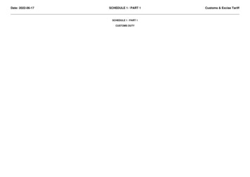
Arch Clin Med Case Rep 2022; 6 (1): 91-104DOI: 10.26502/acmcr.96550455bone mass, BMD and trabecular microarchitecture (BV/TV,Production, Le Genest-St-Isle, France). After one week oftrabecular thickness and porosity) [23,24]. Jump exercise isacclimation, animals were randomly assigned to one of 5known to generate high mechanical stress and significantgroups: control / sedentary rats (S), impact exercises from aanabolic effects [25]. Biomechanical bone properties areheight of 30cm (F30), 45cm (F45) or 60cm (F60), and arelated to the amount of strain applied [26] and the loadingtreadmill group (T). DXA analysis was conducted at time 0rate [27]. It has also been shown that impact exercise(W0) under anesthesia with ketamine-xylazine (80-significantly enhance femoral breaking load and cross-10mg/kg, intraperitoneally, Panpharma). At the end of thesectional moment of inertia [28]. Free-fall impacts arestudy, all animals were euthanized with an intraperitonealsimilarly an effective stimulus for enhancing boneinjection of pentobarbital sodium chloride (0.1mL/100g offormation through an increase in ultimate breaking force inbody weight, Ceva santé animal) [25]. Animals werethe shaft of the forelimbs but not the hindlimbs [29].maintained in observation for a few minutes followingInterestingly, only a few differences in BMD and Crosscardiac exsanguination. The blood was centrifuged and theSectional Area (CSA) have been observed when comparingserum was frozen at 80 C. Left femurs were dissected freedrops from different heights [29].of connective and fat tissues and stored at 20 C and 4 Crespectively.Accordingly, the purpose of the current study was tocompare various forms of low and high impact exercise and2.2. Exercise protocolstheir effects on a comprehensive evaluation of boneTreadmill running (T): This protocol corresponded to astrength, morphology, and biochemistry. To achieve ourpreviously established and adapted method [30] of runninggoal, we compared treadmill running exercise (T) and 3at 8m/min, 1h per day, 5 days/week for 8 weeks (Figure 1).free-fall impact (F) regimens from 30, 45 and 60cm in anFree fall impact rats performed impact exercises for 8effort to determine an optimal dose of exercise to influenceweeks.bone heath status.familiarized to the impact exercise protocol: initial heightDuringtheacclimationperiod,ratswerewas 25cm, and this was gradually increased to 30cm, 45cm2.Materials and Methodsor 60cm. Rats were submitted to 10 drops per day at a2.1. Animals and in vivo experimental designfrequency of 1 drop per 10sec, 5 days/week for 8 weeksAll experiments were approved by the animal protectionfrom a height of 30cm (F30), 45cm (F45) or 60cm (F60)committee of the University of Orleans (CEEA VdL: 2011-(Figure 1). As previously described, the rats were lifted11-2). Fifty female Wistar rats, aged 6 weeks-old, werehorizontally for the drop thus causing them to land on allpurchased from Janvier Animal Production (Janvier Animalfour feet [31].Archives of Clinical and Medical Case Reports93
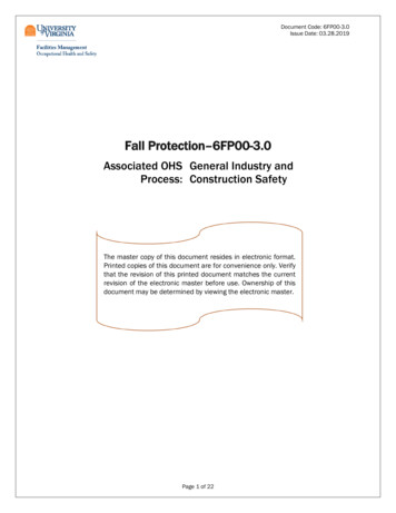
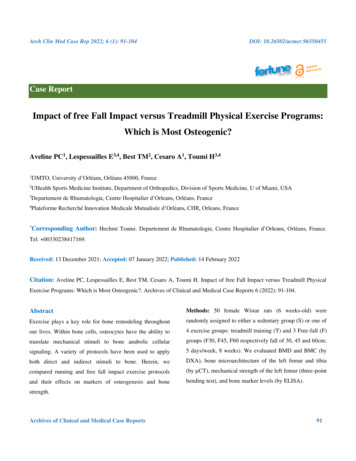
Arch Clin Med Case Rep 2022; 6 (1): 91-104DOI: 10.26502/acmcr.96550455Figure 1: Densitometric characteristics and composition. A) Whole body BMC increases (difference to W0). B) Whole bodyBMD increases (difference to W0). C) Body composition: fat mass, lean mass and weight. The rats in O groups wereovariectomized. Rats in E groups were submitted to 10 impacts per day, 5 days a week during 8 weeks. Rats in Sr groupsreceived 625 mg/kg/day of SrRan. The critical p-value were p 0.05: *, p 0.01: **, p 0.001: ***, NS: non significant and a: vsSh, b: vs ShE, c: vs ShSr, d: vs ShESr, e: vs O, f: vs OE, g: vs OSr and h: vs OESr.2.3. Bone densitometry measurements (DXA)before sacrifice. The root-mean square coefficient ofBMD, BMC and body composition were measured by DXAvariation (CV) was 1.2% for WB BMD measurement. The(Discovery, Hologic, Bedford, USA) using a specific smallroot-mean square CV was determined from three repeatedanimal body composition mode [32]. Both measures weremeasures with repositioning 10 rats as previously describedmade once at week 0 (W0), week 4 (W4) and week 8 (W8)[33].Archives of Clinical and Medical Case Reports94
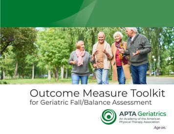
Arch Clin Med Case Rep 2022; 6 (1): 91-104DOI: 10.26502/acmcr.965504552.4. Morphological and topological characteristics ofparameters were recorded: ultimate strength (N), stiffnessthe trabecular bone(N/mm), yield point stress (N/mm²), moment of inertiaTrabecular microarchitecture of the distal metaphysis of the(mm4) and Young’s modulus (MPa). This protocol wasleft femur was evaluated post-mortem using a µCTadapted from Maurel et al. [32].(Skyscan 1072, Skyscan, Belgium) as previously described[32-34]. Briefly, the X-ray source was set at 80kV and2.7. Biochemical analysis100µA with an isometric pixel size 15.49µm. 225 slicesBone turnover markers were analyzed at W0 and W8 (endwere selected for each sample and analyzed by NRecon andof the protocol). Osteocalcin and bone alkaline phosphataseCTan softwares (Skyscan 1072, Skyscan, Belgium). The(ALP) were evaluated as markers of bone formation withfollowing parameters were measured: Bone Volume/TissueN-terminal telopeptide of type I collagen (NTx) for boneVolume BV/TV (%), Trabecular Number Tb.N (1/mm), andresorption. ELISA kits were obtained from EIAab (China)Trabecular Thickness Tb.Th (mm).and used according to the manufacturer’s instructions.2.5. Morphological characteristics of cortical bone2.8. Statistical analysisCortical bone of all left femurs was analyzed with the sameAll numerical variables were expressed as mean SEM. Aacquisition characteristics as trabecular bone. Corticalp-value of p 0.05 was chosen for significance. Statisticalporosity Ct.Po (%) (BV/TV equivalent) and pore numberanalysis was performed using GraphPad Prism softwarePo.N (1/mm) (Tb/N equivalent) were measured as(GraphPad Prism, USA). Physical exercise effects werepreviously described [35].analyzed using a nonparametric Mann-Whitney test tocompare groups. The effects of time were analyzed with a2.6. Bone mechanical testing1-way repeated measures ANOVA.Mechanical properties of all left femurs were assessed by athree-point bending test with a universal testing machine3.(Instron 3343, Instron, Australia). Femurs were secured on3.1. BMC and BMD (Table 1, Figure 2)the two lower supports separated by a distance of 20mm. AtWhole body BMC and BMD (difference to W0) increaseda loading rate of 1mm/min the following mechanicalsignificantly in all groups at W4 and W8.Archives of Clinical and Medical Case ReportsResults95
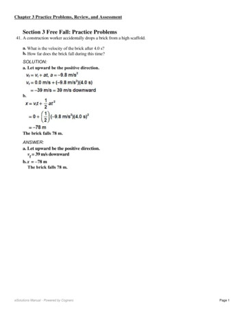
Arch Clin Med Case Rep 2022; 6 (1): 91-104DOI: 10.26502/acmcr.96550455Figure 2: Microarchitecture of the trabecular and cortical bone at the left femurs of Wistar female rats. The rats in O groupswere ovariectomized. Rats in E groups were submitted to 10 impacts per day, 5 days a week during 8 weeks. Rats in Sr groupsreceived 625 mg/kg/day of SrRan. The critical p-value were p 0.05: *, p 0.01: **, p 0.001: ***, NS: non significant and a: vsSh, b: vs ShE, c: vs ShSr, d: vs ShESr, e: vs O, f: vs OE, g: vs OSr and h: vs OESr.Archives of Clinical and Medical Case Reports96
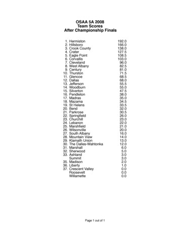
Arch Clin Med Case Rep 2022; 6 (1): 91-104DOI: 10.26502/acmcr.965504553.2. Body weight, lean and fat mass (Table 1)all EXE groups compared to sedentary rats. Note that noWeight decreased significantly only in the F30 compared todifferences were observed for lean body mass.the S group (p 0.050). Fat mass decreased significantly inTable 1: Microarchitecture of the trabecular bone on vertebras of Wistar female rats. The rats in O groups wereovariectomized. Rats in E groups were submitted to 10 impacts per day, 5 days a week during 8 weeks. Rats in Sr groupsreceived 625 mg/kg/day of SrRan. The critical p-value were p 0.05: *, p 0.01: **, p 0.001: ***, NS: non significant and a: vsSh, b: vs ShE, c: vs ShSr, d: vs ShESr, e: vs O, f: vs OE, g: vs OSr and h: vs OESr.3.3. Bone microarchitecture of the left femur (Figure 3)Tb.N was also significantly higher in F60 compared to F45.BV/TV and Tb.Th increased significantly in the F45 andNo difference was observed between S, F30 and T. Tb.SpF60 groups compared to S, F30 and T. Tb.N increaseddecreased significantly in F60 compared to all groups.significantly in F45 and F60 compared to all other groups.There was no difference for Ct.Po, Ct.Th and Po.N.Archives of Clinical and Medical Case Reports97
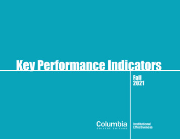
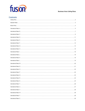
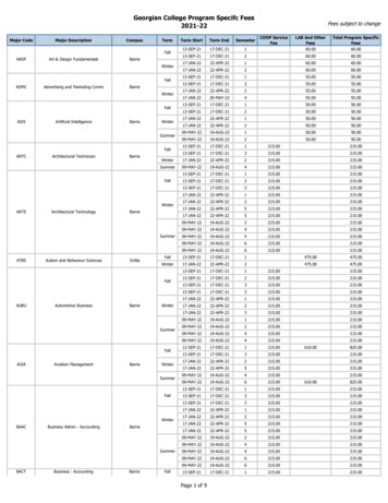
Arch Clin Med Case Rep 2022; 6 (1): 91-104DOI: 10.26502/acmcr.96550455Figure 3: Osteocyte analysis. A) Osteocyte apoptosis represented by % of cleaved caspase-3 immunostaining. B) Lipid cellimmunostaining. The rats in O groups were ovariectomized. Rats in E groups were submitted to 10 impacts per day, 5 days aweek during 8 weeks. Rats in Sr groups received 625 mg/kg/day of SrRan. The critical p-value were p 0.05: *, p 0.01: **,p 0.001: ***, NS: non significant and a: vs Sh, b: vs ShE, c: vs ShSr, d: vs ShESr, e: vs O, f: vs OE, g: vs OSr and h: vs OESr.3.4. Biomechanical analysis of the left femur (Table 2)of inertia decreased significantly in F45 groups compared toYield point stress increased significantly in F45 comparedS and T (p 0.032 and p 0.036 respectively). Noto S, F60 and T (p 0.016, p 0.024 and p 0.005difference was observed between the other groups. CSArespectively). No difference was observed between S, F30,decreased significantly in F30 compared to S and F60 (p F60 and T. Young’s modulus significantly increased in F450.022 and p 0.009 respectively). Concerning ultimatecompared to S and T (p 0.071 and p 0.041). Nostrength and stiffness, no difference was observed betweendifference was observed between the other groups. Momentall groups.Archives of Clinical and Medical Case Reports98
Arch Clin Med Case Rep 2022; 6 (1): 91-104DOI: 10.26502/acmcr.96550455ShUltimate strength(N)Left FemurCross-sectional area(mm²)Moment of inertia4(mm )Yield point stress3.944 0.258c d e f g h ***3.814 0.282c d e f g h ***4.207 0.459f h ***, e g *3.996 0.537f h ***, e g **, d *n 12143.813 17.755h*4.754 0.312a b ***, h *4.164 0.570f h ***, e g *4.733 0.715bfh*221.410 25.469c **, d h *223.396 18.969c h **, d *252.840 18.029b **, a e f *248.460 20.232abef*8994.939 1790.523f **, c h *9500.987 1572.135f h ***, e **10308.371 1080.342f h ***, e **9064.169 1536.809f **, c e h *n 12144.433 11.040bh*Ultimate strength(N)Cross-sectional areaLeft Femur4.524 0.360a b ***, h **, f g *ShESr258.325 42.611NSO4.556 0.176a b ***, h **, f g *(mm²)Moment of inertia4.818 0.435b **, a c f h *(mm 4 )Yield point stressOEOSrOESrn 12145.163 13.110bh*n 12153.405 11.234b ***, a **n 10157.778 13.046a b ***, d e f *4.996 0.0440a b ***, c e *4.941 0.368a b ***, c e *5.274 0.528a b ***, c e **, d *5.555 0.751a b c ***, d e g *4.872 0.585b **, a c f h *5.776 0.857a b c ***, d e g *256.185 20.202cf*232.273 18.045b c ***, a g **, e *264.452 21.385f **243.035 34.205c **, b *223.143 25.247cdh*227.152 25.271cdh*241.152 25.082NS256.580 25.469b **, a e f *7746.458 667.965c ***, b **, d *7032.411 1146.176b c ***, a d **, g *8446.560 1135.202c **, f *7558.501 949.115b c ***, a d *(N/mm²)Stiffness(N/mm)(Mpa)n 12149.433 13.724ab*290.530 33.398f ***, h **, e *(N/mm)Young's ModulusShSr279.731 33.865f ***, h *Stiffness(Mpa)n 12133.189 10.635g h ***, c e f *269.313 34.599f **(N/mm²)Young's ModulusShEn 12134.193 11.313g **, c h *Table 2: Biomechanical testing at the left femurs of Wistar female rats. The rats in O groups were ovariectomized. Rats in Egroups were submitted to 10 impacts per day, 5 days a week during 8 weeks. Rats in Sr groups received 625 mg/kg/day ofSrRan. The critical p-value were p 0.05: *, p 0.01: **, p 0.001: ***, NS: non significant and a: vs Sh, b: vs ShE, c: vs ShSr,d: vs ShESr, e: vs O, f: vs OE, g: vs OSr and h: vs OESr.3.5. Bone remodeling markers (Table 3)compared to all groups. NTX level decreased significantlyALP and OCN levels increased significantly in F45in F45 and F60 compared to S and T.compared to all groups. ALP level was lower in TArchives of Clinical and Medical Case Reports99
Arch Clin Med Case Rep 2022; 6 (1): 91-104DOI: 10.26502/acmcr.96550455Table 3: Bone remodelling markers of Wistar female rats. Alkaline phosphatise (ALP) and ostéocalcine (OCN) were boneformation markers and NTX a bone resorption marker. The rats in O groups were ovariectomized. Rats in E groups weresubmitted to 10 impacts per day, 5 days a week during 8 weeks. Rats in Sr groups received 625 mg/kg/day of SrRan. Thecritical p-value were p 0.05: *, p 0.01: **, p 0.001: ***, NS: non significant and a: vs Sh, b: vs ShE, c: vs ShSr, d: vs ShESr,e: vs O, f: vs OE, g: vs OSr and h: vs OESr.4.exercise. Second, bone formation was affected by theDiscussionMechanical stress is an essential factor for maintainingamount of applied mechanical stress. Both the F45 and F60overall bone health. During loading, bone receives diverseconditionsforms of mechanical stimuli resulting in complex cellularremodeling compared to the F30 group. Biomechanicalresponses.responses favored the F45 training greateradvantagesforbonebiological signals within osteocytes that result in variousdownstream signals and alteration/maintenance of boneFollowing 8 weeks of training, all forms of exercisehomeostasis. In this study, the aim was to explore bonedecreased fat mass compared to the sedentary , there was no exercise effect on lean bodyresponses to different forms and rates of physical exercise.mass. Weight decreased significantly only for the F30We compared treadmill running to free fall impact andcompared to the S condition (p 0.050). This was likelycompared gradual increased heights of the latter (30, 45 anddue to a higher decrease in fat mass in this group compared60cm). Our results demonstrated that free fall impactto the other groups (-20%; Table 1).biomechanical,andexercises were more effective for enhancing bonemicroarchitecture, formation and strength than treadmillArchives of Clinical and Medical Case Reports100
Arch Clin Med Case Rep 2022; 6 (1): 91-104DOI: 10.26502/acmcr.96550455Whole body BMC and BMD significantly increased at W8newly forming and pre-existing bone matrix probablycompared to W0 for all groups. These findings arerelated to type I collagen, which is known to affect the post-consistent with several previous reports. It has been shownyield behavior of bone [37]. The significant increase inthat rats completing a 4 week interval-training program hadfemur bone composition (increase in BMD, BV/TV andhigher femur BMD values compared to non-exercisedTb.Th) impacts bone network structure, which probablycontrols [30]. In a tail-suspension-induced osteopeniaexplain the changes in femur biomechanical properties.model, Ju et al. have investigated the differential effects ofBiochemical changes might have also interfered with bonejump exercise (10 jumps/day, 5 days/week, 40 cm of heightmatrix modification results from impact exercises notably atfor 5 weeks) compared to continuous running exercise onthefemoral BMD. They demonstrated that both jumping andmicrostructure. Both ALP and osteocalcin levels wererunning exercises significantly improved total femoralsignificantly higher for all free fall groups compared to theBMD [36]. At W8, BMC and BMD were significantlyT group. Interestingly, ALP and osteocalcin concentrationhigher in the F45 and 60 groups compared to the S and Twere significantly higher in F45 compared to all groups.groups. Similar results were noted for trabecular boneThe increase in ALP and osteocalcin reflects greatermicroarchitecture of the left femur (BV/TV, Tb.Th, andosteoblastic activity induced by acute exercise but theTb.N). Ju et al. have shown that jump exercise (10response is known to be dependent on exercise intensity andjumps/day, 5 days/week for 5 weeks, 40 cm of height)modality but also to the age and sex [38]. The higher valuescontributed to an increase of Tb.Th and BV/TV (but not forof the F45 group as compared to the other exercise groupsTb.N) in a tail suspension-induced osteopenia model.might reflect that ground reaction forces induced by the freeHowever, the continuous running exercises (25 m/min, 60fall impact (45 cm) would be more efficacious than thosemin a day, 5 days/week, for 5 weeks) increased BV/TV andproduced in other exercise groups in terms of boneTb.N (but not Tb.Th). Consequently, different effects offormation [39]. However, the precise physiological functionthese two exercises on trabecular bone microarchitectureof ALP and osteocalcin in osteogenesis is still unclear.were observed. Tb.Th was increased with jump exerciseOsteocalcin a bone formation marker and identified as a lateand Tb.N alone with treadmill running exercise [36]. In ourmarker of osteoblastic activity. However it has a short half-protocol, Tb.Th increased in all free fall exercises and nolife, accumulate in patients with severe renal failure andmodification was observed in treadmill exercise group. YetVitamin K dependent [37]. Some studies highlight thatTb.TN significantly increased only in F45 and F60 and notosteocalcin is involved in bone mineralization rather thanin F30. Moreover, a significant advantage was observed inmatrix and ALP is involved in osteoid formation and boneTb.TN in F60 compared to F45.mineralization [40]. Regarding NTX, recognized as a bonecollagenfiberlevel andthus improves boneresorption marker, they are generated from the aminoIt was also observed in the current study that yield pointterminus of the type 1 collagen by cleavage of N-terminalstress increased significantly in the F45 compared to the S,region by cathepsin K during the resorption phase of boneF60 and T groups while Young’s modulus increased in theturnover. After 8 weeks of training NTX expression wasF45 compared to the S and T conditions. Changes in thesignificantly decreased in the F45 compared to the S and TArchives of Clinical and Medical Case Reports101
Arch Clin Med Case Rep 2022; 6 (1): 91-104DOI: 10.26502/acmcr.96550455groups. Although we do not assess directly the RANKLAcknowledgmentsexpression in our experiment [41], as the NTX production isThe authors thank Dr. Laurent Benhamou for his supporta reflect of the resorption process, our results suggest thatand Eric Dolleans from I3MTO laboratory, for animalthe mechanical signals induced by the F45 exercise onexperiment.osteoclast gave a better limitation of bone resorption thatthose produced in the other groups. Thus,it may beConflicts of Interesthypothesized that changes in biochemical (NTX, ALP andThe authors declare no conflict of interest.osteocalcin) are sensitive to the amount of mechanicalloadings on both osteoblast and osteoaclast (treadmill andF30 exercises are too low and F60 to high).Reference1.Bass S, Pearce G, Bradney M, et al. Exercise beforePuberty May Confer Residual Benefits in Bone5.ConclusionDensity in Adulthood: Studies in Active PrepubertalBoth treadmill and impact training yielded positive benefitsand Retired Female Gymnasts. J. Bone Miner. Res.on BMD and BMC. However, free-fall exercise producedOff. J Am Soc Bone Miner Res 13 (1998): 500-507.greater effects than treadmill running for several parameters2.Blimkie CJ, Rice S, Webber CE, et al. Effects ofof bone health. Both biochemical and biomechanical boneResistance Training on Bone Mineral Content andparameters were sensitive to the level of applied mechanicalDensity in Adolescent Females. Can. J Physiolstress suggesting that this variable may be important inPharmacol 74 (1996): 1025-1033.exercise prescription programs.3.Kannus P, Haapasalo H, Sankelo M, et al. Effect ofStarting Age of Physical Activity on Bone Mass in theAuthor ContributionsDominant Arm of Tennis and Squash Players. AnnAveline PC, Lespessailles E, Best TM, Cesaro A, Toumi H.Intern Med 123 (1995): 27-31.4.Karlsson MK, Vergnaud P, Delmas PD, et al.FundingIndicators of Bone Formation in Weight Lifters.This research was funded by Servier France.Calcif. Tissue Int 56 (1995): 177-180.5.Bone Remodeling. Metabolism 60 (2011): 373-388.Institutional Review Board StatementThe study was conducted according to the guidelines of theMaïmoun L, Sultan C. Effects of Physical Activity on6.Bonewald LF. The Amazing Osteocyte. J. BoneDeclaration of Helsinki, and approved by the InstitutionalMiner. Res. Off. J Am Soc Bone Miner Res 26 (2011):Review Board of the of the uni-ver-sity of Orleans and all229-238.experiments were approved by the animal protection7.Robergs RA, Icenogle MV, Hudson TL, et al.committee of the Univer-sity of Orleans, France (agreementTemporal Inhomogeneity in Brachial Artery Bloodn : CEEA VdL; delivered: 2011-11-2).Flow during Forearm Exercise. Med. Sci. Sports Exerc29 (1997): 1021-1027.Archives of Clinical and Medical Case Reports102
Arch Clin Med Case Rep 2022; 6 (1): 91-1048.9.DOI: 10.26502/acmcr.96550455Klein-Nulend J, Nijweide PJ, Burger EH. Osteocyte18. Hagihara Y, Nakajima A, Fukuda S, et al. Runningand Bone Structure. Curr. Osteoporos. Rep 1 (2003):Exercise for Short Duration Increases Bone Mineral5-10.Density of Loaded Long Bones in Young GrowingKlein-Nulend J, Bacabac RG, Veldhuijzen JP, et al.Rats. Tohoku J Exp Med 219 (2009): 139-143.Microgravity and Bone Cell Mechanosensitivity. Adv.19. Iwamoto J, Takeda T, Sato Y. Interventions to PreventSpace Res. Off. J. Comm. Space Res. COSPAR 32Bone Loss in Astronauts during Space Flight. Keio J(2003): 1551-1559.Med 54 (2005): 55-59.10. Verborgt O, Tatton NA, Majeska RJ, et al. Spatial20. Iwamoto J, Shimamura C, Takeda T, et al. Effects ofDistribution of Bax and Bcl-2 in Osteocytes afterTreadmill Exercise on Bone Mass, Bone Metabolism,Boneand Calciotropic Hormones in Young Growing Rats.Fatigue:ComplementaryRolesinBoneRemodeling Regulation? J. Bone Miner. Res. Off. J.Am. Soc. Bone Miner. Res 17 (2002): 907-914.J. Bone Miner. Metab 22 (2004): 26-31.21. Iwamoto J, Yeh, JK, Aloia JF. Differential Effect of11. Dallas SL, Prideaux M, Bonewald LF. The Osteocyte:An Endocrine Cell . and More. Endocr. Rev 34(2013): 658-690.Treadmill Exercise on Three Cancellous Bone Sites inthe Young Growing Rat. Bone 24 (1999): 163-169.22. Joo YI, Sone T, Fukunaga M, et al. Effects of12. Thompson WR, Rubin CT, Rubin J. MechanicalEndurance Exercise on Three-Dimensional TrabecularRegulation of Signaling Pathways in Bone. Gene 503Bone Microarchitecture in Young Growing Rats. Bone(2012): 179-193.33 (2003): 485-493.13. Robling AG, Niziolek PJ, Baldridge LA, et al.23. Honda A, Sogo N, Nagasawa S, et al. High-ImpactMechanical Stimulation of Bone in Vivo ReducesExerciseOsteocyte Expression of Sost/Sclerostin. J Biol ChemOvariectomized Rats with the Same Outcome as Sham283 (2008): 5866-5875.Rats. J. Appl. Physiol. Bethesda Md 95 (2003): 1032-14. penic1037.Downregulation and Local Wnt Signaling Are24. Honda A, Umemura Y, Nagasawa S. Effect of High-Required for the Osteogenic Response to MechanicalImpact and Low-Repetition Training on Bones inLoading. Bone 50 (2012): 209-217.Ovariectomized Rats. J. Bone Miner. Res. Off. J. Am.15. Klein-Nulend J, Bakker AD, Bacabac RG, et al.Mechanosensation and Transduction in Osteocytes.Bone 54 (2013): 182-190.16. Toumi H, Higashiyama I, Suzuki D, et al. RegionalVariations in Human Patellar Trabecular Architectureand the Structure of the Proximal Patellar TendonEnthesis. J Anat 208 (2006): 47-57.17. Hamrick MW. A Role for Myokines in Muscle-BoneInteractions. Exerc. Sport Sci Rev 39 (2011): 43-47.Archives of Clinical and Medical Case ReportsSoc. Bone Miner Res 16 (2001): 1688-1693.25. Umemura Y, Ishiko T, Yamauchi T, et al. Five Jumpsper Day Increase Bone Mass and Breaking Force inRats. J. Bone Miner. Res. Off. J Am Soc Bone MinerRes 12 (1997): 1480-1485.26. Rubin CT, Lanyon LE. Regulation of Bone Formationby Applied Dynamic Loads. J. Bone Joint Surg. Am66 (1984): 397-402.27. Turner AS, Alvis M, VanderVliet MM, et al.103
Arch Clin Med Case Rep 2022; 6 (1): 91-104DOI: 10.26502/acmcr.96550455Heterogeneity of Bone Mineral Density in Ewes. ProcGuidelines for Assessment of Bone Microstructure inOrthop Res Soc New Orleans 441 (1994).Rodents Using Micro-Computed Tomography. J Bone28. Järvinen TLN, Kannus P, Sievänen H, et al.Randomized Controlled Study of Effects of SuddenImpact Loading on Rat Femur. J Bone Miner Res 13(1998): 1475-1482.Miner Res Off J Am Soc Bon
2UHealth Sports Medicine Institute, Department of Orthopedics, Division of Sports Medicine, U of Miami, USA 3Departement de Rhumatologie, Centre Hospitalier d'Orleans, Orléans, France 4Plateforme Recherché Innovation Medicale Mutualisée d'Orléans, CHR, Orleans, France *Corresponding Author: Hechmi Toumi. Departement de Rhumatologie .