Transcription
Zhang et al. Stem Cell Research & 2020) 11:180RESEARCHOpen AccessCo-culture of hWJMSCs and pACs in doublebiomimetic ACECM oriented scaffoldenhances mechanical properties andaccelerates articular cartilage regenerationin a caprine modelYu Zhang1,2,3,4, Chunxiang Hao5, Weimin Guo1,2,3, Xiaoyu Peng6, Mingjie Wang1,2,3, Zhen Yang7, Xu Li7,Xueliang Zhang8, Mingxue Chen1,2,3, Xiang Sui1,2,3, Jiang Peng1,2,3, Shibi Lu1,2,3, Shuyun Liu1,2,3*,Quanyi Guo1,2,3* and Qing Jiang4*AbstractBackground: The dedifferentiation of chondrocytes and the unstable chondrogenic differentiation status ofpluripotent mesenchymal stem cells (MSCs) are immense issues in cell-based articular cartilage repair andregenerative strategies. Here, to improve the cartilage characteristics of seed cells, a double biomimetic acellularcartilage extracellular matrix (ACECM)-oriented scaffold was used to mimic the cartilage microenvironment forhuman umbilical cord Wharton’s jelly-derived MSCs (hWJMSCs) and primary cartilage cells (pACs) to regeneratehyaline cartilage.Methods: A double biomimetic ACECM-oriented scaffold was created from the cartilage extracellular matrix of pigarticular cartilage using pulverization decellularization freeze-drying procedures. hWJMSCs and pACs were cocultured at ratios of 50:50 (co-culture group, ACCC), 0:100 (ACAC group) and 100:0 (ACWJ group) in the ACECMoriented scaffold, and the co-culture system was implanted in a caprine model for 6 months or 9 months to repairfull-thickness articular cartilage defects. The control groups, which had no cells, comprised the blank control (BC)group and the ACECM-oriented scaffold (AC) group. Gross morphology and magnetic resonance imaging (MRI) aswell as histological and biomechanical evaluations were used to characterize the cartilage of the repair area.(Continued on next page)* Correspondence: clear ann@163.com; doctorguo 301@163.com;qingj@nju.edu.cn1Institute of Orthopaedics, Chinese PLA General Hospital, 28 Fuxing Road,Haidian District, Beijing 100853, China4Department of Sports Medicine and Adult Reconstructive Surgery, NanjingDrum Tower Hospital, The Affiliated Hospital of Nanjing University MedicalSchool, 321 Zhongshan Road, Gulou District, Nanjing 210008, ChinaFull list of author information is available at the end of the article The Author(s). 2020 Open Access This article is licensed under a Creative Commons Attribution 4.0 International License,which permits use, sharing, adaptation, distribution and reproduction in any medium or format, as long as you giveappropriate credit to the original author(s) and the source, provide a link to the Creative Commons licence, and indicate ifchanges were made. The images or other third party material in this article are included in the article's Creative Commonslicence, unless indicated otherwise in a credit line to the material. If material is not included in the article's Creative Commonslicence and your intended use is not permitted by statutory regulation or exceeds the permitted use, you will need to obtainpermission directly from the copyright holder. To view a copy of this licence, visit http://creativecommons.org/licenses/by/4.0/.The Creative Commons Public Domain Dedication waiver ) applies to thedata made available in this article, unless otherwise stated in a credit line to the data.
Zhang et al. Stem Cell Research & Therapy(2020) 11:180Page 2 of 13(Continued from previous page)Results: Relative to the control groups, both the gross morphology and histological staining results demonstratedthat the neotissue of the ACCC group was more similar to native cartilage and better integrated with thesurrounding tissue. Measurements of glycosaminoglycan content and Young’s modulus showed that the repairareas had more abundant cartilage-specific content and significantly higher mechanical strength in the ACCCgroup than in the control groups, especially at 9 months. On MRI, the T2-weighted signal of the repair area washomogeneous, and the oedema signal disappeared almost completely in the ACCC group at 9 months. HLA-ABCimmunofluorescence staining demonstrated that hWJMSCs participated in the repair and regeneration of articularcartilage and escaped surveillance and clearance by the caprine immune system.Conclusion: The structure and components of double biomimetic ACECM-oriented scaffolds provided a cartilagelike microenvironment for co-cultured seed cells and enhanced the biomechanics and compositions of neotissue.This co-culture system has the potential to overcome the dedifferentiation of passage chondrocytes and theunstable chondrogenic differentiation status of MSCs.Keywords: Acellular cartilage extracellular matrix-oriented scaffold, Human umbilical cord Wharton’s jelly-derivedmesenchymal stem cell, Primary cartilage cell, Co-culture systemBackgroundArticular cartilage injury, usually caused by engaging inexercise, professional training and games, trauma due toaccident, or being overweight, has become increasinglycommon among young populations and professionalathletes. A retrospective study of 5233 knee arthroscopies by Lukasik et al. reported that over 57% of patientswere diagnosed with a cartilage lesion [1]. In addition,the overall prevalence of articular cartilage injury in athletes (36%) is more than twice the rate in the generalpopulation [2]. However, cartilage rarely self-heals because it lacks vascular and lymph tissue to provide nutrients [3]; thus, osteoarthritis generally develops within 5–20 years [4, 5]. Cell-based treatment strategies are promising methods to repair and regenerate articular cartilageand to stop the progression of osteoarthritis [6, 7].However, the histological characteristics of neotissue areusually highly inferior to those of native articular cartilagemainly because of the unstable chondrogenic differentiation status of mesenchymal stem cells (MSCs), which results in the presence of hypertrophic or calcifiedchondrocytes [8–11]. In addition, for autologous chondrocyte implantation (ACI) or matrix -induced chondrocyteimplantation (MACI), chondrocytes also face some problems, including limited cartilage source to isolate primaryarticular cartilage cells (pACs) [8, 12] and dedifferentiationafter several passages [13]. In recent decades, it has beenemphasized that the environment plays a critical role inthe regulation of cell behaviour [14–16]. Co-culture hasbeen explored to promote the chondrogenic induction ofMSCs and to maintain the native phenotype of chondrocytes by bio-stimulation via cell-cell interactions in coculture systems [14, 17, 18]. In addition, co-culture reduces the required amount of cartilage tissue, decreasesthe in vitro culture time and lowers the degree of chondrocyte degeneration [13]. However, some studies showedthat co-culture merely promoted a unidirectional trophiceffect of MSCs on chondrocytes [19, 20]. These differences are probably attributable to the complex microenvironment, which (i) comprises a milieu of biochemical,biomechanical and bioelectrical signals derived from surrounding cells, the extracellular matrix (ECM) and solublefactors and (ii) regulates cell metabolism and function.In vitro, a co-culture system was constructed usingunpassaged pACs possessing their native phenotype andan ACECM-oriented scaffold retaining native cartilagetissue components and oriented structures [21–23];hWJMSCs were displayed as an alternative cells sourcefor cartilage tissue engineering because of distinct chondrogenic potential, highly proliferative activity and safetyfor patients [24, 25]. In vitro, hWJMSCs were cocultured with chondrocytes and successfully differentiated into chondrocytes, as shown by the significant upregulation of collagen II and aggrecan genes [26]. Ingross morphology, engineered tissue was translucent andresilient, similar to hyaline cartilage tissue. Despite thesefindings, the regeneration and repair conditions after theimplantation of engineered tissue into cartilage defectsin vivo remain unknown. Here, we evaluated whetherthe co-culture of hWJMSCs and pACs in a double biomimetic ACECM-oriented scaffold could achieve a jointsurface that mimics the native state as closely as possibleand return the articular cartilage to its natural biomechanics and compositions.Materials and methodsPreparation of seed cells and construction of tissueengineered cartilage in vitroThe collection of three healthy human umbilical cordsfor this study was approved by the Ethics Committee ofthe General Hospital of the People’s Liberation Army(PLA), and written informed consent was provided by all
Zhang et al. Stem Cell Research & Therapy(2020) 11:180mothers. The detailed protocols for the isolation andculture of hWJMSCs and pACs have been previously described [26, 27]. According to previously described protocols, hWJMSCs were isolated using micromassmesenchymal tissue adhesion on a culture dish containing Dulbecco’s modified Eagle’s medium/-Ham’s F 12(Sigma, USA) nutrient medium (DMEM/F12) (containing 10% foetal bovine serum (FBS) (Corning, USA), 100mg/ml penicillin, 10 mg/ml streptomycin and 250 mg/mlamphotericin B). At passage 3, a subset of the isolateswas prepared for co-culture with pACs. pACs were isolated from slices of goat shoulder articular cartilage bycutting them into microscopic-pieces and digesting themusing 0.15% type II collagenase for 2 h at 37 C. Then,the individual cells and cell sheet were collected by centrifugation at 250 g for 5 min at room temperature. Thecells were resuspended in DMEM/F12 culture mediumsupplemented with 10% FBS and antibiotics (100 mg/mlpenicillin, 10 mg/ml streptomycin and 250 mg/mlamphotericin B) and cultured in 25-cm2 culture flaskswith 5% CO2 in a 37 C incubator. The pACs were collected for construction of the co-culture system at approximately 80% confluence.A three-dimensional (3D)-oriented scaffold was madefrom acellular ECM using the freeze-drying techniquepreviously described [21]. After cross-linking by dehydrothermal and water-soluble carbodiimide treatment,oriented scaffolds with a diameter of 6 mm and a thickness of 1 mm were produced and sterilized by 60Co-γ irradiation (at 5 mrad). A total of 107 pACs andhWJMSCs were mixed in three different ratios: 100:0, 0:100 and 50:50 in 100 μl DMEM/F12 medium. Then, thecell suspension was seeded into a 3D-oriented scaffoldand incubated for 2 h at 37 C with 5% CO2 to promotecell adhesion to the scaffolds. Next, cell-scaffold complexes were transferred to 6-well cell culture plates, and5 ml of culture medium (DMEM/F12, 10% FBS, 100 mg/ml penicillin, 10 mg/ml streptomycin and 250 mg/mlamphotericin B) was added. Tissue-engineered cartilagewas acquired after cultivation for 3 weeks at 37 C under5% CO2. The gross morphology was evaluated accordingto size, colour and shape.Cell viability and distribution assessment in the 3Doriented scaffoldsDead/live staining was used to analyse cell viability afterculturing in the scaffolds for 3 days. Briefly, cell-scaffoldcomplexes were stained with FDA (fluorescein diacetate,5 μg/ml, Sigma) and PI (propidium iodide, 5 μg/ml, Sigma)for 5 min and rinsed 3 times with PBS (phosphate-buffered solution, pH 7.4). The samples were immediately visualized using a laser scanning confocal microscope(Olympus FV 1200, Japan) [26]. Red and green fluorescence indicate dead and live cells, respectively.Page 3 of 13To observe the distribution of seed cells in the 3D-orientedscaffold, scanning electron microscopy (SEM) was applied toview the samples in the sagittal plane and in cross sectionsafter culturing for 3 weeks. All samples were fixed in 2.5%glutaraldehyde, dehydrated in a series of graded ethanol to100% ethanol, treated with hexamethyldisilazane and sputtercoated with gold-palladium before observation [21].In vivo experiments using the engineered tissueTo verify the effectiveness and safety of engineered tissue,the ACCC (pACs: hWJMSCs, 50:50) group was used to repair full-thickness articular cartilage defects of the femoralcondyles in a caprine model. The animal experimental procedures followed the standards for the care and use of laboratory animals and were approved by the Institutional AnimalCare and Use Committee of the Chinese PLA General Hospital. Three healthy goats were randomly assigned to each ofthe following groups per time point: a blank control group(BC, full-thickness articular cartilage defects without anytreatment), an ACECM-oriented scaffold group (AC, treatment with ACECM-oriented scaffolds), a pACs ACECMoriented scaffold group (ACAC, treatment with pACs andACECM-oriented scaffolds), a hWJMSCs ACECM-orientedscaffold group (ACWJ, treatment with hWJMSCs andACECM-oriented scaffolds) and a ACCC group (ACCC,treatment with hWJMSCs, pACs and ACECM-oriented scaffolds). Goats (white, male, 30 5 kg,) were treated withsumianxin II (0.1 mL/kg) and ketamine (4 mg/kg) by intramuscular injection. After location in the supine position onoperating table, sterilizing knee joint and spreading thedrapes, the medical parapatellar longitudinal skin incision is8–10-cm long. The capsule of the knee is exposed, andlongitudinal-shaped capsulotomy is performed. Lateral dislocation of the patella and knee flexion at 120 was performed, and then the medical and lateral femoral condylesare exposed. Full-thickness articular cartilage defects in femoral condyles were made using a 6.5-mm-diameter cornealtrephine under sterile conditions, while preserving the subchondral bone. Penicillin sodium was administered for 3 daysto prevent infection. The goats were allowed to move freelyin a comfortable environment after surgery.7.0 T high-resolution magnetic resonance imagingevaluationAt 6 or 9 months after surgery, all goats were euthanized byintravenous pentobarbital overdose. MRI images of the entire fresh knee joints were obtained immediately using avolume coil on a laboratory 7.0 T MRI scanner (BrukerBiospec, Germany). T2-weighted spin-echo images with fatsuppression were obtained in the sagittal, frontal and transverse planes. The imaging parameters were as follows: slicethickness (1.0 mm), echo time (36 ms), repetition time(5109 ms), flip angle (90 ), acquisition time (18 min) andfield of view (75 75 mm). All images were scored
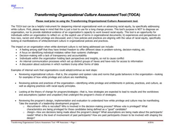
Zhang et al. Stem Cell Research & Therapy(2020) 11:180independently according to the International Cartilage Repair Society (ICRS) Whole-Organ MRI Score (WORMS) ofthe knee in OA by an experienced radiologist specialized inmusculoskeletal medicine and blinded to the group assignments [28].Gross morphology and scoringAfter MRI, the whole knee joints, including the femoralcondyles, the corresponding meniscus and tibia plateau,and the surrounding synovial membrane, were exposed,photographed and grossly evaluated. The repair tissueswere examined macroscopically and scored by threeblinded observers using the ICRS scoring system for cartilage repair, including the degree of repair, integration,surface regularity and overall judgement [29].Histological assessment and scoringCartilage repair areas were fixed in 4% buffered formalinfor 1 week, and decalcified in 10% EDTA for 4 weeks.After dehydration, repair tissues were embedded in paraffin and sectioned into 8-μm slices. Histology sectionswere stained with haematoxylin and eosin (H&E) andsafranin “O” [27]. Three experienced pathologists scoredthe repair tissue in a blinded manner using the ICRSscoring system, which includes cartilage thickness, surface regularity, integration with adjacent host cartilage,cell morphology and matrix staining [30].Immunohistochemical stainingAfter deparaffinization, rehydration and antigen retrievalusing boiled citric acid, tissue slices were incubated inprimary antibodies for collagen type I (ab23446, Abcam,England) and collagen type II (ab34712, Abcam, England) at 4 C overnight. Then, the primary antibody waswashed away with PBS, and a MaxVision DetectionSystem DAB kit (MaiXin Bio, China) was used to measure the colour according to the manufacturer’s protocol.Nanoindentation test and glycosaminoglycan analysesTo assess the biomechanical properties of the repair tissue,Young’s elastic modulus was measured using the In SituNanomechanical Test System (Hysitron, USA) in mediumat room temperature. The radius of curvature of thecospherical diamond probe tip was 100 μm. The trapezoidalload function was used on each indent site with loading(10 s), holding (5 s) and unloading (10 s). The cylindricalloading device was positioned perpendicular to the repairarea and advanced forward by 2000 nm at 200 nm/s.The repair tissue was immediately extracted by the corneal trephine. The samples were crushed, and Grocott’sMethenamine Silver (GMS) Kit (Genmed Scientifics Inc.,USA) was used to test the glycosaminoglycan (GAG) content according to manufacturer’s instructions. The GAGPage 4 of 13content (μg/sample) was calculated as a component of thehistological analysis.HLA-ABC immunofluorescence stainingTo determine whether hWJMSCs participated in the repair and regeneration of articular cartilage, a cell trackingtechnique was applied via HLA-ABC immunofluorescencestaining. Briefly, the slices were dewaxed and dehydratedand the antigen was retrieved. Then, the slices were placedin 1/100 HLA-ABC primary antibody (DCABH-9508,Abcam, England) at 4 C overnight. After washing awaythe primary antibody, the slices were incubated for 1 hwith green fluorescent secondary antibody and for 5 minwith Hoechst 33258 nuclear stain, and images were captured using an Olympus fluorescence microscope.Statistical analysisData were analysed using SPSS 18.0 (SPSS Inc., Chicago,IL, USA). The quantified values are reported as themeans standard deviation. One-way analysis of variance (ANOVA) with the Student-Newman-Keuls (SNK)test was used to determine significant differences between the two groups, n 3. A value of P 0.05 wasconsidered to indicate a significant difference.ResultsGross morphology of the tissue-engineered cartilage, cellviability and distribution in the 3D-oriented scaffoldAfter co-culture in ACECM-oriented scaffolds for 3weeks in vitro, gross morphology characteristics of thetissue-engineered cartilage, including appearance, sizeand colour, were evaluated. In the cell-scaffold groups(including the ACAC, ACWJ and ACCC groups), thepores were almost filled with cells and ECM, and thesize of the constructs was larger than that of the ACgroup. The constructs in the cell-scaffold groups appeared glossier and more semi-transparent than those inthe AC group, and they had an appearance similar to native cartilage tissue (Fig. 1a).After seeding into scaffolds for 3 days, cell viability wasassessed using dead/live staining. Confocal microscopyshowed that green fluorescent cells (living cells) werespread out in the 3D-oriented scaffold and were the maincomponent in each group, even though some red fluorescent cells (dead cells) were dispersed in the scaffold. Thus,the 3D ACECM-oriented scaffold did not display significant cytotoxicity (Fig. 1b).To explore the microstructure of the cell-scaffold, SEMwas used to analyse the distribution of cells and the extracellular topological structure in the scaffold. The scaffoldexhibited a honeycomb structure on cross-sections andstacked compartments on the sagittal plane. Among thecell-scaffold groups, the ACWJ group showed a high celldensity, while the cells were sparse in the ACAC group. In
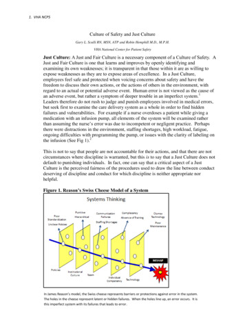
Zhang et al. Stem Cell Research & Therapy(2020) 11:180Page 5 of 13Fig. 1 Hyaline cartilage-like gross morphology and directional distribution of cells and ECM after co-culture in ACECM-oriented scaffold. a After culture for3 weeks, the 3D ACECM-oriented scaffolds were filled with cells and extracellular matrix in the ACAC, ACWJ and ACCC groups, and the appearance of theengineered tissue was semitransparent and glossy. The scale of black bar is 1 cm. b Dead/live staining under 3D confocal fluorescence microscopy. The3D-oriented scaffolds are mainly filled with green fluorescent cells (living cells), with just a few red fluorescence cells (dead cells) present after cultivation for3 days. c SEM results. The upper row shows the high-power (HP) field, and the lower row shows the low-power (LP) field; AC group, the upper and lowerpictures are cross-sectional and sagittal plane views, respectively. In the ACAC, ACWJ and ACCC groups, the HP field shows cells distributed in holes, andthe LP field shows the topological structure of the ECM; black arrows indicate the topological microstructure of ECM secreted by cellsview of the topological microstructure of ECM secretedby cells, the ACCC group presented thick and continuousconnectivity among pores, while the ACWJ and ACACgroups had short and slim morphology, which is critical toguarantee strong mechanical properties, such as pressureand shear forces (Fig. 1c).In vivo experimentsOne goat died during surgery because of an anaestheticoverdose, and another goat’s knee (in the ACWJ group)became infected on the 8th day after the operation but waslater cured. Excluding the cases of infection and death, theremaining goats were included in the results analysis.Neotissue in the ACCC group with mature cartilage MRIsignal and complete subchondral boneMRI of the fresh whole knee joints of the ACCC groupat 6 months showed that the damaged joints were almostcompletely filled with neotissue, and their surfaces weresmooth. However, some cavities were still found in the
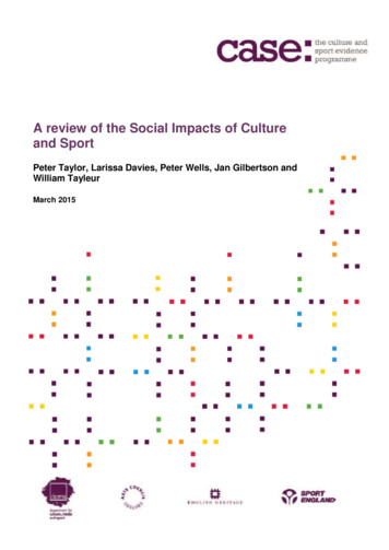
Zhang et al. Stem Cell Research & Therapy(2020) 11:180repair area in the ACAC group and the ACWJ group,and obvious defects were still present in the AC groupand the BC group. At 9 months, the neotissue was uniform and similar to that of native cartilage, and theoedema signal of subchondral bone had completely disappeared. The defects in the ACWJ group, the AC groupand the ACAC group were filled with neotissue, but theMRI results showed that some joint effusion or immature cartilage signal existed (Fig. 2a).Regarding the WORMS results of the knee, the cartilagesignal and morphology scores were better in the ACWJgroup (6 months, 2.0 1.0; 9 months, 1.3 0.6) and theACCC group (6 months, 1.3 1.5; 9 months, 1.6 0.6)than in the AC group or the BC group. The ACAC groupexhibited improved neotissue quality at 9 months compared with that at 6 months. With respect to subchondralbone attrition, the BC group and the AC group weremarkedly concave (5.3 0.6; 6.3 1.2) and slightly concave(2.8 0.7; 1.7 0.4) at 6 months and 9 months, respectively. Compared with the other groups, the marrow abnormality scores were lowest in the ACCC group, with 25% abnormal regions. The total WORMS results revealedthe following: the best neotissue quality and repair wasfound in the knees from the ACCC group and the ACWJgroup, followed by the ACAC group and the AC group.The BC group had the worst WORMS results, with noany repair or regeneration observed (Fig. 2b).The ACCC group achieved faster repair and higher ICRSMECR scores than the other groupsAt 6 months, the cartilage defects were not completelyrepaired with neotissue in any group. However, the ACCCgroup was the only group in which a full-thickness cartilage defect was not observed. At 9 months, the repair areawas plump and glossy because of the regeneration of neotissue in the ACCC group and the ACWJ group, but alocal area of partial-thickness defects intermixed withareas of normal thickness still existed in the AC groupand the ACAC group. The BC group showed almost norepair, or the depth of the defects was quite large (Fig. 3a).The ICRS-MECR results showed that the repair andregeneration in the ACCC group (6 months, 10.0 1.0;9 months, 11.0 1.7) was significantly better than that inthe ACAC group (6 months, 7.0 2.0; 9 months, 9.7 0.6), the AC group (6 months, 4.7 1.2; 9 months, 7.3 2.1) and the BC group (6 months, 1.1 0.4; 9 months,1.5 0.7), which highlights the intense integration withthe border zone, the large degree of defect repair andthe hyaline macroscopic appearance (Fig. 3b).Histological level revealed abundant hyaline cartilagecomponents in the ACCC groupThe H&E results showed that at 6 months, neotissuepadded the defects and covered and tightly aligned withPage 6 of 13the subchondral bone, and the thickness of the neotissuewas similar to that of the surrounding cartilage in theACCC group, whereas in the BC group, the subchondralbone was naked and concave. The neotissue in the ACgroup was thinner and less evenly distributed than inthe cell-scaffold groups, including the ACAC group, theACWJ group and the ACCC group. At 9 months, a distribution of mixed columnar clusters, a smooth surfaceand normal subchondral bone could be clearly observedin the ACCC group. The cartilage in the AC group didnot integrate with the surrounding normal cartilage, andthe cartilage in the ACAC group had an uneven surface(Fig. 4a).Safranin “O” staining revealed that the ECM of neotissue had a sharp outline and showed different thicknessesand various grades of maturity. At 6 months, the ACWJgroup and the AC group showed immature neotissuethat appeared as loose fibrocartilage-like tissue. Compared with the ACCC group, other groups showed infrequent cartilage lacuna formation and dense matrixmasses in the repair area at 9 months (Fig. 4b).Immunohistochemical staining at 6 months showedthat the ACCC group was negative for collagen Iwhereas the AC group and the ACWJ group were not.And collagen I at 9 months in the ACCC group wasslightly higher than AC group. The ACCC group wassignificantly positive for collagen II at both 6 monthsand 9 months, while the control groups but the ACgroup showed negative staining results. Compared withthe AC group, collagen II was less expressed in theACCC group at 6 months, but there was no significantdifference at 9 months (Fig. 4 c, d).According to the ICRS-VHAS, the ACCC group (6months, 13.3 1.2; 9 months, 14.7 2.1), the ACACgroup (6 months, 11.7 1.5; 9 months, 12.7 1.2) andthe ACWJ group (6 months, 11.7 1.5; 9 months, 13.0 2.0) achieved better tissue repair than the BC group (6months, 0.7 0.6; 9 months, 1.0 1.0). The significantadvantages of the ACCC group over the other cellscaffold complex groups included the formation of mature hyaline cartilage-like ECM and a columnar or clustered cell distribution (Fig. 4e).Biomechanical and glycosaminoglycan quantitativeanalysesAs a critical functional indicator of articular cartilage,Young’s modulus was accurately measured using thenanoindentation technique. At 6 months, the Young’smodulus of the ACCC group (3.9 0.9) was significantlyhigher than those of the other groups, including theACWJ group (2.2 0.6), the ACAC group (2.1 0.4) andthe AC group (1.7 0.3). The difference between theACCC group and cell-scaffold groups increased at 9months (Fig. 5a).
Zhang et al. Stem Cell Research & Therapy(2020) 11:180Page 7 of 13Fig. 2 The ACCC group achieved early mature hyaline cartilage regeneration. a T2-weight spin-echo MRI with fat suppression in the sagittalplane and the coronal plane for each group at 6 months (6 m) and 9 months (9 m). Arrows indicate the defect region. b The scores were derivedfor five groups using the International Cartilage Repair Society (ICRS) Whole-Organ MRI Score (ICRS-WORMS) of the knee in OA. WORMS indicatesthe average score of the whole knee at 6 months (6 m) and 9 months (9 m). WORMS-MA, WORMS-Marrow abnormality: WORMS-BA, WORMSbone attrition; WORMS-CA, WORMS-cartilage; T test was used for the data analysis and all data are expressed as the mean standard deviation(n 6). Black circle, black square, black triangle and black star indicate significant differences between the ACCC group and the BC, AC, ACAC, andACWJ groups, respectively. P 0.05 indicates a statistically significant differenceThe total GAG content per sample showed no significant difference among the cell-scaffold groups, but itwas more abundant in the ACCC group at 6 months.The GAG content of the ACCC group was higher thanthat of the AC group at 9 months, while no significantdifference was found between the ACAC group and theACWJ group (Fig. 5b).hWJMSCs participated in the repair and regeneration ofcartilage defectsAs a human leukocyte antigen I molecular marker, HLAABC immunofluorescence staining can provide evidencedemonstrating that hWJMSCs are one part of the neotissueand evade from surveillance of the immune system. Greenfluorescence can be observed in the repair area of theACCC group, while it is absent in the normal area, indicating that hWJMSCs were present in the repair area and werean important component of neotissue (Fig. 6).DiscussionA co-culture system provides a potential strategy for therepair and regeneration of cartilageArticular cartilage injury is a common sports-related disease that severely affects subsequent achievements andcareer planning. A co-culture system provides a win-winstrategy to eliminate issues related to chondrocytes and
Zhang et al. Stem Cell Research & Therapy(2020) 11:180Page 8 of 13Fig. 3 The repair area was smooth and full in the ACCC group. Gross morphology of the femoral condyle, the counterpart meniscus and the tibiaplatform and the results of International Cartilage Repair Society (ICRS) scoring system. a The red-dotted circles show the defects and the repairarea; the black arrows and the black bars indicate the torn meniscus and a 1-cm scale, respectively. b ICRS scoring system for macroscopicevaluation of cartilage repair, ICRS-ORA, ICRS-overall repair assessment; ICRS-MA, ICRS-macroscopic appearance; ICRS-IBZ, ICRS-integration toborder zone; ICRS-DDR, ICRS-degree of defect repair; n 6, data are expressed as the mean standard deviation; black circles, black squares, blacktriangles and black stars indicate significant differences between the ACCC and the BC, AC, ACAC and ACWJ groups, respectively. P 0.05indicates a s
In vitro, a co-culture system was constructed using unpassaged pACs possessing their native phenotype and an ACECM-oriented scaffold retaining native cartilage tissue components and oriented structures [21-23]; hWJMSCs were displayed as an alternative cells source for cartilage tissue engineering because of distinct chon-