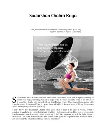
Transcription
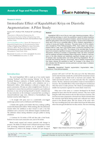
Open AccessAnnals of Yoga and Physical TherapyResearch ArticleImmediate Effect of Kapalabhati Kriya on DiastolicAugmentation: A Pilot StudySarate SG1*, Pallem NM1, Pathak SD2 and BhogalRS21Department of Biomedical Engineering, SriSivasubramaniya Nadar College of Engineering, India2Department of Scientific Research, Kaivalyadhama YogaInstitute, India*Corresponding author: Sarate SG, Department ofBiomedical Engineering, Sri Sivasubramaniya NadarCollege of Engineering (Affiliated to Anna University,Chennai), B806, Marg Pushpadruma Apartments,Old Mahabalipuram Road, Kalavakkam, District:Chengalpattu, Tamil Nadu, IndiaReceived: November 25, 2020; Accepted: December14, 2020; Published: December 21, 2020AbstractKapalabhati (KB) is one of the six main yogic cleansing processes. KB is ayogic breathing technique in which the practitioner resorts to bellow breathingthrough an active exhalation and passive inhalation, wherein the lower part of theabdomen gets pulled in with every active exhalation. The aim of this fundamentalstudy was to find out an immediate effect of KB on Diastolic Augmentation ina group of young and healthy volunteers. The pulse waves of all the subjectswere recorded by Photo Plethysmography (PPG). Each subject was made topractice KB for 3 sets, each of 50 strokes while a continuous recording of thepulse wave was made using PPG. The analysis of the recorded data of pulsewave forms showed a significant increase in the Diastolic peak amplitude duringKB practice, showing an increase in Augmentation Index (AI) when comparedwith the readings recorded immediately before KB practice. Student’s t-test wasdone on the data obtained and statistically significant changes were seen inAI between data recorded before and while performing Kapalabhati. We canconclude that practicing KB may, convincingly, lead to Diastolic Augmentationthat clearly reduces the workload on heart. An increase in the values of AIalso indicates that KB improves coronary perfusion and also helps in collateralformation of capillaries.Keywords: Kapalabhati; Diastolic augmentation; Augmentation index;Compliance; Pulse plethysmographyIntroductionThe word Kapalabhati (KB) is made up of two words: kapalameaning “forehead” and bhati meaning “shining” [1,2]. The termForehead Shining is indicative of blissful meditation like feeling.The technique of KB involves gentle, active exhalation just as ablacksmith’s bellow, while inhalation is allowed to happen with apassive automation. KB steps are rather straightforward. However, itis important to ensure that one performs it properly as per traditionaldirections or may end up hurting himself or herself. A detailed stepby step instruction on the technique is presented here according toclassical/traditional yogic texts, followed at Kaivalyadhama YogaInstitute Lonavla, India (Figure 1):1. Sit straight with spine erect in a comfortable cross-leggedseated yogic posture such as Sukhasana and Padmasana.2. Exhale with a gentle, active bellow-like force, making a puffingsound of the blacksmith’s bellow. While exhaling, allow the abdominalmuscles below the umbilicus to draw inward in such a way thatexhalation and a concavity in the abdomen happen simultaneously.The abdomen relaxes during inhalation and gets pulled in duringexhalation.pressure (ΔP) and C ΔV/ΔP. The aorta up to the iliac bifurcationand its main branches namely the carotid and the innominate arteriesare compliant vessels, which work as reservoirs and conduits. Theradial and femoral arteries are more muscular than the compliantarteries. The iliac, subclavian and auxiliary vessels are intermediate instructure. The extensible fibres of the vessel wall govern its behaviourwhen that artery is under pressure. The volume accommodated for asmaller rise in pressure is greater for a more elastic vessel [3,4].Pulse wave of the artery is a contour developed by the bloodpumped by heart when the left ventricle contracts. This wave travelsalong the walls of the arteries in the tree-like structure of arteries. Thiswave has two main components-a forward wave which is formed whenthe ventricles expel blood during systole and a reflected wave whichreturns in the diastolic phase, after the closure of the aortic valve.The wave travelling down the aorta reflects back from the bifurcationwhere the aorta gets divided into two iliac arteries. The wave returning3. Continue for 50 strokes of KB and then take a brief break.Perform two more such consecutive sets, each of 50 strokes.Blood vessels expand and contract passively with changes inpressure. The ability of a blood vessel to distend and increase volumewith increasing transmural pressure can be quantified as vesselCompliance (C) which is the change in volume (ΔV) per change inAnn Yoga Phys Ther - Volume 4 Issue 1 - 2020ISSN: 2573-8585 www.austinpublishinggroup.comSarate et al. All rights are reservedFigure 1: Steps followed while performing Kapalabhati.Citation: Sarate SG, Pallem NM, Pathak SD and Bhogal RS. Immediate Effect of Kapalabhati Kriya on DiastolicAugmentation: A Pilot Study. Ann Yoga Phys Ther. 2020; 4(1): 1043.
Austin Publishing GroupSarate SGFigure 2: Pulse Wave.In this experimental work, we are presenting the data from twodifferent experiments. In one experiment, BIOPAC MP 45 module wasused to detect the pulse waves on one subject. In second experiment,we used a device called PulseSensor to acquire the PPG signal. Theintra-abdominal pressure is raised transiently while practicing KB. Inthe above-mentioned traditional technique, the whole of abdominalmusculature is not contracting forcefully. In this technique, onlythe abdominal musculature below the umbilicus is getting pulled induring the exhalations. This movement can generate a pressure wavein the aorta to the returning wave and help in coronary filling. Theseexperiments were designed to examine this aspect of KB so that itcan be recommended to patients with cardiovascular diseases as atherapeutic measure.Materials and MethodsFigure 3: Block diagram of experimental setup.to the heart helps in supplying blood to the myocardium by fillingthe coronary arteries during diastole. That is why the velocity of thereturning wave is very important. If the arteries are stiffer, the bloodreturns faster. If the returning wave is faster, then it may enter intothe forward wave of the same cardiac cycle and increases the bloodpressure reading and inadequately fills the coronary arteries [5]. Itclearly shows schematic form of a typical PPG waveform (Figure 2).The diastolic wave can be augmented using devices like IntraAortic Balloon Pump (IABP) or Enhanced External CounterPulsation Technique (EECP). IABP consists of a balloon whichis synchronised with the ECG and inflates in the aorta during thediastole [6,7,8,9]. The EECP machine consists of three cuffs, whichare placed over the calf, thighs and buttocks. These cuffs are alsosynchronised with the ECG and create a squeezing action startingfrom the calf towards the abdomen [10,11,12,13].To monitor the blood volume changes in microvascular bed oftissue, we can use Photo Plethysmography (PPG) which is a simpleand low-cost technique [14,15,16,17,18,19,20]. It is a non-invasivetechnique used to measure heart rate from skin surface. Blood ispumped by the heart to periphery during each cardiac cycle. Thepressure drops by the time blood comes to the skin, but it is sufficient tochange the diameter of the arteries and arterioles in the subcutaneoustissue. The change in volume because of the pressure wave can bedetected with gentle placement of a light emitting diode over theskin, which illuminates the skin and then measures the amount oflight, which gets transmitted or reflected back to a photodiode. In thiswork, we are exploring the therapeutic use of KB like IABP and EECP,without any expenses.Submit your Manuscript www.austinpublishinggroup.comIn the first experiment, the BSL (Biopac Student Learning)system was used. It includes data acquisition hardware MP 45 whichhas built-in universal amplifiers to record and condition variousbio-signals. This data acquisition system receives the signals fromPPG sensor placed on the finger of the individual. The signal wasacquired through Acqcheck 4.0 software. In the second experiment,PulseSensor [21] hardware was used to record the PPG signals. Thesignal which comes from this device is analog voltage signal with apredictable wave shape. This sensor was connected to the computervia NI DAQ USB-6008, which is a data acquisition device that canperform many functions. The data acquired through this device wasvisualised on the computer using NI LabView software. Informedconsent was taken from all subjects prior to the experiments.The first study was performed on seven male subjects. However,it was observed that there were much motion artefacts in most ofthe data and the data from only one subject could be processed. Theissue of motion artefact was solved in the subsequent experiment bydesigning a hand stabiliser, which prevented the body movementsfrom reaching the sensor placed on the fingertip. A group of 15healthy subjects within the age group of 20-21 participated in thesecond study. All subjects had a normal blood pressure and had nohistory of any cardiovascular disease. They were asked to practice KBas per instructions stated above. The sensor was placed on the rightindex finger of each subject. None of the subjects had any previousexperience of KB practice.The pulse sensor consists of three terminals, one for Ground,second for 5V VCC and third one for Output Analog pin. TheSensor was given a power supply of 5V and grounded. The Analogoutput pin was connected as input to the NI DAQ 6008, whichwas integrated inside the LabVIEW software. Sampling rate wasset as 100Hz for the signal acquisition and stored as .xlsx file. Thepulse signal was visualized in the graph indicator of the LabVIEWsoftware. “While” loop was used for the data acquisition block to havea continuous monitoring of the pulse signal obtained from the sensor.For removing noise form the signal, smoothing filter was used.Smoothing filter is a moving average filter, which averages the valuesof the signal and gives a clear waveform without the noise. Smoothingfilter block is available in the LabVIEW software and it was placedjust after the signal acquisition block for the DAQ. Smoothing filterwas useful for the first derivative signal, which had noise with ripples(Figure 3).Ann Yoga Phys Ther 4(1): id1043 (2020) - Page - 02
Austin Publishing GroupSarate SGFigure 6: Superimposition of first derivative of the original signal over theoriginal signal in a signal acquired before performing KB.Figure 4: Block Setup in LabVIEW.Figure 7: Superimposition of first derivative of the original signal over theoriginal signal in a signal acquired while performing KB.amplitude.Figure 5: Plots of PPG waves before and while practicing Kapalabhati withVoltage in mV on X-axis and time in milliseconds on Y-axis.Python programming language was used to visualize and analysethe PPG signals of the subjects. Python packages ‘pandas’ and‘bioread’ were used to load the data files. ‘BaselineRemoval’ packagewas used to remove the baseline drifting of the PPG signal. ‘Matplotlib’package was used to plot and visualize the PPG signals. The segmentsof normal pulse waves and those with increased diastolic peakswere plotted to compare the changes in diastolic peaks. The package‘numpy’ was used to apply first derivative to the PPG signals.The upper panel in (Figure 5) shows the waveform of PPG beforepracticing KB and the lower panel shows the waveform of PPGwhile practicing KB in one of the subjects. It is clearly visible that thediastolic peaks in the plot while practicing KB show higher diastolicpeaks.To detect the systolic and diastolic peaks, the first derivative ofthe acquired signal was taken. The points in the first derivative signal,with zero value, coincide with the peaks in the original signal. Thesystolic and diastolic peaks were obtained by superimposing theoriginal signal with the first derivative plot of the same signal. Theamplitude was noted at all such time points in the original denoisedsignal to get the values of the systolic peak (X) and diastolic peak (Y)amplitudes. The AI was calculated by using the formulaAI Y/XWhere X is Systolic peak amplitude and Y is Diastolic peakSubmit your Manuscript www.austinpublishinggroup.comFigure 6 shows the PPG signal taken before practicing KB,being superimposed on the first derivative plot of the same signal.(Figure 7) shows the PPG signal taken while performing KB, which issuperimposed on the first derivative plot of the same signal.ResultsIn the first experiment, there were least motion artefacts in onesubject. The data collected was analysed using Python programminglanguage. The SciPy package was used to check for normality of thedata using the normal function. The p-value was 0.000635, which isway less than the alpha value of 0.05. Thus, we assumed for all ourdata that the data is normally distributed and used Student’s t testto test the significance of difference between values of AI before andwhile performing KB. The averages of the values of AI were plottedalong with their standard deviations as shown in (Figure 8).t-valueFrom the table, the t Stat value was obtained as -13.255 (Table1). The t Critical values for both the one-tail and two-tail test weregreater than the t Stat value. This indicates that the there was asignificant difference in the AI values of the subject before and whileperforming KB.p-valueThe p-values from both the one-tail and two-tail tests are 1.64E07 and 3.29E-07 respectively. These values are far lesser than thesignificant level alpha, which is generally taken as 0.05. If the p-valueis greater than significance level of 0.05, it means that there is 95%Ann Yoga Phys Ther 4(1): id1043 (2020) - Page - 03
Austin Publishing GroupSarate SGTable 1: Student’s t test Results in First Experiment.t-Test: Paired Two Sample for MeansVariable 1Variable vations1010Pearson Correlation0.142897Hypothesized Mean Difference0df9t Stat-13.255P(T t) one-tail1.64E-07t Critical one-tail1.833113P(T t) two-tail3.29E-07t Critical two-tail2.262157Figure 8: Average of AI values of 1 subject before and while performing KBin the first experiment.Table 2: Student’s t test Results in Second Experiment.t-Test: Paired Two Sample for MeansVariable 1Variable ervations15Pearson Correlation-0.28659Hypothesized Mean Difference0df14t Stat-8.26147P(T t) one-tail4.70E-07t Critical one-tail1.76131P(T t) two-tail9.40E-07t Critical two-tail2.144787chance that there is no significant difference between the two groupson which the t-test was performed. The p-values obtained were lesserthan 0.05, which shows that there was significant difference betweenthe values before and during KB.In the second study, a wrist stabiliser was used to prevent thebody vibrations from reaching the index finger where the sensor wasplaced. The AI values for all the 15 subjects, before and during KB,were calculated. To examine if there is a significant change in the AIvalues before and while performing KB, Student’s t-test was used. Theresults of t-test, for paired two sample for Means, were obtained asbelow using Microsoft Excel.t-valueFrom the table, the t Stat value was obtained as -8.261465381(Table 2). The t Critical values for both the one-tail and two-tail testwere greater than the t Stat value. This indicates that the there was asignificant difference in the AI values of the subjects before and whileperforming KB.p-valueThe p-values from both the one-tail and two-tail test are 4.69971E07 and 9.39941E-07 respectively. These values are far lesser than theSubmit your Manuscript www.austinpublishinggroup.comFigure 9: Plots of all 15 subjects before and while performing KB.Figure 10: Average of all 15 subjects before and while performing KB.significant level α, which is generally taken as 0.05. The p-valuesobtained were lesser than 0.05, which shows that there was significantdifference between the values before and during KB.The AI values of all the 15 subjects were separately plotted asshown in (Figure 9). The values for all 15 subjects were then averagedand plotted collectively, as shown in (Figure 10). With these results,it is observed that practice of KB causes diastolic augmentation in thecapillaries of the finger.DiscussionVarious traditions of Yoga instruct the practice of KB in differentways. In one tradition, the procedure says the practitioner should sitwith erect neck and back, inhale through both nostrils and exhalerapidly by flapping the abdomen during each exhalation at a paceof 60-120 breaths/min [30]. In the technique, which was followed inour experiments, only the lower abdominal muscles are used duringexhalation and that too with a gentle force. The results from our twopilot experiments show that there is significant diastolic augmentationAnn Yoga Phys Ther 4(1): id1043 (2020) - Page - 04
Austin Publishing GroupSarate SGwhile practicing traditional KB used in this study. Diastolicaugmentation, seen in PPG signal, represents the transient increasein pressure in the arteries during diastole. Diastole is the time whenthe heart relaxes and prepares for the next systole. The blood whichremains in the arterial tree at the end of the diastole creates load onthe heart. The heart has to pump the blood from the left ventriclesinto the aorta. When the aortic valve opens up in the ejection phaseof the cardiac cycle, the blood which is already in the arterial treeopposes the blood which is being newly pumped by the ventricle [3].During diastole, if the volume of blood in the arterial tree is pushedforward in the tributaries by pressure created by the reflected wave,then the left ventricle gets lesser resistance in upcoming systole.Thus, the load on the heart gets reduced. We have found in ourstudy that while performing KB the diastolic pressure wave getsenhanced i.e., augmented. This augmented pressure wave duringdiastole distributes blood to all organs of the body, thus improving theoverall perfusion of all organs. Also, the coronary arteries get a bettervolumetric filling if the diastolic augmentation takes place [6-13].Heart receives its own adequate blood supply during the diastole only,as during systole the contracted myocardium squeezes the coronariesand empties them. Hence, during diastole, the reflected wave pushesblood into the coronaries through the ostia. Thus, the study showsthat KB provides all the benefits of diastolic augmentation and canbe used as a therapeutic process. Various studies on KB have shownthat there is sympathetic stimulation of the heart with practice of KB.With our results, it can be seen that there is diastolic augmentation ofthe arterial system using PPG. Along with the diastolic augmentationof the arterial system, the gentle strokes on lower abdomen also exertforce on the inferior vena cava, thereby improving the venous returnto the heart. The increased venous return to the right atrium must beresponsible for the increase in the LF/HF ratio which is a parametershowing increase in sympathetic activity in the sympathovagal toneof the heart [27,28,29]. It is evident from our study that the mostprobable cause of increase in LF/HF ratio is the Frank-Starlingmechanism [3] and not the increase in sympathovagal tone. It may bepossible that the increased venous return to the right atrium causesthe increase in the heart rate and increases the LF/HF ratio of theHRV. Increased venous return may increase heart rate by 10 to 15 %above the existing baseline rate, before practicing KB. Thus, it mayrise from normal 72-84 to 84-96 beats per min. In well controlledhypertensive patients with anti-hypertensive drugs, baseline pulserate is normal up to 80/min and so KB should not have any adverseeffect.As KB causes diastolic augmentation, it is also evident that it willgive all the good effects of other techniques like EECP. The diastolicaugmentation improves the perfusion of brain, heart as well askidneys. Due to reduction in afterload on the heart, the stroke volumeof the heart also improves. This improves the left ventricular ejectionfraction as well. The increased perfusion of various organs stimulatesthe endothelial cells. They produce nitric oxide and VascularEndothelial Growth Factor (VEGF) [12,22,-25] which are known tohelp in angiogenesis. This helps in development of collateral bloodvessels in the arterial tree. Thus, all vital organs benefit from thepractice of KB. As observed in studies on EECP [10-13], KB must alsobe reducing the endothelin and Brain Natriuretic Polypeptide (BNP)which are released in ventricular stress. All these benefits of EECPSubmit your Manuscript www.austinpublishinggroup.comshould further be investigated for KB.ConclusionWhen KB is practiced with the instructions used in ourexperiments, it shows significant increase in the AI values whileperforming KB. The statistical analysis of our data collected from15 healthy individuals shows that KB produces significant diastolicaugmentation. These results indicate that KB improves coronaryfilling, as well as, perfusion to all vital organs in the body as it causesdiastolic augmentation. Thus, KB can be used as an alternative toexpensive treatment modalities like EECP and give good results. Amore extensive study is needed to further explore the effects of KB.Thus, we can conclude that KB can be a good economic alternative toEECP, which is used in treating angina.At present, hypertensive and Ischaemic Heart Disease patientsare not advised to perform KB with an apprehension that KB mightincrease load on heart. In one study it was observed that KB doesnot cause Hyperventilation Syndrome [26]. It is clear that KB onlymarginally increases the heart rate. But, by not allowing patients topractice KB one deprives them of the benefits of improved health ofheart musculature which is obtainable due to improved coronaryperfusion as a result of diastolic augmentation. This also reducesthe heart load due to redistribution of blood to all organs and in theprocess improves their functions, which in turn, will improve theoverall health in general. Besides being deprived of cardiovascularsystem benefits, one is also deprived of the most important effects andbenefits of KB on respiratory system [30], if patients are not allowedto practice KB. To name a few benefits; 1. Improving the overalllung capacity by opening up alveoli in passive regions of the lungs,2. Providing adequate oxygenation to all organs so as to make themwork efficiently, 3. Forceful exhalation improving the blood supply ofthe respiratory tract mucosa. This enhances phagocytic action againstpathogens, thus improving immunity [31].We would like to conclude that if one practises classical KB, asdescribed above, in relation to method and also quantity, one mayimmensely be benefitted. However, controlled studies on a largerscale would alone fully authenticate the results of this study and lendcredence to our plausible claim that the classical mode of KB can bepracticed by all with advantage. It is conjectured, through this study,that the gentle nature of the traditional KB practice may show moreenhanced meditative feelings, thus facilitating meditation a far betterway.AcknowledgementWe are grateful to Dr. A. Kavitha, Professor and Headof Department, Department of Biomedical Engineering SriSivasubramaniya Nadar College of Engineering, for giving uspermission to use the lab facilities at the Department. We also extendour gratitude to Mr. Sivananthan and Mr. Sasidharan who helpedus in the labs through our experiments. The authors also thank ShriO.P.Tiwari Secretary Kaivalyadhama and Shri Subodh Tiwari CEOKaivalyadhama for encouraging the research study.References1. Sahay GS, Bhogal RS. Kapalabhati Techniques in Selected Classical andModern Yoga Texts-A. Yoga Mimamsa. 2012; 4:326-347.2. Swami NS. Prana and Pranayama. 1st edn. Bihar School of Yoga: YogaAnn Yoga Phys Ther 4(1): id1043 (2020) - Page - 05
Austin Publishing GroupSarate SGPublications Trust, Munger, Bihar, India. 2009; 143-145.3. Hall JE, Hall ME. Guyton and Hall Textbook of Medical Physiology. 14editon. Elsevier. 2020.th4. Richard EK. Cardiovascular Physiology Concepts. 2020.5. Ranganathan N, Sivaciyan V, Saksena F. The art and science of cardiacphysical examination. 2nd edn. Totowa NJ, editors: Humana Press. 2006.6. Gurbel PA, Anderson RD, MacCord CS, Scott H, Komjathy SF, PoultonJ, et.al. Arterial diastolic pressure augmentation by intra-aortic ballooncounterpulsation enhances the onset of coronary artery reperfusion bythrombolytic therapy. Circulation: Journal of the American Heart Asoociation.1994; 89: 361-365.7. Krishna M, Zacharowski K. Principles of intra-aortic balloon pumpcounterpulsation. Continuing Education in Anaesthesia. Critical Care & Pain.2009; 9: 24-28.8. Ozdemdir O, Alyan O, Kacmaz F, Kaptan Z, Ozbakir C, Geyik B, et.al.Evaluation of Effects of Intra aortic Balloon Counterpulsation on AutonomicNervous System Functions by Heart Rate Variability Analysis. AnnNoninvasive Electrocardiol. 2007; 12: 38-43.9. Risk MR, Ramirez AJ, Fischer EIC. Hemodynamic Improvement andHeart Rate Variability during Aortic Counterpulsation. Conference paper inComputers in Cardiology. IEEE. 2004; 31: 453-455.10. Wang LQ, Persson BG, Bengmark BG. Collateral Formation after RepeatedTransient Dearterization of the Rat Liver. HPB Surgery. 1992; 6: 105-113.11. Dierk W, Tragner P, Wawer A, Porst H, Werner GD, Gross P. Enhancedexternal counterpulsation: a new technique to augment renal function in livercirrhosis. Nephrol Dial Transplant. 2005.12. Arthur MF. Enhanced External Counterpulsation: Mechanism of Action. ClinCardiol Suppl. 2002; 25: 11-15.13. Wu X, Zhang Y, Yuan H. The Optimization Study on Time Sequence ofEnhanced External Counter-Pulsation in AEI-CPR. Journal of Computers.2009; 4: 1243-1248.14. Elgendi M. On the Analysis of Photoplethysmogram Signals. CurrentCardiology Reviews. 2012; 8: 14-25.15. Yadhuraj SR, Sudarshan BG, Prassana Kumar SC. GUI Using MATLABfor PTT and HRV Analysis for Detecting Early Changes in PhysiologicalVariables. International Journal of Advanced Scientific Research andManagement. 2019; 4: 221-226.17. Yousef Q, Reaz MBI, Ali MAM. The Analysis of PPG Morphology: Investigatingthe Effects of Aging on Arterial Compliance. Measurement Science Review.2012; 12: 266-271.18. Tamura T, Maeda Y, Sekine M, Yoshida M. Wearable PhotoplethysmographicSensors-Past and Present. Electronics. 2014; 3: 282-302.19. Rundo F, Conoci S, Ortis A, Battiato S. An Advanced Bio-Inspired PhotoPlethysmography (PPG) and ECG Pattern Recognition System for MedicalAssessment. Sensors. 2018; 18: 405.20. Allen J. Photoplethysmography and its application in clinical physiologicalmeasurement. Physiol Meas. March. 2007; 28: R1-R39.21. PulseSensor Playground Toolbox.22. Tammela T, Enholm B, Alitalo K, Paavonen K. The biology of vascularendothelial growth factors. Cardiovascular Research. 2005; 65: 550-563.23. Olsson AK, Dimberg A, Kreuger J, Claesson-Welsh L. VEGF receptorsignaling-in control of vascular function. Nature Reviews. 2006; 7: 359-371.24. Tabibiazar R, Rockson SG. Angiogenesis and the ischaemic heart. EuropeanHeart Journal. 2001; 22: 903-918.25. Cotton JM, Thomas MR, Dunmore BJ, Salisbury J, Shah AM, Brindle NPJ.Angiogenesis in chronically ischaemic human heart following percutaneousmyocardial revascularisation. British Medical Journal. Heart. 2002; 87: 281283.26. Wenger MA, Bagchi BK. Studies of Autonomic Functions in Practitionersof Yoga in India. Behavioral Science. Journal of the Society for GeneralSystems Research. 1961; 6: 312-323.27. Yadav A, Singh S, Singh KP. Role of Pranayama breathing in rehabilitationof coronary artery disease patients-A pilot study. Indian Journal of TraditionalKnowledge. 2009; 8: 455-458.28. Puthige R, Ramakrishnana AG, Nagendra HR, Telles S. Indian Journal ofPhysiology and Pharmacology. 1998; 42: 467-472.29. Gupta R. Acute Effect of Kapalbhati Yoga on Cardiac Autonomic ControlUsing Heart Rate Variability Analysis in Healthy Male Individuals. Journal ofHuman Physiology. 2020; 2: 16-22.30. Saoji AA, Raghavendra BR, Manjunath NK. Effects of yogic breath regulation:A narrative review of scientific evidence. Journal of Ayurveda and IntegrativeMedicine. 2019; 10: 50-58.31. Pathak SD. Yoga through Doctor’s Eye. Sudha Publication. 2016.16. Wowern Ev, Ostling G, Nilsson PM, Olofsson P. Digital Photoplethysmographyfor Assessment of Arterial Stiffness: Repeatability and Comparison withApplanation Tonometry. PLoS One. 2015; 10.Submit your Manuscript www.austinpublishinggroup.comAnn Yoga Phys Ther 4(1): id1043 (2020) - Page - 06
output pin was connected as input to the NI DAQ 6008, which was integrated inside the LabVIEW software. Sampling rate was set as 100Hz for the signal acquisition and stored as .xlsx file. The pulse signal was visualized in the graph indicator of the LabVIEW software. "While" loop was used for the data acquisition block to have