Transcription
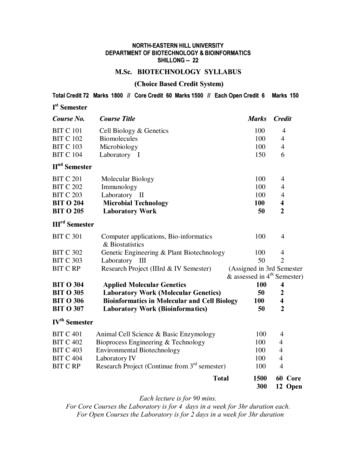
International Journal of Molecular and Clinical Microbiology 11(2) (2021) 1495-1508International Journal ofMolecular and Clinical MicrobiologyRecombinant production of chimeric protein Mfp-3-GvpA in yeast PichiapastorisNazanin Bolghari1, Hadi Habibollahi1, Masoumeh Anvari1 and Hosein Shahsavarani1,2*1. Department of Biology, Rasht branch, Islamic Azad University, Rasht, Iran.2. Department of Cell and Molecular Sciences, Faculty of Life Sciences and Biotechnology, Shahid Beheshti Univ., Tehran,Iran.ART ICLE INFOArticle history:Received 22 December 2020Accepted 24 August 2021Available online 1 December 2021keywords:Mytilus edulis,Mfp-3,GvpA,Pichia pastoris,Protein expirationABSTRACTMussels secrete protein-based polymers mainly mussel foot proteins (Mfps),enabling them to anchor to various surfaces in a saline, intertidal zone. Using Mfpproteins as novel water-resistant adhesive materials have been suggested forbiomedical purposes due to their unique features including large abundance ofcatechol aligned with amphiphilic and ionic properties. Despite the more promisingcharacteristics of foot proteins, they have not been widely exploited for biomedicalor even industrial applications, since farming the mussels are not commerciallyviable due to ecological issues and their highly territorial nature. Present studyaimed to examine engineering of recombinant synthesis of Mfp3 protein fused toanother marine based curli protein, gas vesicle protein A(GvpA) in nonpathogenicyeast, Pichia pastoris, expression system. To this end, sequences of the Mfp3 andGvpA were extracted from the NCBI database and were inserted into the pPICZ Avector in order to obtain an efficient expression level of chimeric protein. Obtainedvectors were transformed into E. coli TOP10F' for multiplication and thenlinearized plasmid transformed into Pichia pastoris for protein expression. The bestexpression level obtained after 96 hours' incubation with methanol induction. Inconclusion, active recombinant adhesive Mfp3-GvpA fused protein weresuccessfully expressed in P. pastoris suggesting potential expression system forfuture bio-adhesive or any fused protein production. .1. IntroductionThe production of bio adhesives with therequired characteristics (biocompatibility, nontoxicity in the human body, and strong, stableadhesion) has long been considered by scientists.One of the most important points in theproduction of bio adhesives is the resistance ofthis material in situations similar to thephysiological conditions of the human body(humid conditions with low pH( . Actually,every produced bio adhesive has toxicprecursors that in many cases lead to tissuerejection or cancer (Amini et al., 2017; Cha etal., 2008; Ferdosian et al., 2017).Mussel Mytilus edulis has long been ofinterest to scientists because it has the ability toproduce sticky proteins with incredibleproperties, including resistance to wet conditionsand low pH (Even et al., 2008; Zhao and Waite,2006). Mytilus edulis can produce eight types ofdifferent sticky proteins that help each other toproduce viscose substances. These proteinsinclude mfp-1, mfp-2, mfp-3, mfp-4, mfp-5,
N. Bolghari et al./International Journal of Molecular and Clinical Microbiology 11(2) (2021) 1495-15081496PCOL-D, and PCOLNG (Danner et al., 2012;2.2. Transformation of Gene in to the E. coliLu et al., 2012).TOP10F ′ cellMfp-3 and mfp-5 proteins are of particularThe vector was transferred to the competentimportance because they have a higher adhesioncell of E. coli TOP10F′ (TOP10F is a type of E.strength than other proteins produced by Mytiluscoli bacteria that is very useful for geneedulis. The high adhesion strength of thesetransfer). In this study, this cell was preparedproteins is because of their large amount offrom Invitrogen in Luria-Bertani agar mediumDOPA (3 and 4 dihydroxyphenylalanine) (Evenwith zeocin antibiotic (final concentration of 50et al., 2008; Hosseini et al., 2017; Warner andμg / mL) by the heat shock method (Froger andWaite, 1999). As the smallest protein in Mytilus,Hall, 2007). Then, plasmid was extracted frommfp-3 contains 20-25% of DOPA and a highthe bacterial cell with a high concentration usingamount of arginine, which changes to 4Gen all plasmid extraction kit (Dadgar et al.,hydroxy-arginine and 2, 4 tryptophan in post2015).translational (Barclay et al., 2017; Hwang et al.,Concentrated plasmids were linearized by2012; Yajing Kan et al., 2014; Li et al., 2020).Sac1 enzyme by the overnight method (Urdea etThe production of mfp-3 recombinant stickyal., 1988). The linear gene was then cleaned upprotein in bacterial hosts has been done byby the Gen all cleanup kit to remove salts anddifferent methods, but research has shown thatRNAs (Dadgar et al., 2015). Afterwards, PCRusing curli proteins with the sequence of musselwasperformedwithprimerswhosefoot adhesive proteins can increase the gth (Chao Zhong et al., 2014).program is listed in Table 2.Research on the effect of different pHs onThen, electrophoresis gel (1% agarose) andmussel adhesive proteins shows that theDNA ladder (100-10kda bp) (SMBIO DM3200)adhesion of mussel foot protein in acidicwere utilized to confirm the presence of theconditions is much higher than in neutral orgene. After staining the gel with DNA STAINalkaline conditions. Because under acidic(Gen Bio) for checking the presence of the gene,conditions, DOPA is immediately oxidized tothe gel was transferred to the gel documentationdopaquinones (Ezzeldin et al., 2012). In thesystem for the final testing.study conducted by Cereghino et al., bacterialcells were used as hosts, but due to the inability2.3. Pichia pastoris competent cellsto be modified after the translation ofprokaryotic cells, the addition of the tyrosinaseTo prepare the Pichia pastoris competentenzyme was required to convert all tyrosine tocell,a single clone of Pichia pastoris strainDOPA. The production of recombinant proteings115(Invitrogen) was first cultured in 5 ml ofin prokaryotic hosts was low and a large amountYPD MEDIOM medium and placed overnight.of culture medium was required to produceWhen the OD of the desired culture mediumprotein (Cregg et al., 2000; Damasceno et al.,reached OD 600, it showed the ideal growth of2012).yeasts. At this stage, 800 μl of pre-culturesample was introduced into 125 cc of culture2. Materials and Methodsmedium. After 24 hours, when OD 0.5 was2.1. Sequence selection and design of genereached, another 300 μl of pre-culture mediumstructureswas added after another overnight OD 1.3.At this stage, the whole culture medium wasAfter selecting the sequence of mfp-3, thecentrifuged at 1500 rpm for five minutes at 4 Csequences of GvpA, JS Linker, His-Tag, and(Eppendorf). Next, the supernatant wasantibiotic resistance (Zeocin) were added to thediscarded, and 15 ml of cold sterile water wasabove-mentioned sequence, and the sequenceadded to the remaining sediment. Soft pipingwas optimized for expression in the yeast. Then,was performed to dissolve the sediment well.the sequence was ordered to Biomatik companyThe centrifuge was performed again at 1500 rpmin Canada, and the company synthesized it andin five minutes and 4 C; then the supernatantput it inside the pPICZA vector.was discarded, and 7.5 ml of cold sterile waterwas poured and pipetted to dissolve the
N. Bolghari et al./International Journal of Molecular and Clinical Microbiology 11(2) (2021) 1495-1508precipitate well. Afterwards, the previouscentrifuge process was repeated. Next, 1 ml ofcold sorbitol (1 M) was poured and pipettedagain until the precipitate was completelydissolved and centrifuged. The supernatant wasdiscarded and 250 μl of cold sorbitol was added,pipetted and the samples were transferred to ice.The yeast cell stage was ready to receive aforeign gene (Competent cell) (to prepare thecompetent cell, the Invitrogen protocol was used(dos Santos et al., 2016; Higgins, 1998).2.4. Transformation geneThe concentrated plasmid was transferred tothe competent cell of the Pichia pastoris byelectroporation. At this stage, electroporationdevice (pulsar-Eppendorf) was used (power2000 watts and time of 500 microseconds).Yeast cells containing possible genes werefurther examined for five days in YPDS culturemedium (1% yeast extract, 2% peptone, 2%dextrose, 1 M sorbitol, 2% agar) with zeocin(100 μg mL-1 g) at 28 Degrees Celsius.(LinCereghino et al., 2005).2.5. PCR test to prove the existence of the targetgeneTo confirms the presence of the gene in theyeast, the program of Table 1, gelelectrophoresis (1% agarose) and DNA ladder(bp100-10kda) were used. After staining the gelwith DNA stain, the gel was transferred to thedocumentation gel system.2.6. Yeast cell growthTo evaluate the growth of yeast cells afterthe addition of methanol in hours 0, 12, 24, 48,72 and 96, 2 cc was taken from the growingsample. and samples were examined for ODObviously over time and adding the rightamount of methanol. We have observed thelogarithmic growth of yeast cells As shown inFigure 10 (Higgins and cregg,1998).2.7. Protein expression in Pichia pastorisThe Invitrogen protocol was used for geneexpression and protein production (Higgins andCregg, 1998). For this purpose, Pichia pastoriswas first cultured in Buffered complex glycerolmedium (BMGY) as the standard complex1497medium for Pichia pastoris cultivationcontaining peptone, yeast extract, yeast nitrogenbase (YNB), and a phosphate buffer. After 18hours, the whole culture was centrifuged, andyeast precipitate was transferred to Bufferedcomplex methanol medium (BMMY) as thestandard complex medium for Pichia pastoriscultivation containing peptone, yeast extract,yeast nitrogen base (YNB), and a phosphatebuffer. This culture medium contained methanol.Pure methanol was added to 0.5% of the totalculture medium daily because Pichia pastoris isa methylotrophic yeast. At 0, 24, 48, 72, and 96,2 cc of the culture medium was sampled andcentrifuged, and the precipitate was kept at anegative 20 degrees Celsius.2.8. Lysis of Pichia pastoris cellsPrecipitate samples collected over five dayswere lysed by glass bead (acid washed) (size:0.5 microns). Cell lysis steps: First, 100microliters were added to each BB buffer (50mM Sodium phosphate plus 1 mM pmsf and 1mM EDTA2NA), and 0.5 ml of glycerol to avolume of 100 cc was adjusted for pH (PH 7.4). Then, glass bead was added to the weightof the precipitate, and the microtubule wasplaced on a shaker for 30 seconds to break thestrong and thick wall of Pichia pastoris. After30 seconds, the microtubes were transferred tothe ice for 30 seconds. This cycle was repeatedeight times. After centrifugation at 10000 rpmfor five minutes, the supernatant was stored. 50μl was considered for SDS PAGE test and 50 μlfor western blot test (Jan et al., 2016).2.9. SDS PAGE analysisSDS PAGE test was used to check thepresence of protein for this purpose, a loadingbuffer was added to the sample obtained fromthe previous step (After boiling the samples, itwas added to the SDS cassette). In this test, thebottom gel was 10%, and the top gel was 5%.After the required time (2.5 to 3 hours) andcomplete opening of the protein ladder, the gelwas removed from the SDS-PAGE cassette andstained with Kumasi Blue for 20 minutes. In theSDS-PAGE stage, in addition to the specializedband, many bands may be seen, indicating thepresence of yeast proteins. If a specialized bandis seen in the right place, the western-blot test
N. Bolghari et al./International Journal of Molecular and Clinical Microbiology 11(2) (2021) 1495-15081498should be used for more specialized examinationa binding buffer solution was added to the(Jan et al., 2016).precipitate. After dissolving the precipitate, glassbead was added and stirred for 30 seconds, then2.10. Western blot analysisplaced on ice for 30 seconds. This cycle wasrepeated eight times. After centrifugation (atThe western blot test is an accurate test to10000 rpm for 10 minutes at 4 C), cellprove the presence of a specialized protein. Insupernatant was isolated and passed through athis method, protein bands that are separated0.22 sterile syringe filter (Gøgsig et al., 2012).from each other on polyacrylamide gel areAbsorption chromatography with nickel resintransferred from the gel to a membrane that canwas used to purify the protein. First, the cellbind and stabilize proteins. In this study,soup was transferred to the chromatographicnitrocellulose paper was used because thecolumn with the binding buffer solution (50 mMsequence of the designed target gene used theNa2H2po4 with 300 mM Nacl and 10 mMHis-Tag sequence. Anti His-Tag antibody wasimidazole was reduced to 100 cc and the pHused to prove the presence of the desired proteinadjusted) and placed on a shaker with ice for onein the western blot test. If the desired protein ishour. After this time, specialized proteins wereexpressed, a single band will appear next to theexpected to bind to nickel resins due to theirladder protein and in the correct position on theHis-Tag sequence. Then, the whole solution wasnitrocellulose paper (Mahmood and Yang,passed through the column, and 6 cc of washing2012).buffer solution was added to it. After 10minutes, this solution was passed through the2.11. Evaluation of protein production in yeastcolumn and fresh washing buffer solution wasadded to the column again. After 10 minutes,Because the pPICZA vector was used in thisthis solution was also removed. This cycle wastest and the pPICZA vector is an Intracellularperformed three times and at the end, 2 cc ofsecretion vector, and because we did not have asolution elution buffer containing high amountsecretory signal sequence in our gene sequenceof imidazole was added to the column. Since theto check the Intracellular secretion of the vectoraffinity of imidazole is higher than that ofand prove the absence of the desired protein inproteins containing His-Tak sequence to nickelthe culture medium. The amount of proteinresin, it caused the release of mfp-3-GvpAproduced by yeast was assessed once usingprotein. After 20 minutes, when the solution wassupernatant and once using sediment (lysedpassed through the column at the exit stage ofyeast cell). to evaluate the amount of proteinthis solution, the Bradford test was performed,produced, SDS method was used. As shown inand the solution was removed until the color ofFigures 11 and 12, the amount of protein in thethe Bradford reagent did not change (Yang et al.,culture medium was very small and most of the2009).protein was present in the yeast cell (jan et al.,2016).2.13. SDS page test after purification2.12. Protein purificationFor a large-scale production and preparationof protein mixtures for purification of proteinssuch as the expression stage, pretreatment wasgiven in the BMGY medium. The precipitatewas then transferred to BMMY medium, and0.5% of the total volume methanol was added toculture medium daily. This process continuedfor five days, and then the whole culturemedium was centrifuged (at 10000 rpm for 10minutes at 25 C). The resulting precipitate wascell lysed with glass bead (0.5 μm, Sigma acidwash) and 3 cc of binding buffer solution. First,Thechromatographicsolutionwastransferred to the dialysis bag with kataf 15 toremove imidazole and achieve pure protein.After separating the protein from imidazole, thesample, which only contained the specializedprotein, was evaluated by the SDS page. As onlythe specialized protein was to be present in theresulting solution, we expected to see a singleprotein band in the correct position. The SDSPAGE test was used to evaluate the single-bandposition of the protein (In this test, the bottomgel was 10%, and the top gel was 5%). At thisstage, the protein marker (10-180 kDa) PM 1500(Excel Band) was used.
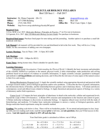
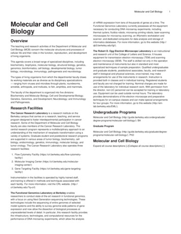
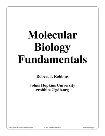
N. Bolghari et al./International Journal of Molecular and Clinical Microbiology 11(2) (2021) 1495-1508Table 1. Specifications of primersPrimerName of PrimerAOX1 Promoter3′GACTGGTTCCAATTGACAAGC5′Forward primerAOX1 Terminator5′GCAAATGGCATTCTGACATCC3′Reverse primerApplicationConfirmation ofthe presence ofgenes149912Table 2. Uses of the thermal PCR programPartTemperatureSection (ºC)TimeNumber of cyclesStart denaturationDenaturation95945 min30 Sec1Connection5530 sec30Elongation7250secEnd of lengthening727 Min13. ResultsFigure1, Structure of Gene mfp3-GvpAFigure 2. Various steps of the study from design of recombinant vectors to quantitative evaluation of expressedtarget protein
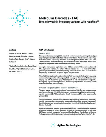
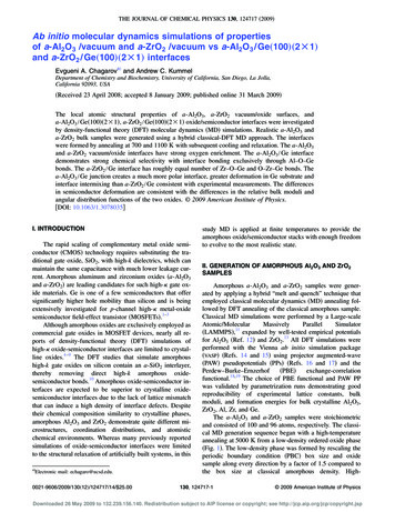
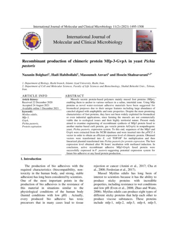
1500N. Bolghari et al./International Journal of Molecular and Clinical Microbiology 11(2) (2021) 1495-1508Figure 3. reports a gene MFP3-GvpA, His-Tag, gs linker, and antibiotic resistance sequence (Zeocin( made byBiomatics Company and placed in the pPICZA vector.Figure 4. Gene transfer to the competent cell of E. coli TOP 10/ and confirmation of the presence of the gene byPCR showing the column a of the leader used in electrophoresis, and column b showing the mfp3-GvpA gene band,observed at 839 bp.
N. Bolghari et al./International Journal of Molecular and Clinical Microbiology 11(2) (2021) 1495-15081501Figure 5. Electrophoresis after enzymatic cut two bands is observed by SAC1 enzyme, the band in the 750 bpregion represents the gene and the band seen in the 2000 bp region represents the pPICZA vectorFigure 6. Gene transfer to Pichia pastoris strain GS115 by electroporation method a) negative control includecells without target genes and culture medium containing antibiotics b) positive control in this test, 300 microlitersof Pichia pastoris cell containing target gene was used and incubated in the presence of Zeocin antibiotic for 5 daysC) positive control in this test, 280 microliters of Pichia pastoris cell containing target gene. Due to the presence ofzeocin antibiotic resistance gene, Pichia pastoris yeast cells grew in YPD culture medium D) from single colonies ina plate containing YPD culture medium containing zeocin antibiotic matrix was given
1502N. Bolghari et al./International Journal of Molecular and Clinical Microbiology 11(2) (2021) 1495-1508Figure 7. Electrophoresis after mfp3-GvpA gene transfer to Pichia pastoris; the colonies grown in the positivecontrol plate were used to evaluate the presence of the gene in the PCR test. shows DNA ladder a) mfp3-GvpA geneobserved in 1100K b) mfp3-GvpA gene negative control.Figure 8. SDS PAGE of mfp3-GvpA protein to show protein expression in Pichia pastoris mfp3-GvpA proteinwas expressed along with other Pichia pastoris proteins on different culture days. In this image, the band observedin the range of 30 kDa, which was seen in all hours of sampling from 36, 60, 72, and 96 hours, indicating thepossible presence of the mfp3-GvpA protein. Also, the presence of bands in non-specialized sites indicates theexpression of Pichia pastoris proteins.
N. Bolghari et al./International Journal of Molecular and Clinical Microbiology 11(2) (2021) 1495-15081503Figure 9. Evaluation of the presence of mfp3-GvpA protein by western blot test; the target protein band wasobserved in the western blot test with a size of about 30 kDaYeast growth chart due to the addition of e2sample3Figure10. Growth rate Yeast chart at different hours Add methanol
1504N. Bolghari et al./International Journal of Molecular and Clinical Microbiology 11(2) (2021) 1495-1508Protein production rate from ample2sample3Figure 11. The rate of protein production using residual sediment from yeast cellsProtein production rate from e1sample2sample3Figure12.The rate of protein production using supernatants given that Vector pPICZA is an Intracellularsecretion vector . The possibility of protein secretion to the out of cell is very low. This test was performed to provethat the vector is Intracellular secretion .
N. Bolghari et al./International Journal of Molecular and Clinical Microbiology 11(2) (2021) 1495-15081505Figure 13. Perform a SDS PAGE test to see a single band that proves the presence of pure protein (MFP3GVPA) at 30 KDa position, indicating the proper purification of mfp-3-GvpA protein.4. DiscussionRecombinant protein production wasperformed using mfp-5 in bacterial host cells(Sun and Waite, 2005; Wang et al., 2019). 6 μgof mfp-1 protein was purified per liter of culturemedium and used to produce cell adhesives. Theresults were better than other cell adhesives,such as cell tak (Hwang et al., 2005).Mfp-3 protein was used to make a newsilicone fiber adhesive that had a high adhesionto dry and wet surfaces but was very difficultand expensive to produce on a large scale (Lin etal., 2007). Mfp-3 protein was examined atdifferent pHs. It was used in the coating ofdental implants, but it was found that thisrecombinant protein had little resistance at thesurface of mica, and at pH 5.5. Mfp3 proteinwas produced by di-tyrosine cross-linking,which was activated despite light rays, but thisprotein had a toxic effect on cell growth (Yu etal., 2013).Bio adhesive was produced using mfp-5dimer. This adhesive had better resistance thanthe mfp-5 monomer (Jeon et al., 2015).The adhesion of most of the adhesivesproduced at low pH was investigated in thisstudy. Overnight protein treatment in thepresence of acetic acid at pH 3.5 and 5.5 had asignificant effect on protein adhesion andstrength. This study showed how cationic ionscould increase the strength of mfp-5 to thesurface of mica. In nature, mfp-6 has anantioxidant role and provides a good substratefor better adhesion than mfp-5 to the surface. Inthis study, hydronium ion was used artificiallyand had a good effect on mfp-5 adhesion (Kan etal., 2014).Cross-joints in the bioadhesive structure wereinvestigated, indicating increased adhesion andadhesive survival. Among the naturalcompounds that can be used as cross-linking arecompounds that bind to the proteins of primarybacteria and Escherichia coli. The mfp-3-CsgArecombinant protein was able to form a stable βstructure in aqueous solution so that mfp wouldbe present in the CsgA amyloid regions in thecenter. Successful expression of mussel footadhesive proteins was evaluated with CsgA.Then, by combining oyster foot proteins withEscherichia coli CsgA amyloid fiber subunit, an
N. Bolghari et al./International Journal of Molecular and Clinical Microbiology 11(2) (2021) 1495-15081506attempt was made to improve the quality andon a large scale and in bioreactors withquantity of adhesion of these proteins (Zhong etcontinues method.al., 2014).ConclusionBiological wood glue production was performedusing recombinant mfps. This adhesive was freeThe results of the study show that theof chemicals and very practical (Ferdosian et al.,recombinant adhesive protein mfp3-GvpA is a2017). Zinc and copper were used to increasevery good candidate for the production of biothe adhesion of mfp-5 and mfp-3 proteins.adhesives. It is well expressed in eukaryoticRelatively stronger polymers were produced,cells of Pichia pastoris and does not require theand the presence of the amino acid cysteine wasaddition of the enzyme tyrosine for expression.shown to improve the adhesion (Forooshani andThis protein can be very cost-effective in largeLee, 2017).scale productions.In this study, the chimeric protein mfp-3GvpA was expressed in the host cell PichiaAcknowledgementspastoris, and the amount of protein producedwas much higher than that of the bacterial host.The authors are grateful to Elmira Sheukhi,The adhesive protein produced was purified.KeyvanShahriari, and Saadi Hosseini, theBecause previous research has used Ecoliworking staff at Pasteur Institute of Iran.derived csga curli proteins as a pathogenicbacterium, this study used the Curli proteinReferecessequence, which is derived from a nonpathogenic bacterium (Archaebacterium) andAmini, S., Kolle, S., Petrone, L., Ahanotu, O.,can therefore be used, expected not to causeSunny, S., Sutanto, C. N., et al., (2017).allergies in humans.Preventing mussel adhesion usingDue to the success of the present study in thelubricant-infused materials. Science.expression and purification of mfp-3-GvpA357: 668-673. doi:10.1126/science.adhesive proteins in Pichia pastoris yeast cells,aai8977.performed for the first time in this host, it isBarclay,T. G., Hegab, H. M., Clarke, S. R.,recommended that future research focus on theseGinic‐Markovic,M. (2017). Versatilemethods to produce protein in larger and highersurfacemodificationusing olydopaminevolumes. Because absorption chromatographyandrelatedpolycatecholamineswas used in this study to purify proteins, it isChemistry,structure,and applications.possible that some proteins will still adhere toAdvancedMaterialsInterfaces.4(19):nickel particles as they leave the column.160-197.Therefore, it is suggested that other purificationCha, H. J., Hwang, D. S., Lim, S. (2008).methods such as ion exchange or HPLC be usedDevelopment of bioadhesives fromto achieve higher concentrations of recombinantmarine mussels. Biotechnol J. 3(5): 631protein. The production of recombinant proteins638. doi:10.1002/biot.200700258mfp3-GvpA in eukaryotic cells such as CHO orCregg,J.M., Cereghino, J. L., Shi, J., Higgins,insect cells is also suggested. To evaluate theD.R. (2000). Recombinant proteinadhesion of these proteins, the use of livingexpressionin Pichia pastoris. Moleculartissues such as cell culture or scratches in miceBiotechnology.16(1): 23-54.or rats is also recommended. It is alsoDadgar,P.F.,Keramatipour,M., Nourbakhsh,recommended to get more protein Instead of aF., Talebi, S., Vodjgani, M. (2015).vector pPICZA use another vector that has aInvestigating the effect of rs3783605sequence of protein secretion signals out of thesingle-nucleotide polymorphism on thecell. Or use the sequence of secretory signals inactivity of VCAM-1 promoter in humanyour gene sequence, because the secretion ofumbilical vein endohelial cells. Iran Jprotein into the culture medium increases theAllergy Asthma Immunol. 14(2): 179lifespan of yeast and thus increases the87.production of the desired protein. Perhaps withthis method, the desired protein can be produced
N. Bolghari et al./International Journal of Molecular and Clinical Microbiology 11(2) (2021) 1495-1508Damasceno, L. M., Huang, C., Jr., Batt, C. A.,(2012). Protein secretion in Pichiapastoris and advances in proteinproduction. Applied Microbiology 1-3654-zIsraelachvili, J. N., Waite, J. H., (2012).Adhesion of mussel foot protein Mefp-5to mica: an underwater 1/bi3002538dos Santos, F. P., Rodrigues, K. B., de Santana,M., Salum, T., Favaro, L. d. L, (2016).BRASIL, B., Expressão heteróloga demonoxigenasesdepolissacarídeosbacterianas em Pichia pastoris. Paperpresented at the Embrapa AgroenergiaArtigo em anais de congresso (ALICE).Even, M. A., Wang, J., Chen, Z. (2008).Structural Information of MusselAdhesive Protein Mefp-3 Acquired atVariousPolymer/Mefp-3SolutionInterfaces. Langmuir : the ACS journalof surfaces and colloids. 24(11): 57955801. doi:10.1021/la800138xEven, M. A., Wang, J., Chen, Z, (2008).Structural information of musseladhesive protein Mefp-3 acquired atvariouspolymer/Mefp-3solutioninterfaces. Langmuir the ACS journalof surfaces and colloids. 24(11): 57955801. doi:10.1021/la800138xEzzeldin, H. M., Klauda, J. B., Solares, S. D.,(2012). Modeling of the major gasvesicle protein, GvpA: from proteinsequence to vesicle wall structure.Journal of structural biology. 179(1):18-28. doi:10.1016/j.jsb.2012.04.015Ferdosian, F., Pan, Z., Gao, G., Zhao, B.,(2017).Bio-Based Adhesives and Evaluation forWoodCompositesApplication.Polymers (Basel). 9(2): 100-123.doi:10.3390/polym9020070Froger, A., Hall, J E. (2007).Transformation ofplasmid DNA into E. coli using the heatshock. JoVE (Journal of VisualizedExperiments. 6: 222-253.Gøgsig, T. M., Kleimark, J., Nilsson Lill, S. O.,Korsager, S., Lindhardt, A. T., Norrby,P.-O., 2012. Mild and efficient nickelcatalyzed Heck reactions with electronrich olefins. Journal of the AmericanChemical Society. 134(1): 443-4521507Higgins, D. R., Cregg, J. M., (1998). Pichiaprotocols (Vol. 103): Springer.Hosseini, S., Zolgharnine, H., Bargahi, A.,Khaledi, H., Salamat, N., Archangi, B.,(2017). Purification of Bysal Mussel(Modiolus sp. PG) Adhesive Protein fp2 from Nortern Seashore of PersianGulf.BPUMS,20(5):481-491.Retrieved from , D. S., Gim, Y., Cha, H. J., (2005).Expression of functional recombinantmussel adhesive protein type 3A inEscherichiacoli.BiotechnologyProgress. 21(3): 965-970.Hwang, D. S., Zeng, H., Lu, Q., Israelachvili, J.,Waite, J. H., (2012). Adhesionmechanism in a DOPA-deficient footprotein from green mussels. Soft Matter.8(20):5640-5648.doi:10.1039/C2SM25173FA., Shah, S. H., Ibrahim, M. I., Ilyas, M.,(2016). Optimization of an efficientSDS-PAGE protocol for rapid proteinanalysis of Brassica rapa. J. Bio. Env.Sci. 9(2): 17-24.Jeon, E. Y., Hwang, B. H., Yang, Y. J., Kim, B.J., Choi, B.-H., Jung, Cha, H. J. (2007).Rapidly light-activated surgical proteinglue inspired by mussel adhesion andinsectstructuralcrosslinking.Biomaterials. 67: 11-19.Kan, Y., Danner, E. W., Israelachvili, J. N.,Chen, Y., Waite, J. H., (2014). BoronateComplexFormationwithDopaContaining Mussel Adhesive ProteinRetards pH-Induced Oxidation andEnables Adhesion to Mica. PLOS ONE.9(10): 108-869.Kan, Y., Danner, E. W., Israelachvili, J. N.,Chen, Y., Waite, J. H. (2014). BoronatecomplexformationwithDopacontaining mussel adhesive proteinretards pH-induced oxidation andenables adhesion to mica. PLOS ONE.9: 146-177.Kord Forooshani, P., Lee, B. P., (2017).bioadhesivematerialsRecentapproaches in designing inspir
the sequence was ordered to Biomatik company in Canada, and the company synthesized it and put it inside the pPICZA vector. 2.2. Transformation of Gene in to the E. coli TOP10F ′ cell The vector was transferred to the competent cell of E. coli TOP10F′ (TOP10F is a type of E. coli bacteria that is very useful for gene transfer).